Special Projections of the Elbow
1/22
There's no tags or description
Looks like no tags are added yet.
Name | Mastery | Learn | Test | Matching | Spaced |
|---|
No study sessions yet.
23 Terms
AP Partial Flexion
these 2 elbow images are in what position (2 for 1!!)
Humerus
in this AP Partial Flexion is the humerus or forearm parallel
Forearm
in this AP Partial Flexion is the humerus or forearm parallel
acute flexion
these 2 images of the elbow are in what position (2 for 1!) CR perpendicular to humerus and then CR perpendicular to forearm
distal humerus
this acute flexion of the elbow image is showing the __ __
proximal forearm
this acute flexion of the elbow image is showing the __ __
AP Partial Flexion, Acute Flexion
what 2 position (2 images each) compromises for an AP elbow radiograph
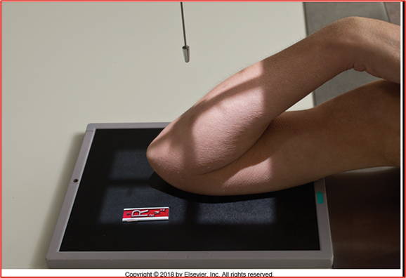
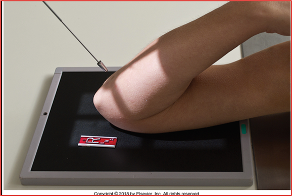
Coyle Method
what position compromises for medial or lateral oblique images of the elbow
radial head
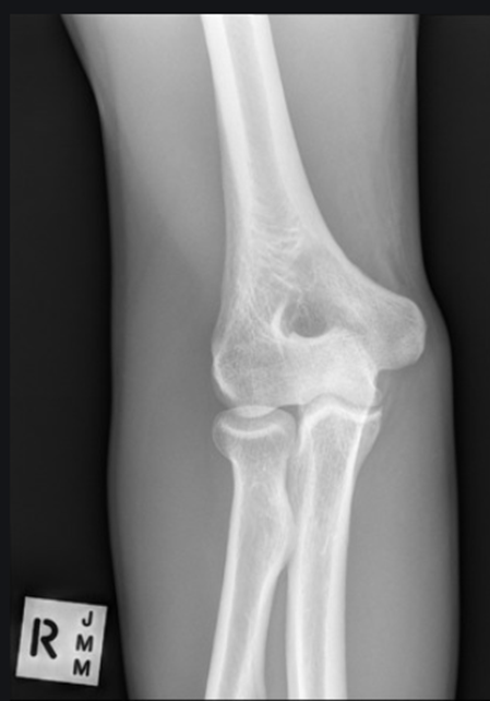
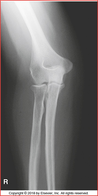
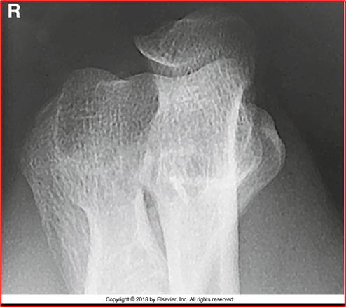
which coyle method is this image showing (toward shoulder)
coronoid process
which coyle method is this image showing (away from shoulder)
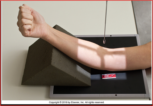
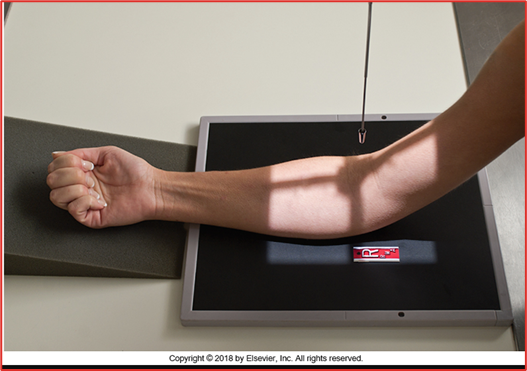
90
for the radial head coyle method the elbow needs to be flexed at __ degrees
80
for the coronoid process coyle method the elbow needs to be flexed at __ degrees
45
for any coyle method the CR needs to be angles at __ degrees (either away from or towards the shoulder)
radial head
this is an image of what coyle method projection
coronoid process
this is an image of what coyle method projection
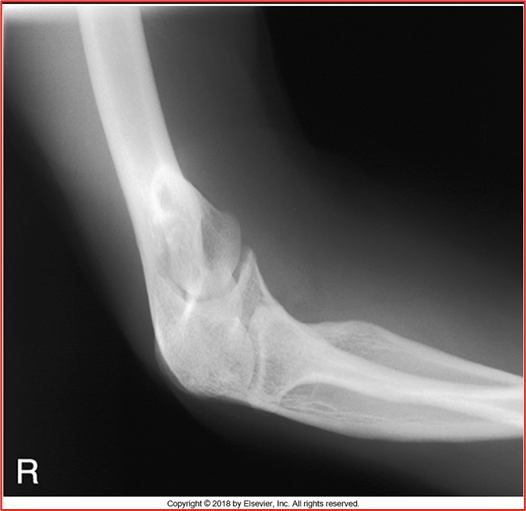
lateral oblique
this image is what projection of the elbow
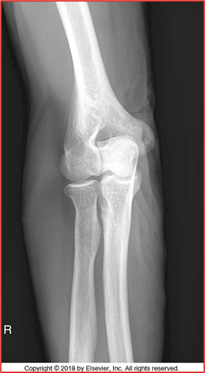
medial oblique
this image is what projection of the elbow
radial head
which coyle method projection compromises for a lateral oblique
coronoid process
which coyle method projection compromises for a medial oblique
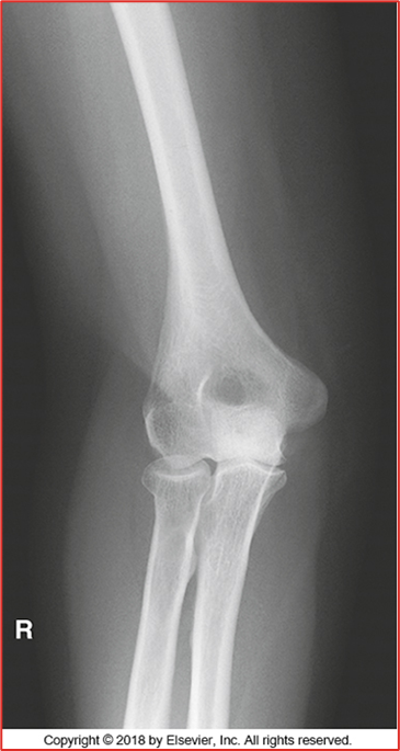
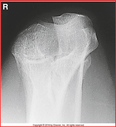
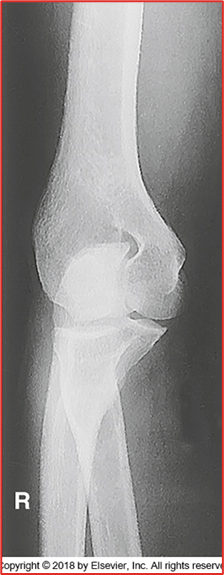
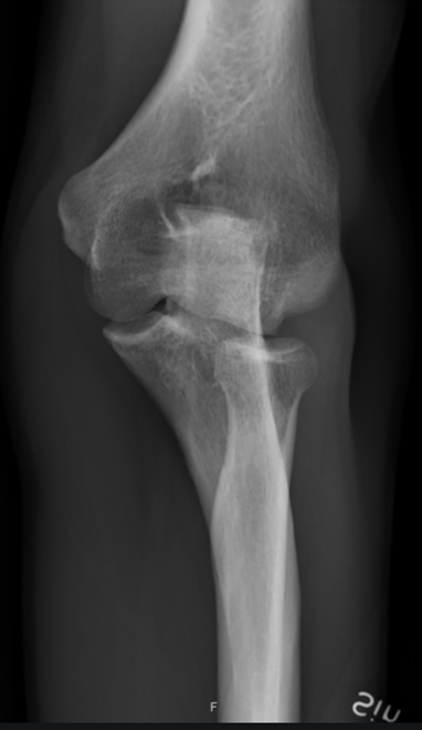
-All anatomy present (humerus, radius, and ulna)
-Only should be partial superimposition of the radius and ulna (this image they are fully separated)
-They are laterally rotated (fix by medially rotating them and feeling the epicondyles)
-Epicondyles not perfectly in profile (due to rotation)
-Olecranon in olecranon fossa
-Open Joint Space = pretty good
-No crowning of radial head (good)
REPEAT
Critique this AP Elbow Image
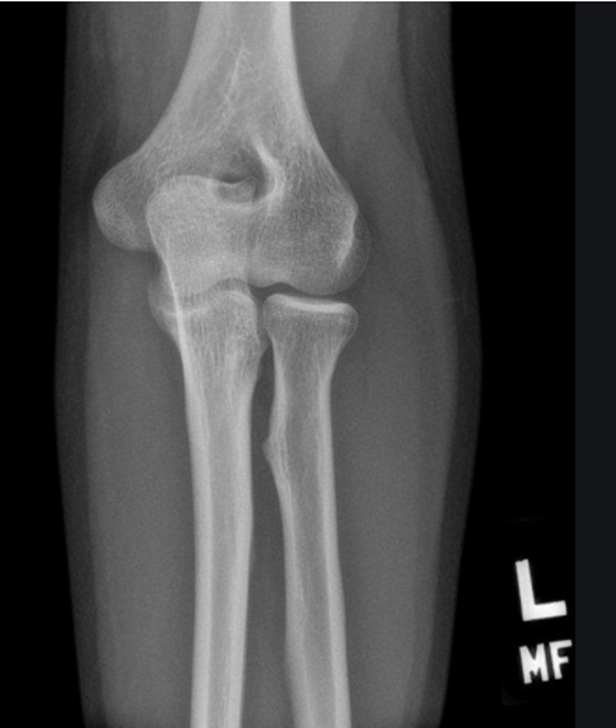
-All anatomy present
-Collimation is tight, want to open up some more
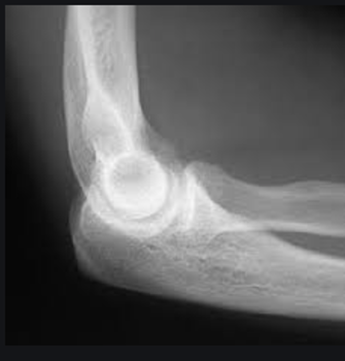
-Totally body positioning was not utilized because you cannot see the 3 concentric arches (trochlear notch, trochlear sulcus, ridges of the capitulum and trochlea)
-Not fully bent 90 degrees but close
-Coronoid process and radial head should be half superimposed (this image is a little more than half)
-Olecranon is in profile
-No marker
-REPEAT cause not in a true lateral position
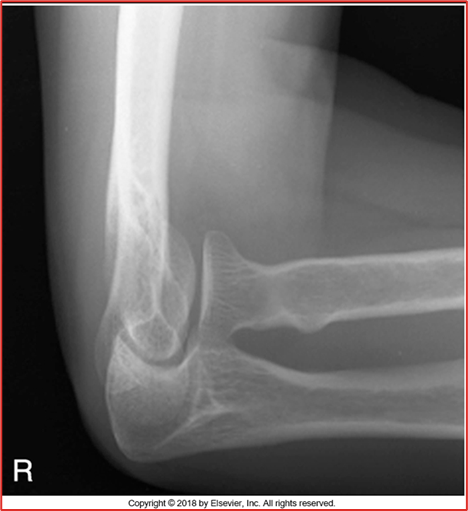
Critique this lateral elbow image
-Not complete superimposition of radial head and ulna (NOT enough rotation) need more medial rotation
-Should see the coronoid process and trochlea in profile
-Olecranon should be in olecranon fossa
-NO Marker
-REPEAT
critique this medial oblique elbow image
-Radius and ulna should be completely separated (almost there but not fully)
-There is crowning of the radial head (should NOT be present, should be flat) meaning they are not fully extended
-Capitulum and radial head present
-REPEAT because of crowning
critique this lateral oblique elbow image