lecture 19 + 20 - therapeutic approaches to reversing sensory loss:
 Call Kai
 Learn
Learn Practice Test
 Spaced Repetition
 Match
Match Flashcards
 Knowt Play
1/20

There's no tags or description
Looks like no tags are added yet.
Name | Mastery | Learn | Test | Matching | Spaced | Call with Kai | Chat |
|---|
No analytics yet
Send a link to your students to track their progress
21 Terms
Why is the eye a good target for cellular therapy, and what is the role of the RPE?
The eye is accessible and visible, allowing cells to be delivered precisely to the retina and monitored.
It is immune privileged because of the blood-retina barrier.
It is enclosed, limiting the spread of complications, and one eye can potentially act as a control for the other.
The macula provides central, high-acuity vision; its centre, the fovea, has a very high density of cones.
Retinal pigment epithelial (RPE) cells form a monolayer beneath photoreceptors and are essential for their survival.
RPE functions include nutrient/ion/water transport, absorbing stray light, recycling retinal, phagocytosing photoreceptor debris, and secreting signalling factors.
RPE also contributes to the blood-retina barrier.
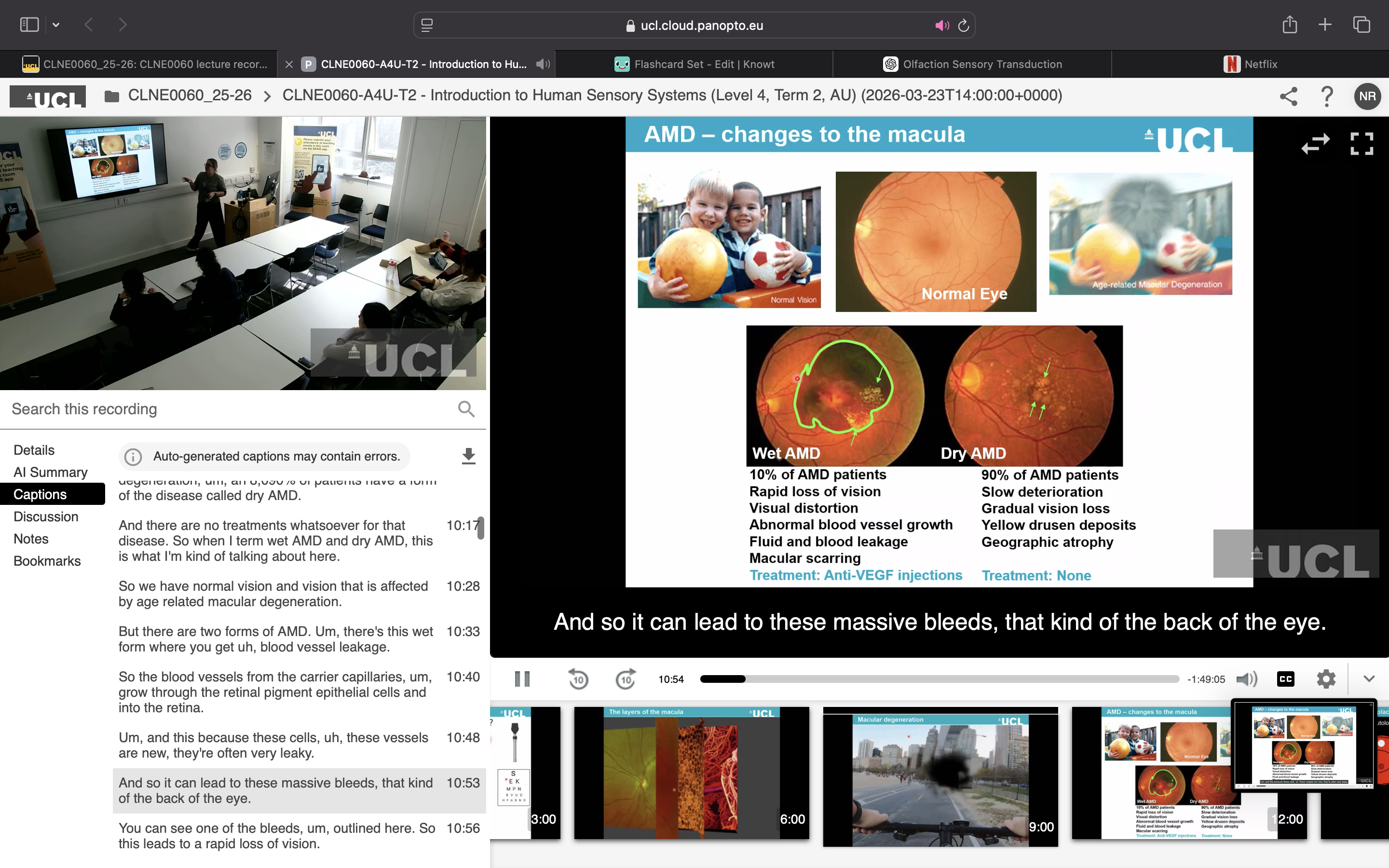
What is age-related macular degeneration (AMD), and what happens in dry vs wet AMD?
AMD causes progressive loss of central vision, affecting activities such as reading, recognising faces and driving; distorted/wavy lines on an Amsler grid can be an early sign.
Dry AMD (~90%): accumulation of drusen (fat/protein deposits) → RPE degeneration → photoreceptor death → gradual geographic atrophy and vision loss. No established treatment was discussed for this form.
Wet AMD (~10%): abnormal blood vessels grow from the choriocapillaries through the RPE (neovascularisation) → leakage/bleeding → scarring and rapid vision loss. Anti-VEGF injections prevent further abnormal vessel growth but do not replace the damaged RPE.
In both forms, RPE degeneration ultimately causes photoreceptor loss.
What evidence showed that replacing RPE cells could treat AMD?
Experimental surgeries showed that RPE replacement can restore vision.
Macular translocation: the retina is physically moved so the macula sits on healthier RPE → about a 5-line improvement in vision in some patients.
Autologous RPE transplantation: healthy RPE from peripheral retina is transplanted beneath the macula → also produced about a 5-line visual improvement, with one patient regaining their driving licence.
However, these surgeries are long, complex and high-risk, and mainly suitable for late-stage disease.
How were functional RPE cells produced from human embryonic stem cells, and how was their function tested?
Human embryonic stem cells are self-renewing and pluripotent, meaning they can be expanded and differentiated into different cell types.
Cells from the inner cell mass of a blastocyst were expanded in culture; removing factors that maintain pluripotency allowed them to differentiate.
RPE cells were identified by their pigmentation, isolated and grown into a cobblestone-like monolayer resembling normal human RPE.
Their identity was confirmed using gene expression (PCR), protein markers and morphology.
Function was tested by showing that the RPE could phagocytose photoreceptor debris.
How was the RPE therapy delivered as a transplantable patch, and what did animal studies show?
Injecting RPE cells alone is problematic because they form a disorganised, immature and non-polarised mass rather than the normal monolayer.
A porous polyester track etched membrane was therefore used as a scaffold, allowing RPE cells to form a mature, polarised monolayer while permitting transport of nutrients, ions and water.
The surgeon creates an opening, lifts the retina with saline, and inserts the RPE-coated patch beneath the macula, where it is held in place after the saline is removed.
RPE on the patch maintained the photoreceptor layer, whereas the patch alone caused photoreceptor loss because it separated the photoreceptors from their RPE.
Thus, the experiments showed that the RPE cells—not simply the patch—are essential for maintaining photoreceptors.
What steps were needed before the RPE patch could be tested in humans?
Safety testing: confirmed the differentiated RPE cells did not form teratomas; undifferentiated embryonic stem cells did.
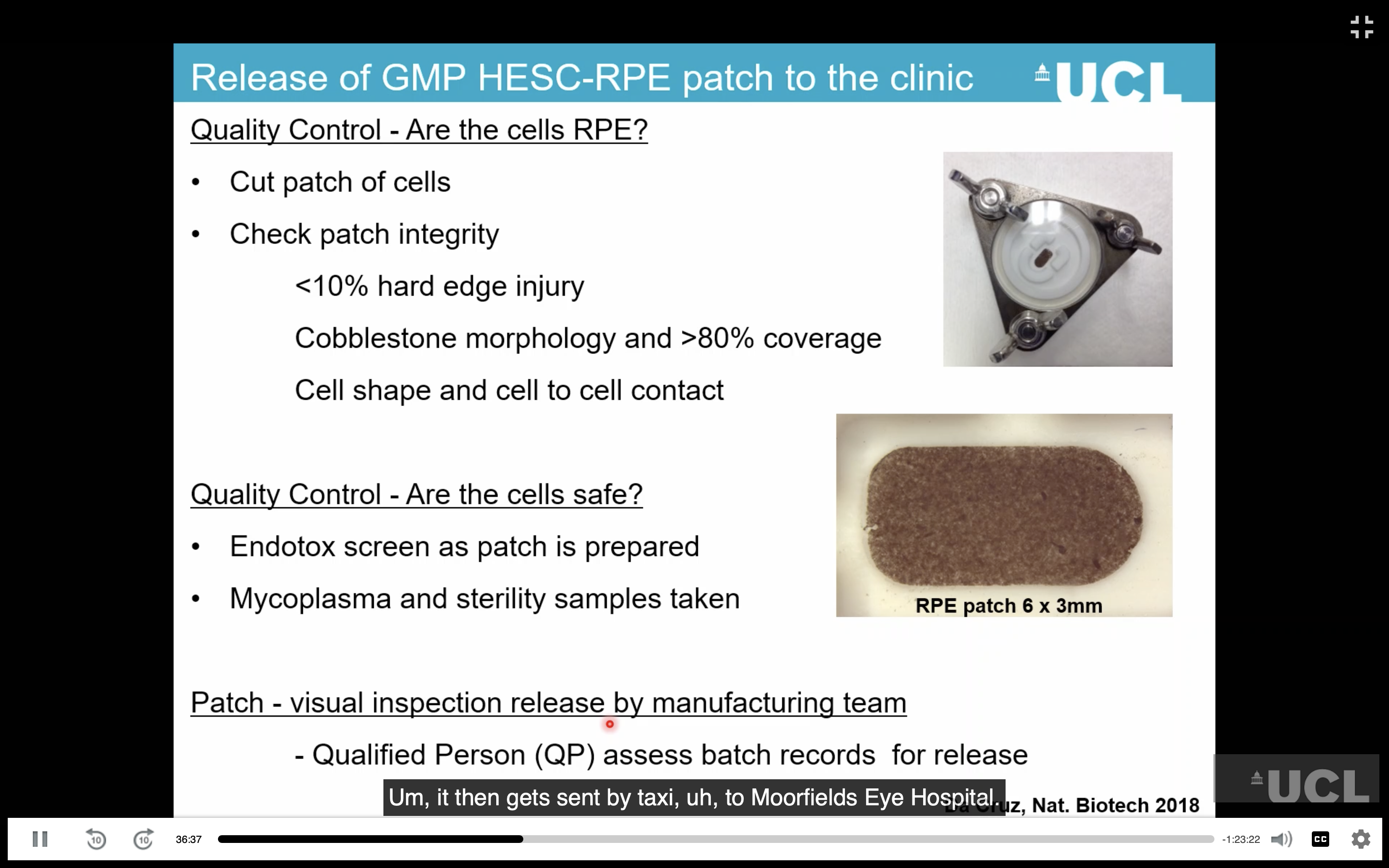
GMP manufacturing: cells were differentiated, expanded and grown on the membrane under Good Manufacturing Practice (GMP) conditions.
The final patch was only 3 × 6 mm, enough to cover the macula/foveal region.
A specialised surgical delivery tool was developed to roll the patch up, place it under the retina and safely release it.
MHRA and ethics approval were required before clinical trials; approval took about 4 years.
Why were patients with wet AMD chosen for the first clinical trial?
Wet AMD causes a rapid loss of vision, but photoreceptors can still be alive shortly after a macular haemorrhage.
The patch could therefore replace damaged RPE and preserve the surviving photoreceptors.
Surgery needed to occur within about 6 weeks for the best chance of saving vision.
What were the clinical outcomes of the RPE patch?
Expected clinically significant improvement = 3-line increase in visual acuity.
Both initial patients achieved a 6-line improvement.
Reading improved:
Patient 1: 5 → 51 → 84 → 69 words/min at 0, 6 months, 1 year and 2 years.
Patient 2: 0 → 26 → 47 → 41 words/min.
OCT/autofluorescence/microperimetry showed the RPE survived, functioned and supported photoreceptors.
No serious adverse reactions or teratomas; complications were mainly surgical.
Vision improved/stabilised over 6 months–2 years, possibly as cells integrated and matured.
What are the limitations and future of the therapy?
Some deterioration occurred after ~5 years, possibly due to ongoing AMD, immune response or RPE migration.
The eye is immune privileged, but long-term immune effects involving microglia remain uncertain.
One patch costs about £4,000 in materials, although overall treatment costs would be higher.
The therapy targets the underlying RPE loss/dysfunction rather than just AMD symptoms.
The therapy has been developed further by Tenpoint Therapeutics.
Next step: larger multicentre clinical trials.
Overall: RPE patches were shown to survive, integrate, support photoreceptors and improve vision.
How are inherited retinal diseases (IRDs) clinically classified?
Progressive: worsen over time;
stationary: do not progress (e.g. congenital stationary night blindness).
Progressive IRDs can be non-syndromic (retina only) or syndromic (retinal disease + other body systems, e.g. Bardet-Biedl, Usher syndrome).
Non-syndromic types include:
Macular dystrophy: affects the macula.
Rod-cone dystrophy: rods affected first → night blindness and tunnel vision.
Cone-rod dystrophy: cones affected first → central visual blurring, later spreading.
Choroideremia: affects the retina and other structures such as the choroid.
IRDs show genetic heterogeneity (different genes can cause the same disease) and phenotypic heterogeneity(different diseases can result from variants in the same gene).
They can be autosomal recessive, autosomal dominant or X-linked.
How do different inheritance patterns affect the mechanism of IRDs?
Autosomal recessive: two faulty copies of the gene→ usually loss of function.
Autosomal dominant: one faulty copy of the gene; mechanisms include haploinsufficiency, toxic gain-of-function and dominant-negative effects.
X-linked: can be recessive or dominant; inheritance differs between males and females because males have only one X chromosome.
Understanding the specific mutation and inheritance is important because it determines which therapy can be used.
How does phototransduction convert light into an electrical signal?
Light causes 11-cis retinal → all-trans retinal, activating rhodopsin.
Activated rhodopsin activates the G-protein transducin → activates cGMP phosphodiesterase.
Phosphodiesterase converts cGMP → GMP, causing cGMP-gated cation channels to close.
Ca²⁺/other cation influx decreases → the photoreceptor hyperpolarises.
This converts light into an electrical signal transmitted to bipolar cells through ribbon synapses.
Photoreceptors continuously release neurotransmitter in darkness; light reduces neurotransmitter release.
Their response is graded rather than an action potential, allowing them to encode small changes in light intensity.
Why is the eye a good target for gene therapy, and what therapies can be used?
The eye is anatomically accessible and compartmentalised, allowing direct delivery and relatively low therapeutic doses.
The blood-retinal barrier makes the eye relatively immune privileged, reducing systemic immune responses.
Non-invasive imaging such as fundus imaging and OCT allows treatment and disease progression to be monitored.
Therapeutic approaches include:
Pharmacotherapy: drugs/small molecules; can be gene- or mutation-specific/agnostic.
Neuroprotection: protects surviving cells through stress management, proteostasis and microglial maintenance.
Optogenetics: introduces light-sensitive genes/optogenes into surviving retinal cells; limited by sensitivity, wavelength and response speed.
Retinal prostheses: electrically stimulate surviving ganglion cells; currently produce visual sensations rather than clear vision.
Stem-cell therapy: replaces lost retinal cells.
Gene therapy: replaces, supplements or modifies defective genes. (RNA or DNA targeted therapies)
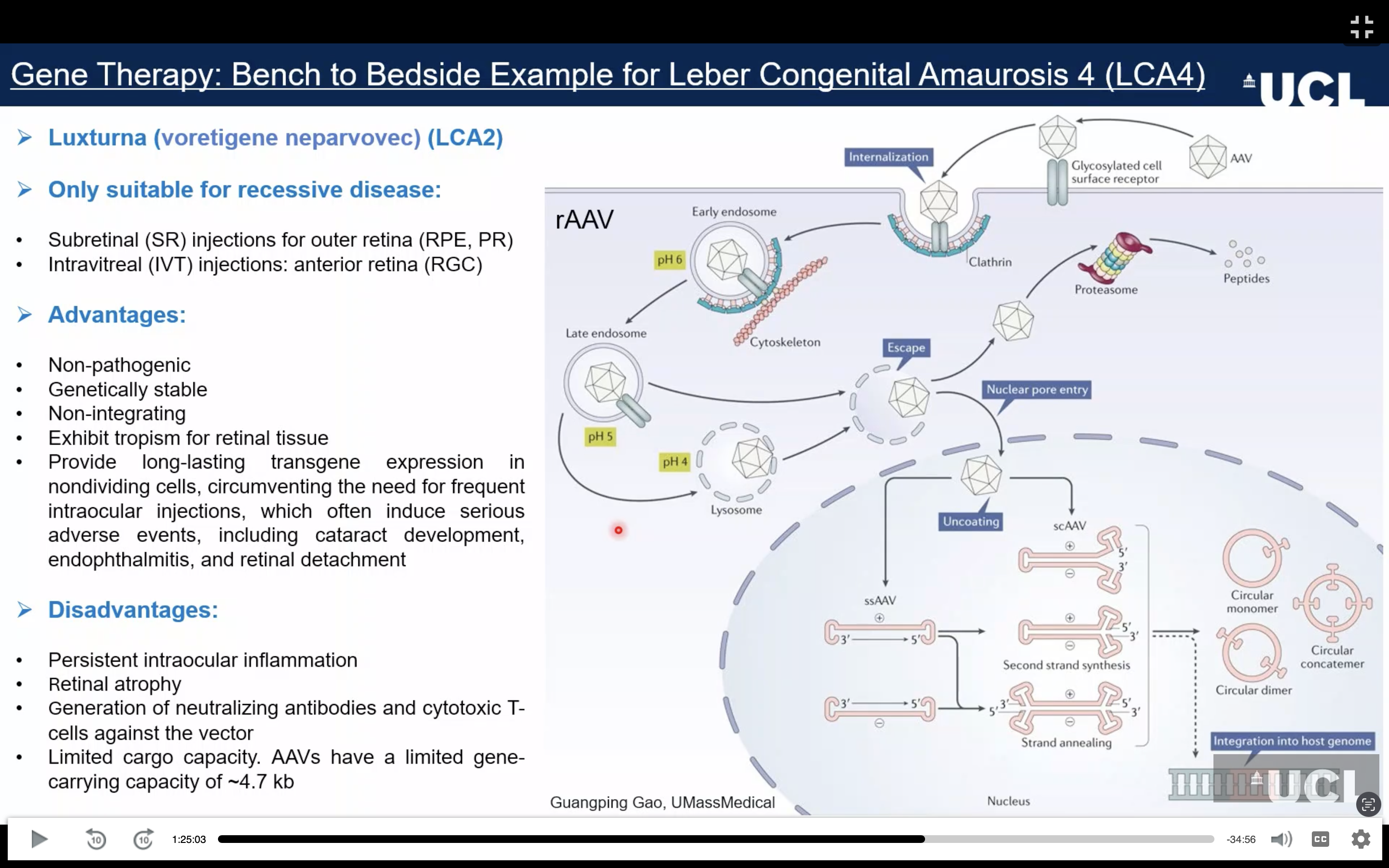
How does AAV gene replacement therapy work, and why is it mainly used for recessive IRDs? +advantages and disadvantages?
Autosomal recessive diseases usually involve loss of function, so supplying a functional gene can restore the missing function.
AAV (adeno-associated virus) is an inactivated viral vector carrying the therapeutic transgene.
AAV enters retinal cells → reaches the nucleus → forms a long-lasting DNA structure that expresses the therapeutic gene.
Self-complementary AAV (scAAV) allows faster gene expression by bypassing the initial conversion to double-stranded DNA.
AAV capsids can be engineered to target RPE cells or photoreceptors and provide long-lasting expression in non-dividing cells.
Example: Luxturna replaces defective RPE65 in LCA2; RPE65 is an enzyme in the visual cycle and is expressed in the RPE. It is delivered by subretinal injection.
What is IPL1-associated LCA and why is early treatment important?
Leber congenital amaurosis (LCA) caused by autosomal recessive loss-of-function variants in IPL1, a photoreceptor-specific gene.
Congenital/very early onset: severe visual impairment or complete blindness from birth.
Children may show roving eye movements and inability to fixate.
There is no electrophysiological retinal response from the earliest measurable age.
Therapeutic window is approximately 1–5 years; after ~5 years, photoreceptor degeneration is too advanced for gene therapy because sufficient surviving cells are needed.
What does IPL1 do, and how does its loss cause photoreceptor degeneration?
IPL1 is located from the connecting cilium to the photoreceptor synapse, including rod/cone inner segments and outer nuclear layer.
It is not present in rod outer segments, where phototransduction occurs.
IPL1 is required for proper assembly of the phosphodiesterase (PDE) complex.
Loss of IPL1 → PDE complex cannot assemble → PDE subunits are degraded.
Without PDE → cyclic GMP cannot be hydrolysed → cyclic GMP-gated channels remain open → continuous Ca²⁺ influx.
This resembles constant exposure to light, causing toxic, rapid degeneration of rods and cones.
How are patient-derived retinal organoids (ROs) made, and what are their features?
Renal epithelial cells from urine → reprogrammed into iPSCs(induced pluripotent stem cells) → differentiated into 3D retinal organoids using different media.
iPSCs form neuro-retinal vesicles, which are isolated and cultured to develop into organoids.
ROs follow the spatial and temporal development of the human neuro-sensory retina.
They form:
Outer nuclear layer: rods + cones
Inner nuclear layer: bipolar, horizontal + amacrine cells
Ganglion cell layer
Photoreceptor inner/outer segments + connecting cilium
Limitations: develop inside-out, lack a normal RPE monolayer, causing less organised photoreceptor outer segments; ganglion cells also become sparse with maturity.
Despite this, ROs provide a human, patient-specific model for studying disease and testing therapies.
How was CRISPR used in IPL1 disease modelling and gene correction?
CRISPR-Cas9 uses guide RNA to target a specific DNA sequence and Cas9 cuts the DNA.
NHEJ repairs the break but can create indels → useful for knocking out IPL1 and modelling the disease.
IPL1 knockout reproduced the patient phenotype: no IPL1 → loss of PDE6 → increased cGMP.
HDR can introduce or correct specific mutations, but is inefficient and does not work well in post-mitotic photoreceptors.
CRISPR can also create isogenic controls: genetically identical cells differing only in the mutation, making it easier to prove that the mutation causes the disease.
Correcting the IPL1 mutation restored IPL1/PDE6 and reduced cGMP.
How was IPL1 gene therapy developed and what did it achieve?
Because IPL1 is <4.7 kb, it can fit inside an AAV vector.
AAV delivered the IPL1 coding sequence under a rhodopsin kinase promoter, so IPL1 was produced specifically in photoreceptors.
In patient organoids, treatment restored IPL1 and PDE6 and reduced the abnormal cGMP accumulation.
The same approach was then given to children 1–2.8 years old in one eye.
After ~3.5 years:
Treated-eye logMAR improved from ~2.7 → ~0.9 = major visual improvement.
Visually evoked brain responses also improved.
Untreated eyes showed no improvement and deteriorated.
Other potential treatments include antisense oligonucleotides (destroy mutant mRNA or correct abnormal splicing) and base/prime editing.
These can be highly precise, but challenges include delivery, low efficiency, technical complexity, off-target effects, ethical concerns and very high cost.
How can antisense oligonucleotides (ASOs) treat genetic diseases?
RNA-targeted therapies can treat both autosomal recessive and dominant diseases.
Some ASOs bind mRNA → form a DNA–RNA hybrid → RNase H1 degrades the mRNA.
Useful for dominant diseases where the harmful allele needs to be removed.
Other ASOs bind a cryptic splice site and act as a steric block, preventing abnormal splicing.
Can restore normal splicing when an intronic variant causes an exon to be incorrectly included/excluded.
What are genome-editing therapies and what are the advantages and limitations of precise editing?
Non-homologous end joining (NHEJ): creates double-strand breaks to remove problematic regions of DNA.
Base editing: makes precise single-base changes without a double-strand break.
Can convert A → G or C → T.
Prime editing: allows precise correction of a wider range of variants.
Advantages: highly precise, versatile, potentially treats many variants and has fewer indels/off-target effects than double-strand-break approaches
Limitations: difficult delivery, low efficiency, technically complex, incomplete editing, off-target effects, ethical concerns and very high cost/accessibility.