combined 1 & 2 lab final
1/85
There's no tags or description
Looks like no tags are added yet.
Name | Mastery | Learn | Test | Matching | Spaced | Call with Kai |
|---|
No analytics yet
Send a link to your students to track their progress
86 Terms
Where is the Myelencephalon?
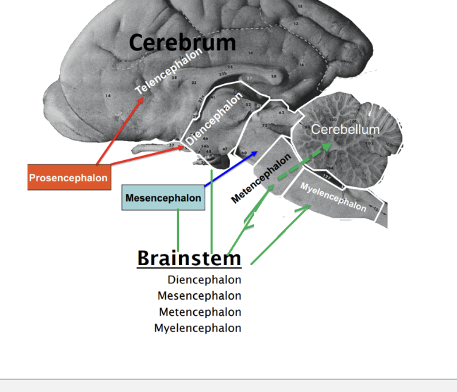
Where is the Metencephalon (pons)?

where is the Mesencephalon?

where is the Diencephalon?

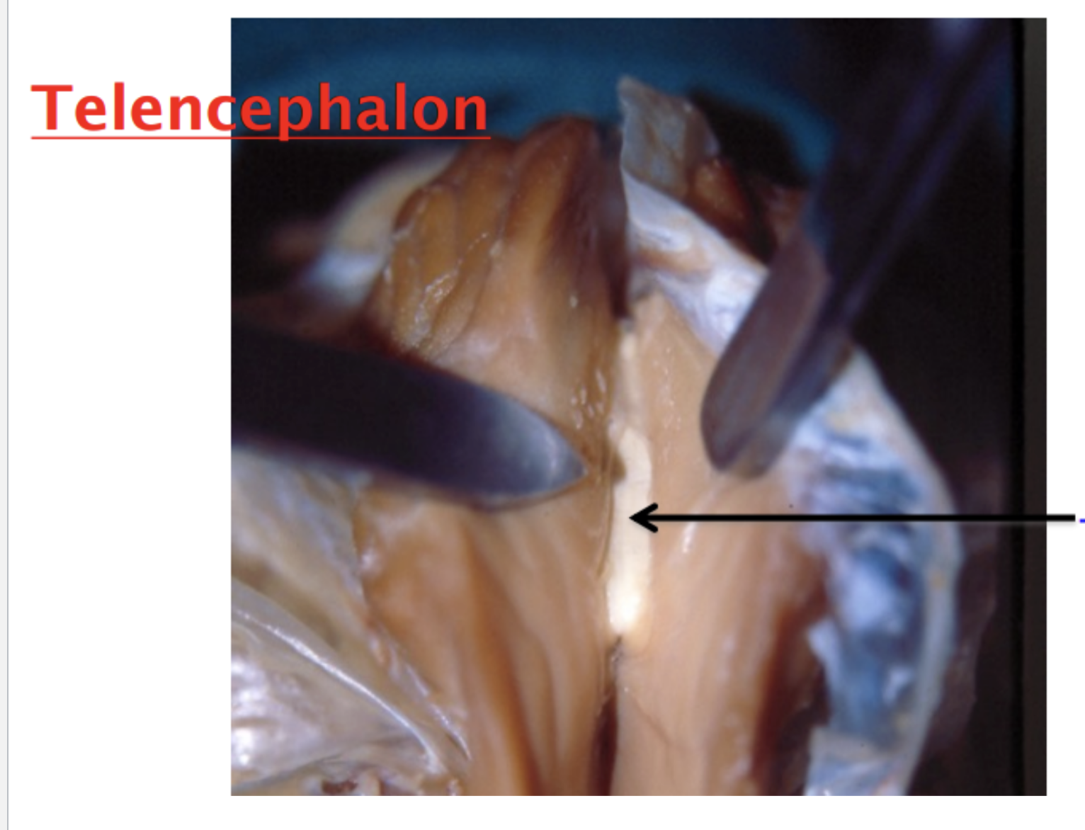
Where is the Telencephalon

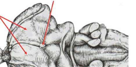
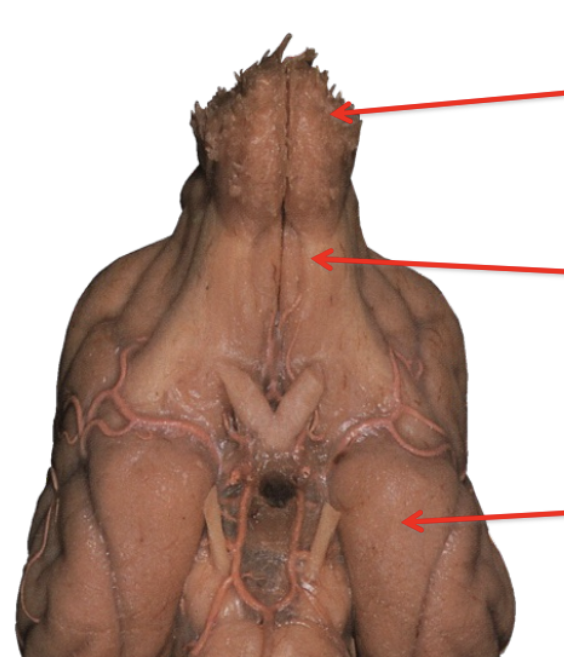
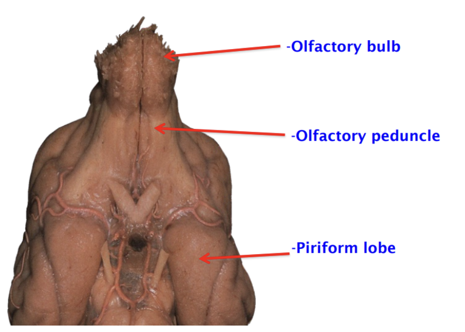
What is being shown in the picture green arrows
Rhomboid fossa
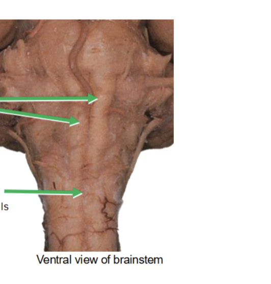
what does the top two green arrows indicate
Pyramids (descending motor fiber)

what is the bottom arrow and what is its important
Decussation of pyramids:
The crossover of descending motor fibers is considered the boundary between the medulla oblongata and the spinal cord. Explains why the left and right side of the brain control different functions

what is the bottom arrow pointing to
Arbor vitae
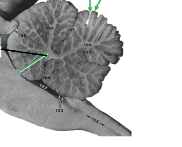
what is the right top arrow pointing to
Folias
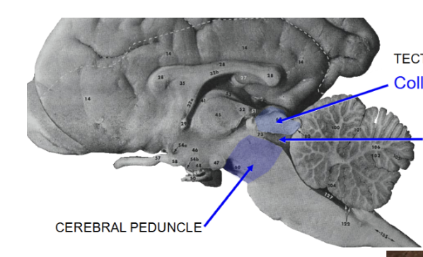
what is the bottom left arrow
Cerebral peduncle
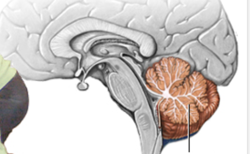
what is this part of the brain
cerebellum
What is this general structure
tectum
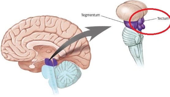

What are the top arrows
Rostral colliculi

What are the bottom arrows
caudal colliculi
what is the arrow in the middle pointing to?
Mesencephalic aqueduct
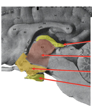
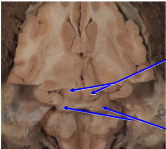
what are these 4 arrows
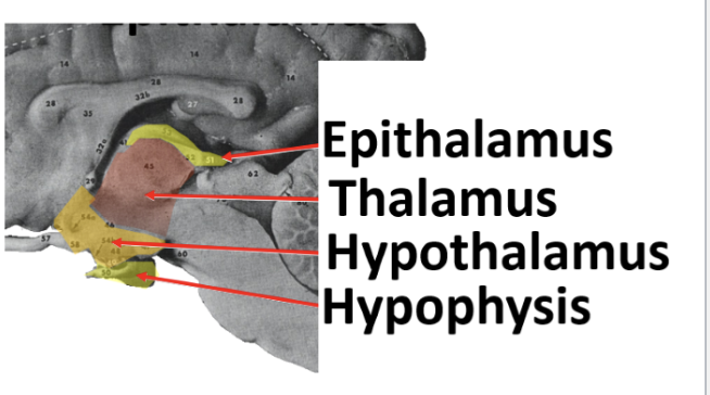
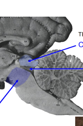
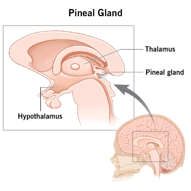
what is the arrow coming from the top pointing to
Pineal gland
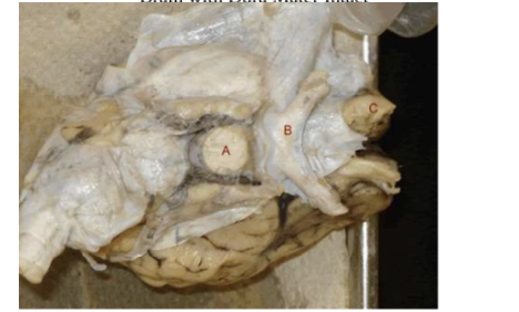
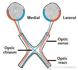
what is B ?
Optic chiasm
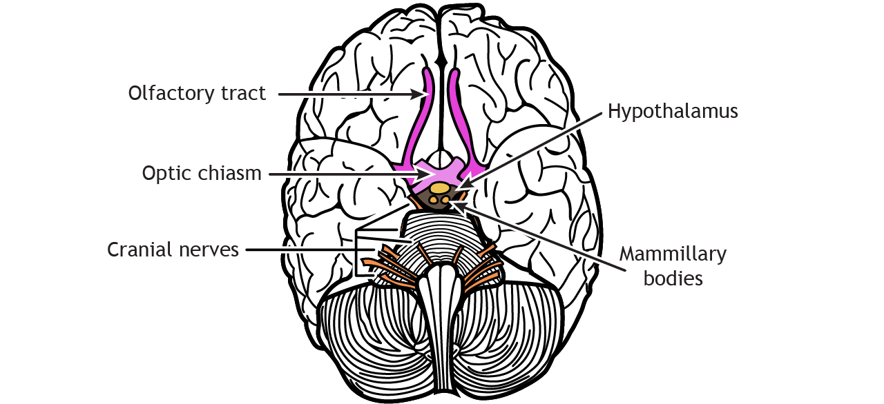
what are the top two arrows pointing to?
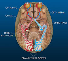
optic tract know where this is
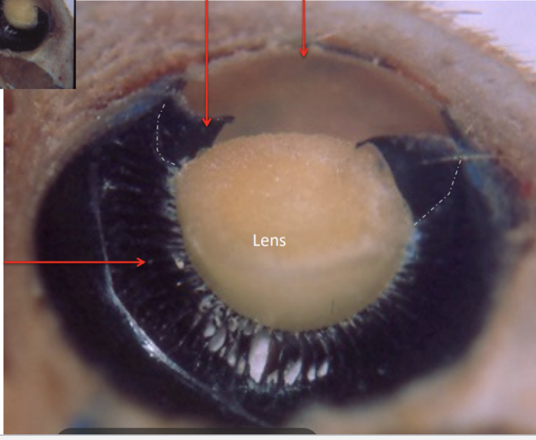
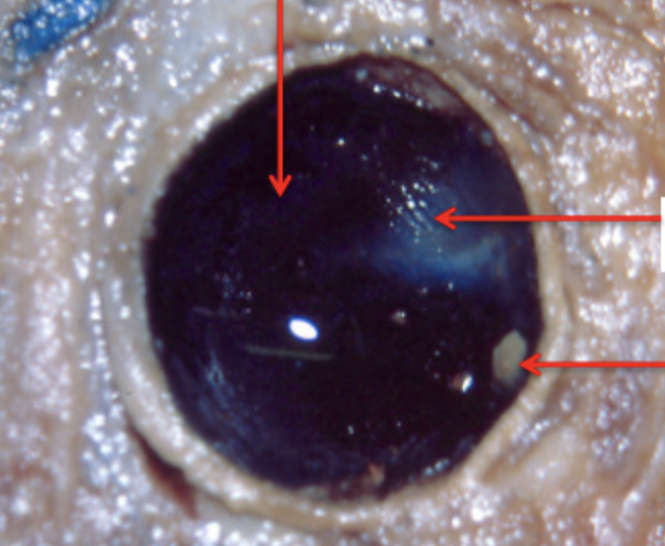
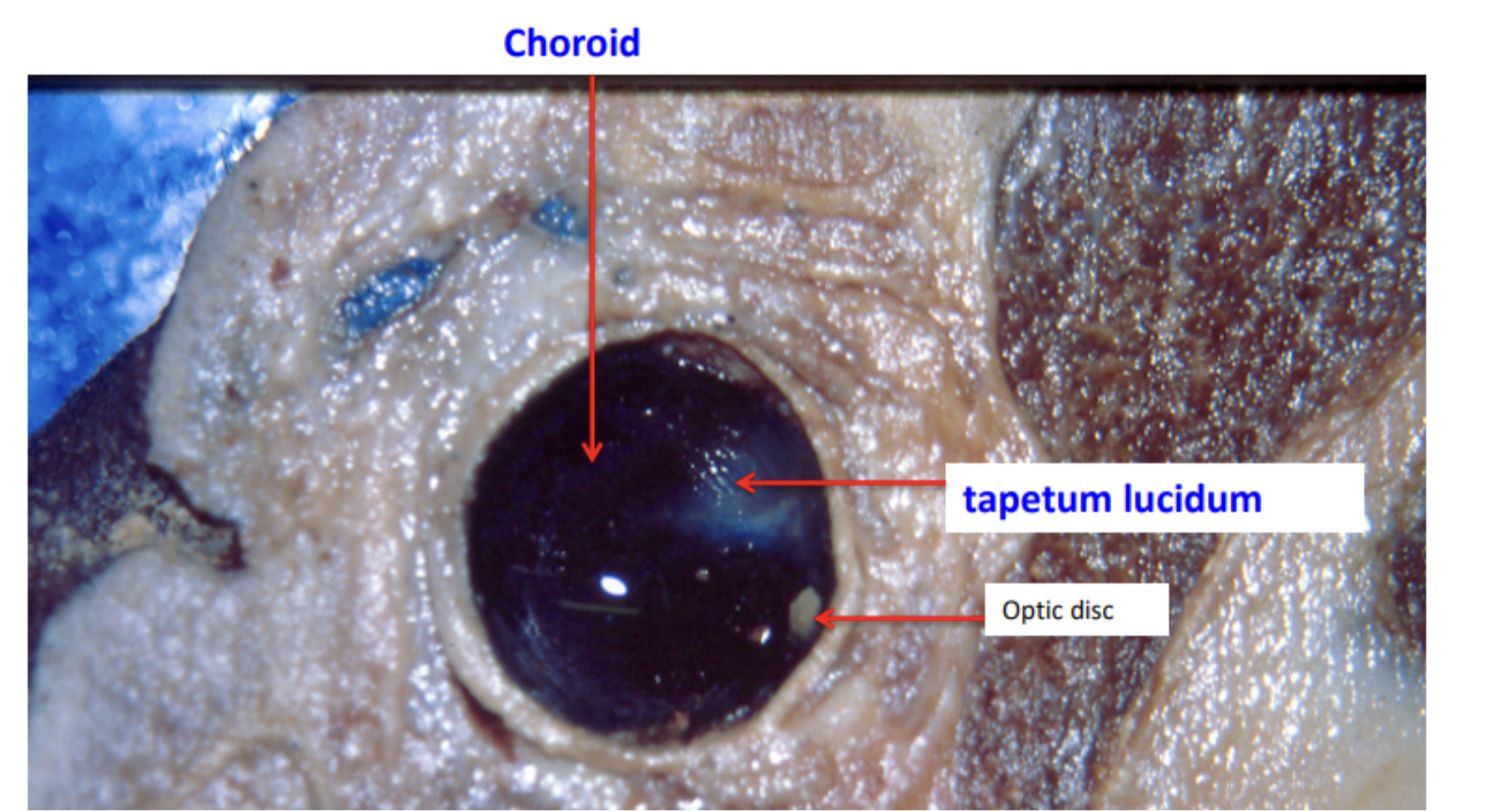
label the iris, cornea and ciliary body
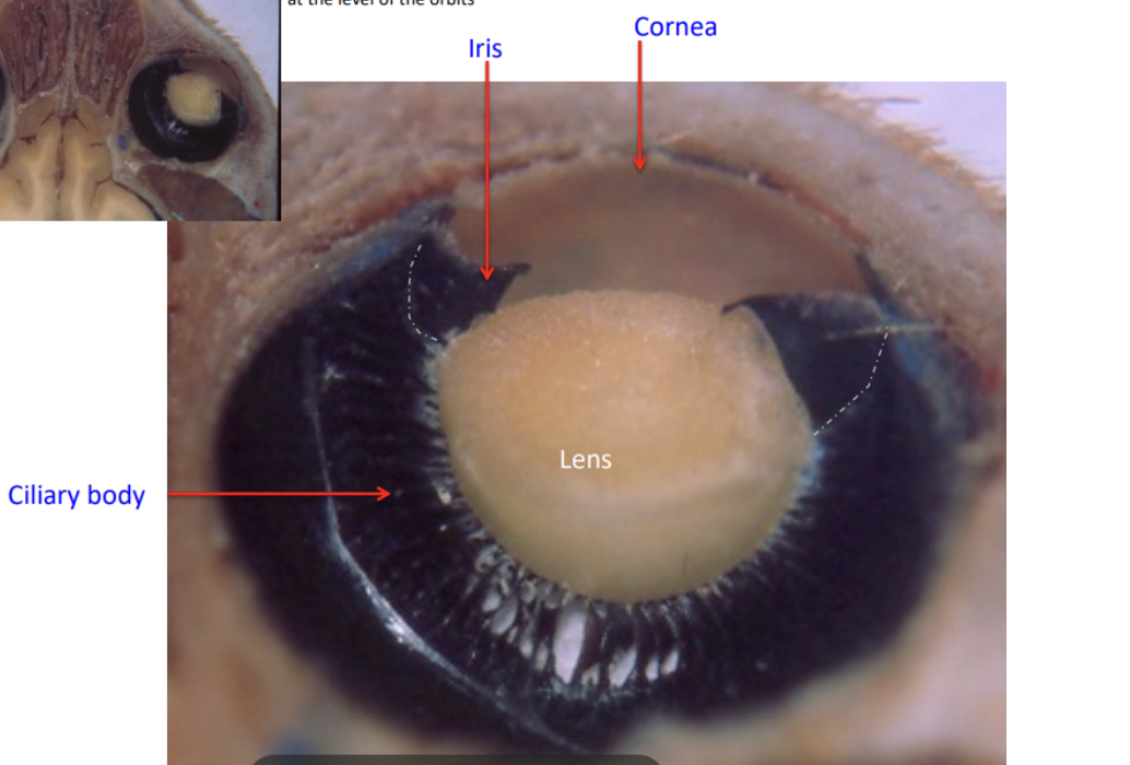
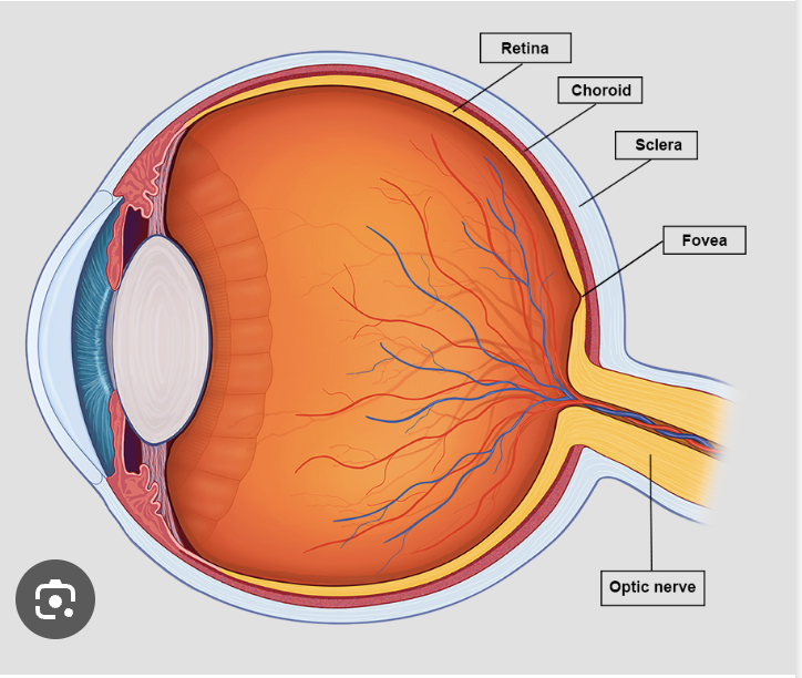
know the sclera , choroid
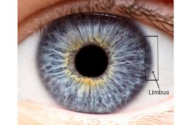
know the limbus
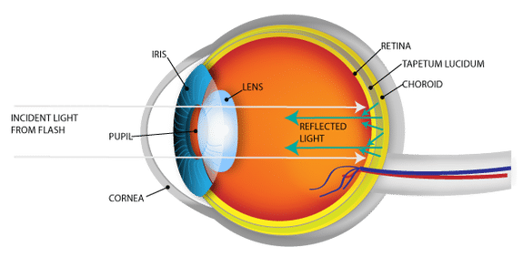
tapetum luciderm, retina
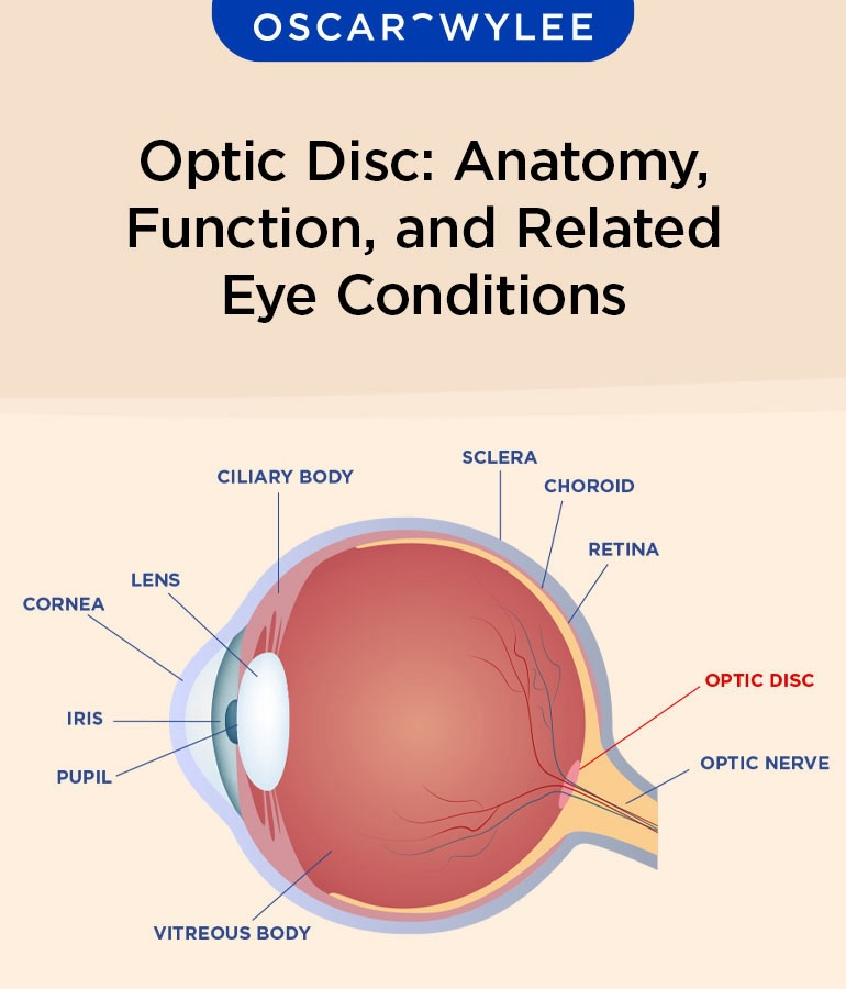
know the optic disk an the optic nerve
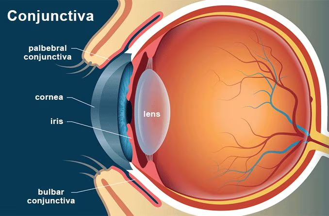
palpable and bulbar conjunctiva
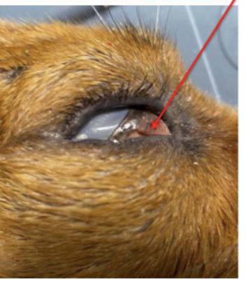
what is this picture pointing to
third eyelid/ nictitating membrane
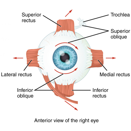
Recti muscles (lateral, dorsal, ventral, medial (rarely can be found))
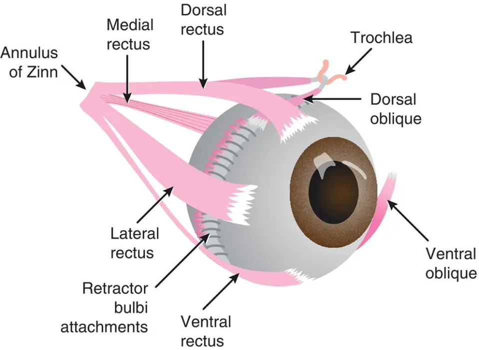
Oblique muscles (dorsal/ventral)
retractor bulbi muscles how many
4 (retracts the eye)
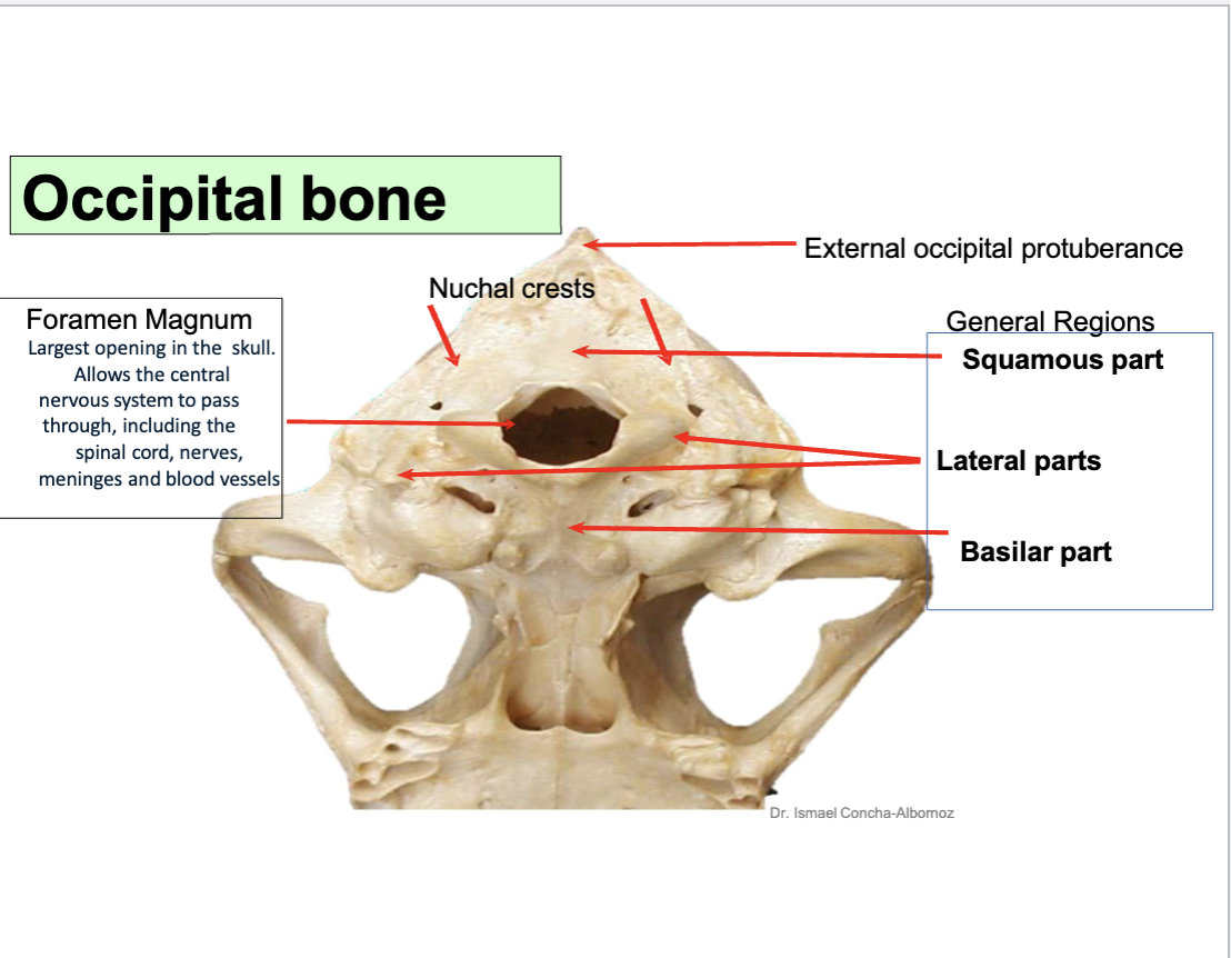
know the regions
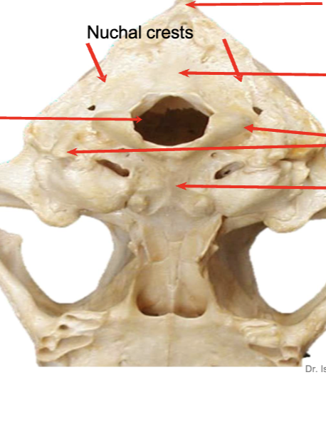
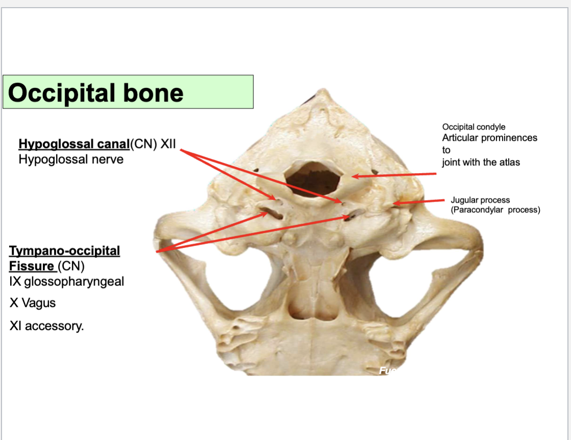
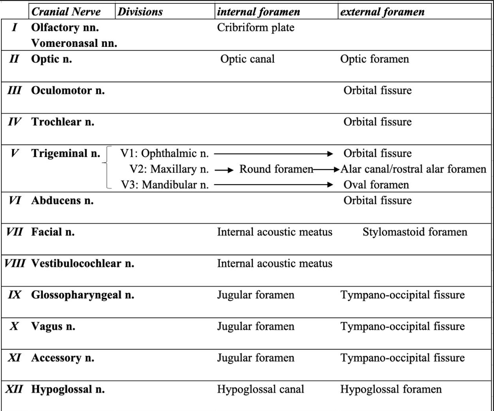
label the Nuchal crest, external occipital protuberence, foreamen magnum
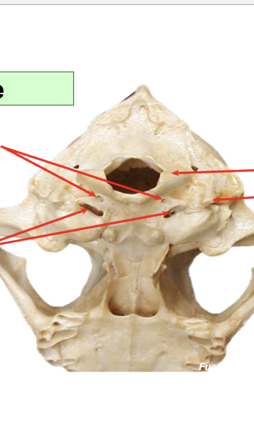
Label the occipital process, jugular process, Hypoglossal canal (CN XII),
Tympano-occipital fissure (CN IX,CN X, CN XI, Internal carotid artery, Internal jugular vein)
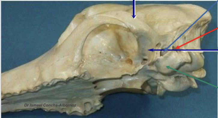
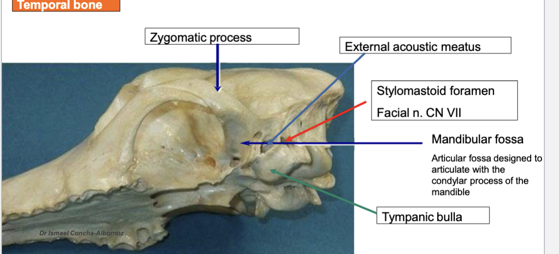
label the zygomatic arch, external acoustic meatus, tympanic bulla, mandibullar fossa
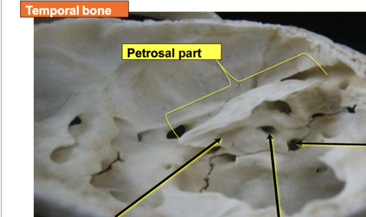
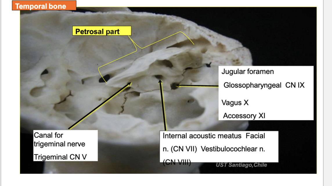
Label the
Internal acoustic meatus (CN VII, CN VIII)
Jugular foramen (CN IX, CN X, CN XI)
know the frontal bone and nasal bone
label the vomer
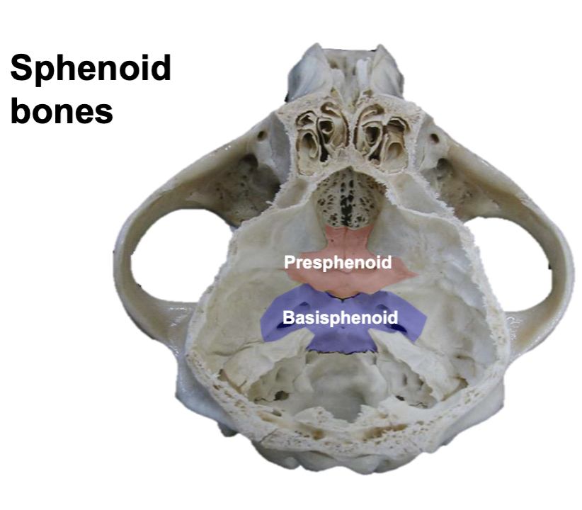
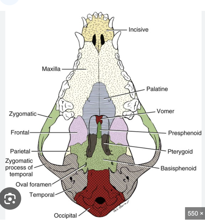
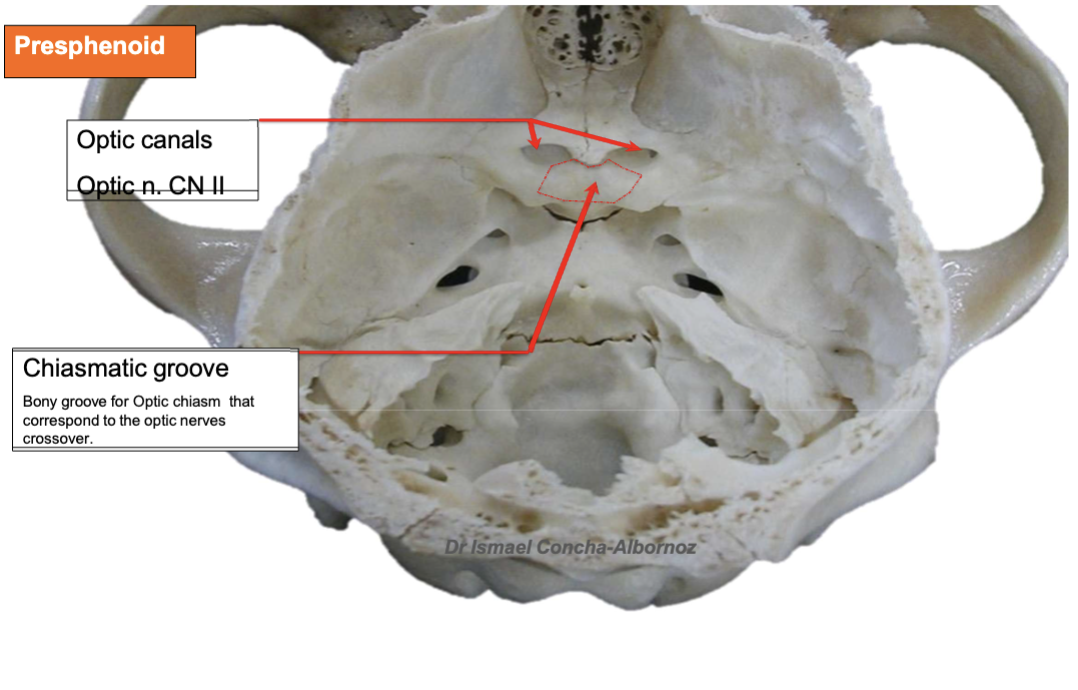
Know the basisphenoid bone and presphenoid
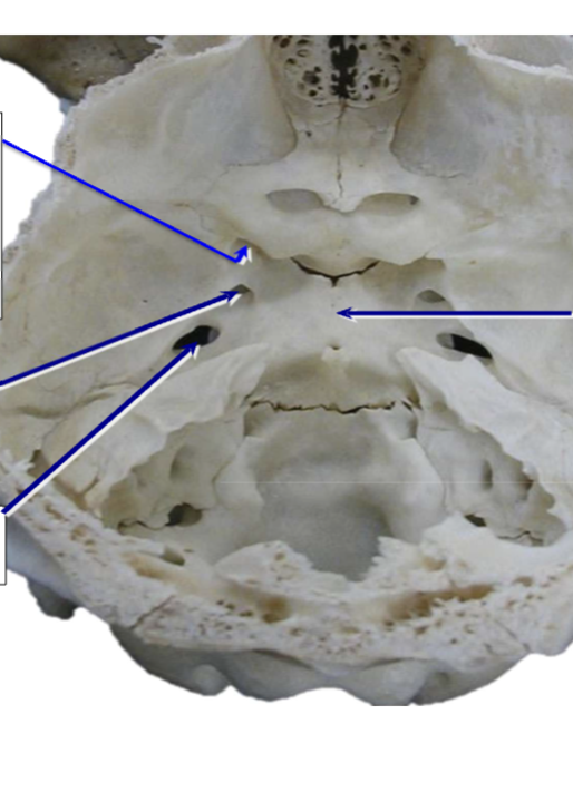
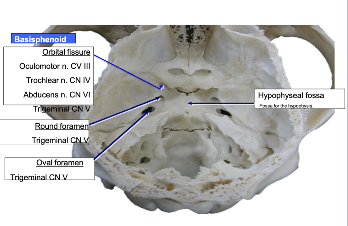
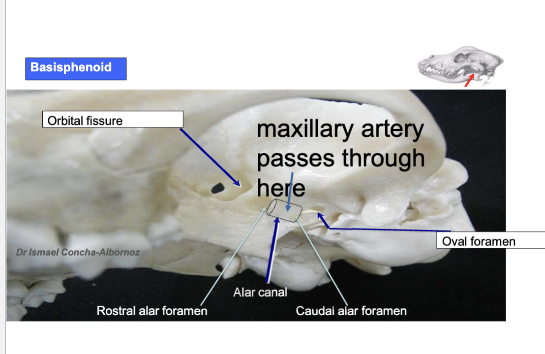
basisphenoid:
label the Orbital fissure (CN III, CN IV, CNV, CNVI)
Round foramen/alar canal/rostral alar foramen (CNV)
Oval foramen (CNV)
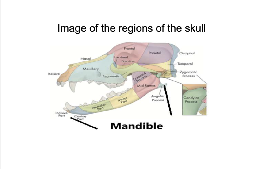
know the lacrimal bone and the maxilla
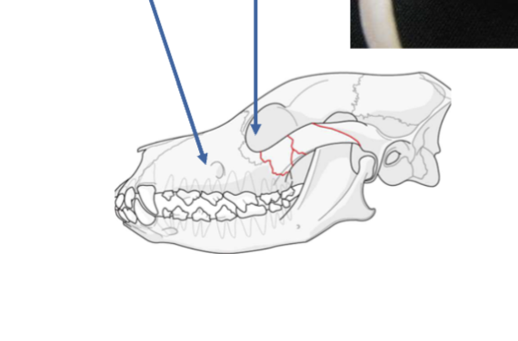
where is the infraorbital canal
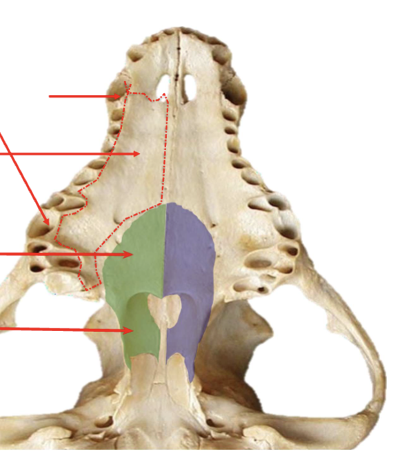
label the palentine process, alveolar process
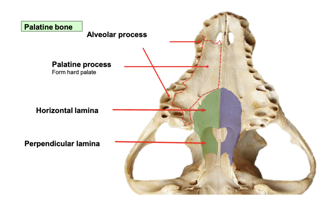
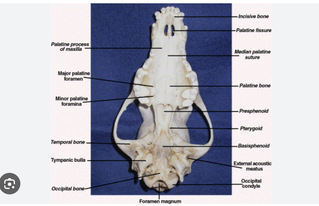
palentine bone
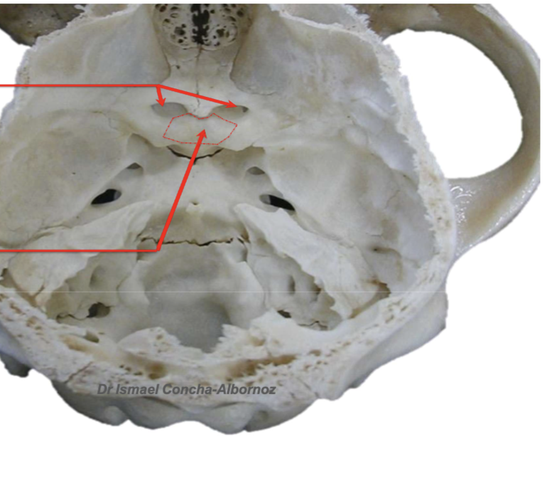
label the optic canal
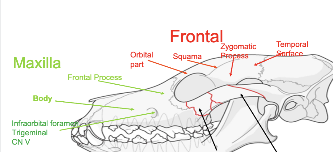
label the frontal process, temporal process, and zygomatic arch
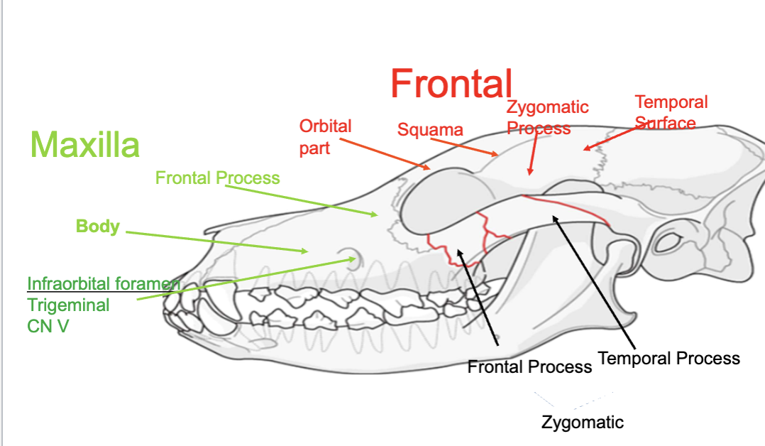
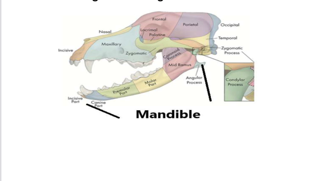
incisive bone
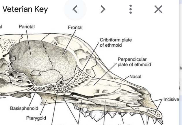
cribiform plate
ethmoid
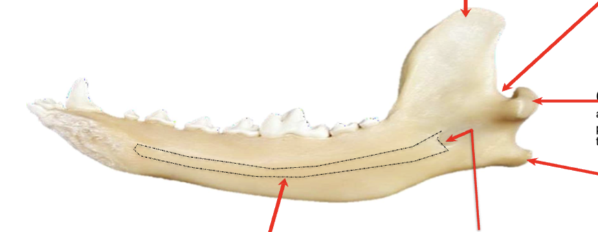
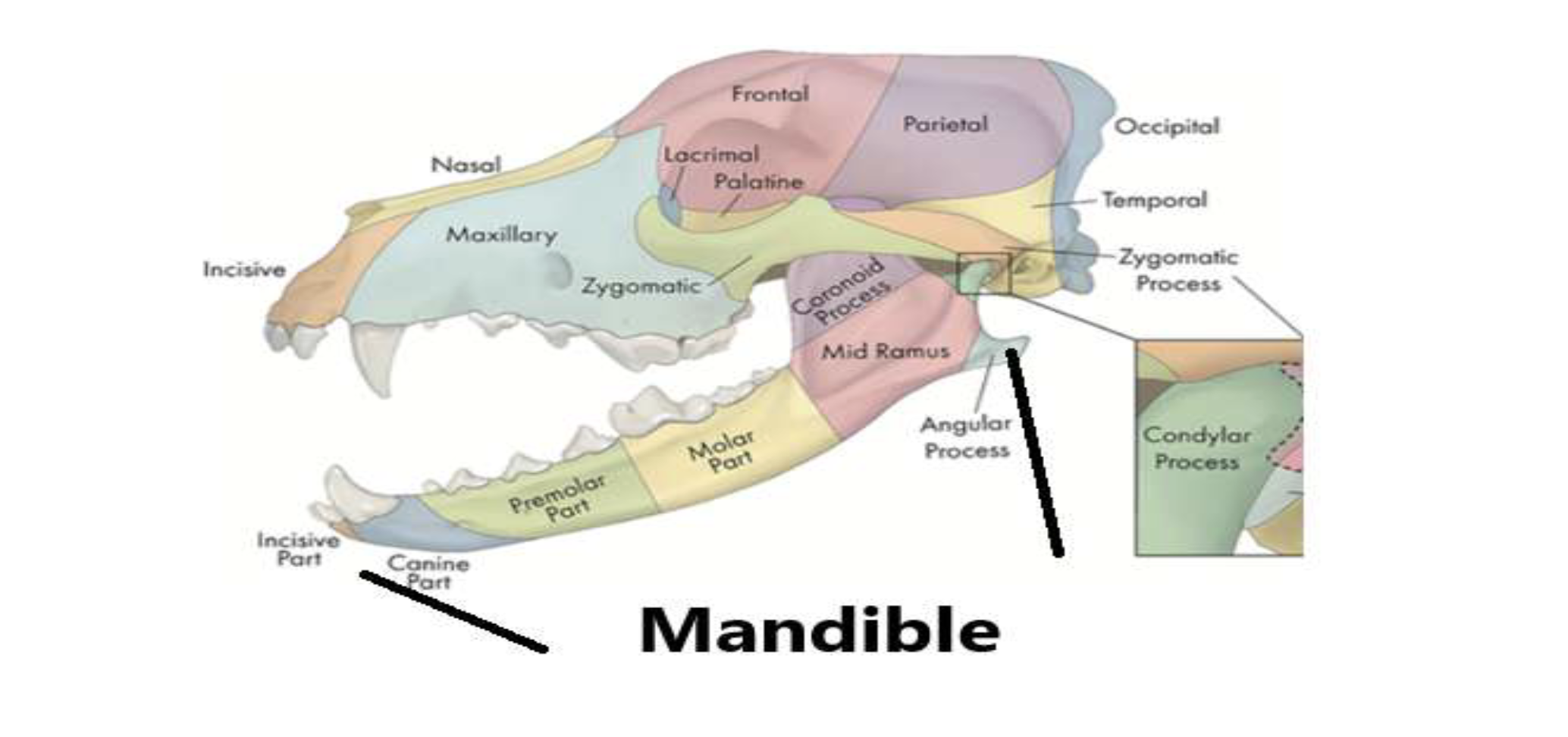
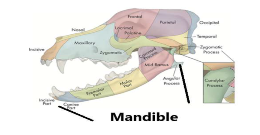
label the condyle, angular process, coronoid process, mandibular foreamen,
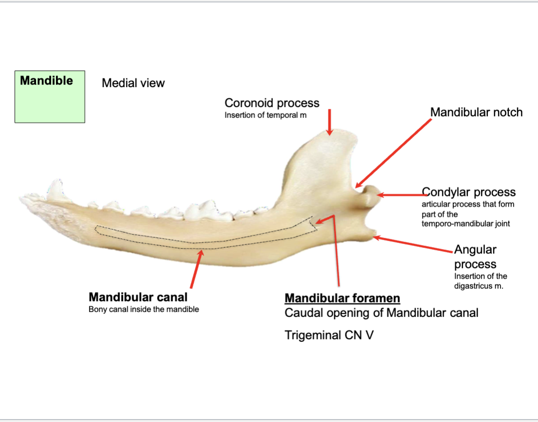
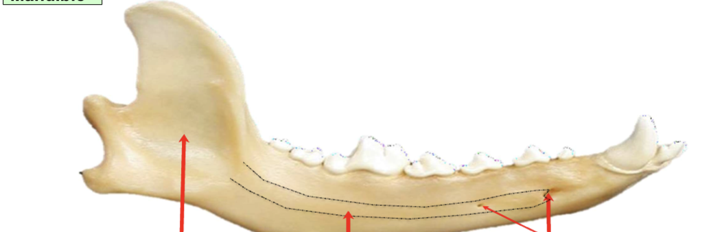
label the mental foreamen
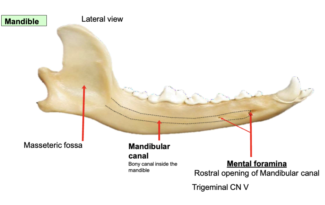
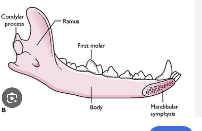
ramus and body

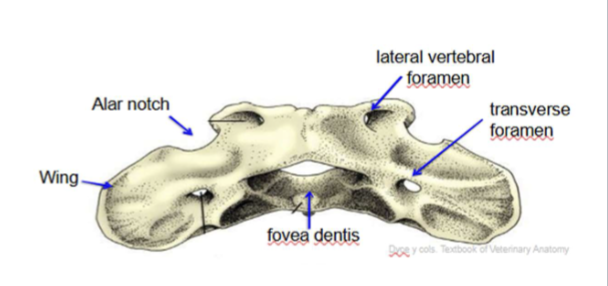
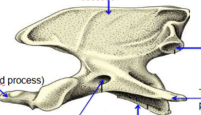
what spinal bone is this
C1 atlas

label the transverse foramen
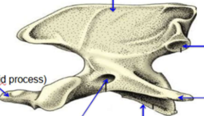
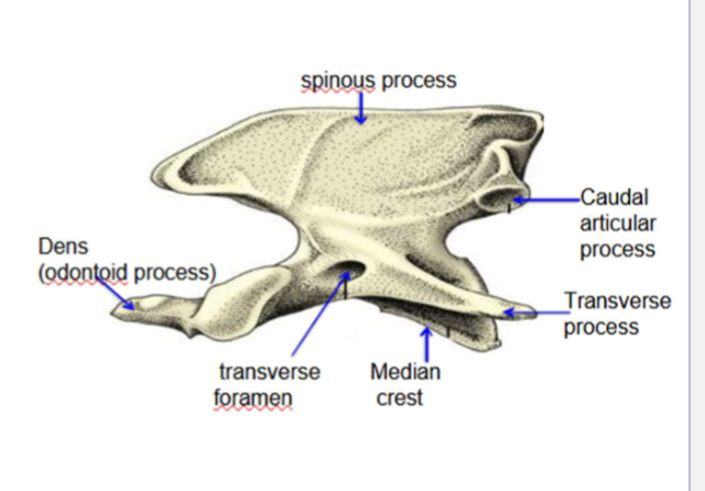
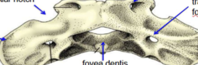
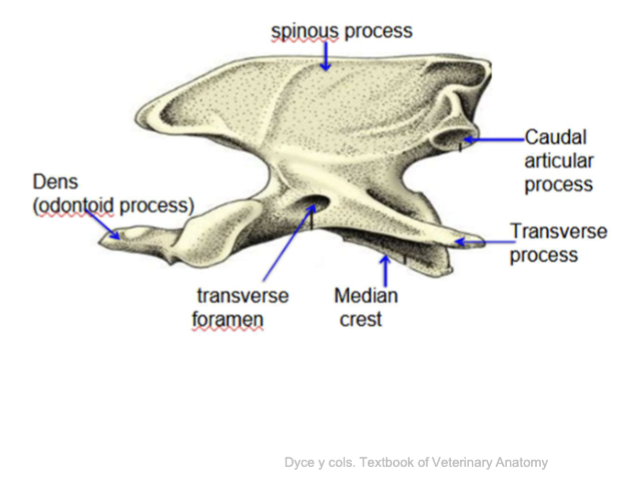
What spinal bone is this
Axis C2

label the transverse process, caudal articular process, transverse foramen, label the spinous process

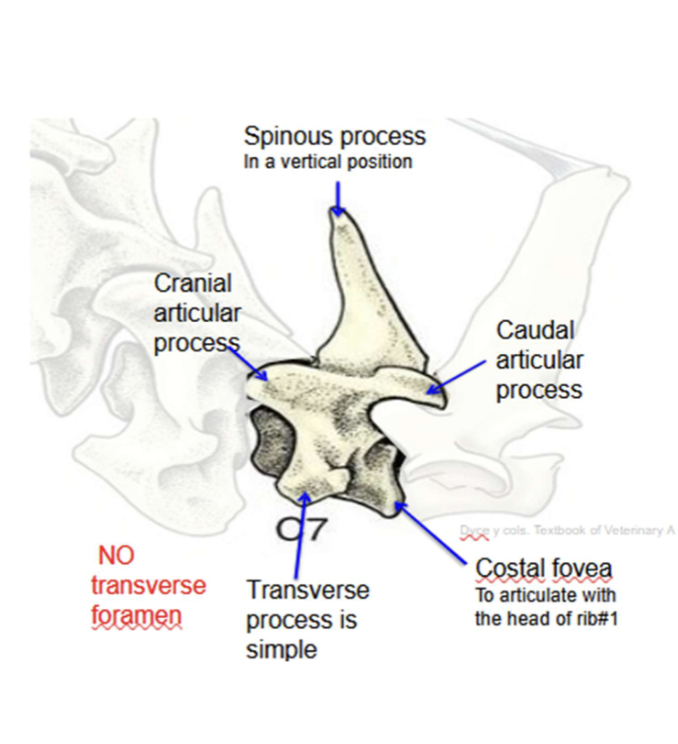
which spinal bone is this
C7
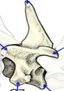
label the spinous process, caudal articular process, cranial articular process, transverse process, NO TRANSVERSE FORAMEN,

which spinal bone is this
thoracic verterbrea
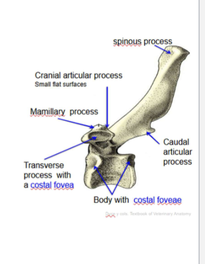
label the spinous process, caudal/ cranial articular surface, transverse process, body
what are the parts of the atlas C1 label wing, alar foramen, dorsal tubercle
alar foramen is the other hole on top next to alar notch. Dorsal tubercle is middle top then there is a ventral middle bottom

where are the parts of the axis or C2 dens, spine

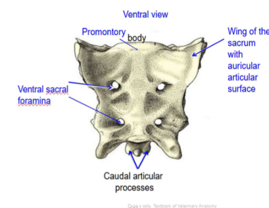
what is this
sacrum
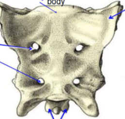
label these parts of the sacrum:
promontory
sacral foramina
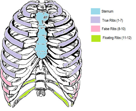
Know rib formula and vertebrae formula
label the head and the tubercle
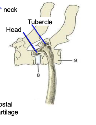
what are the three types of ribs and how do they differ
True attach to the sternum
False attachment with cartilage
floating no attachment
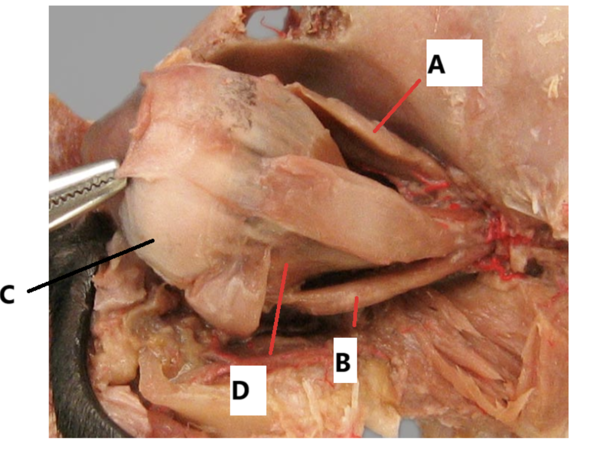
Label the picture
what controls A & B?
A: Dorsal rectus muscles
B:Ventral rectus muscles
C:sclera
D:retractor bulbi muscle
Oculomotor III
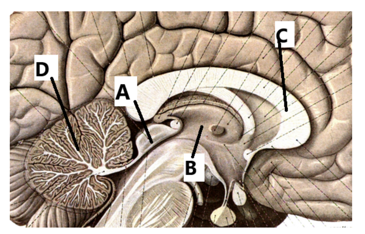
label the picture
A: colliculi
B: thalmus
C: Corpus callosum
D: cerebellum
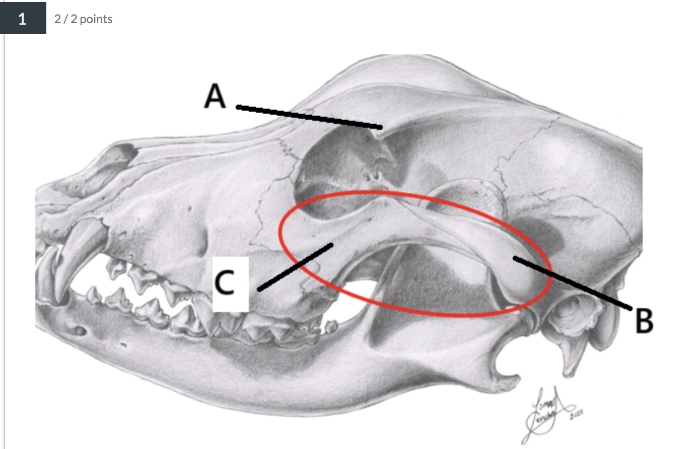
label the picture
A: zygomatic process
B: Temporal process
C: Frontal process
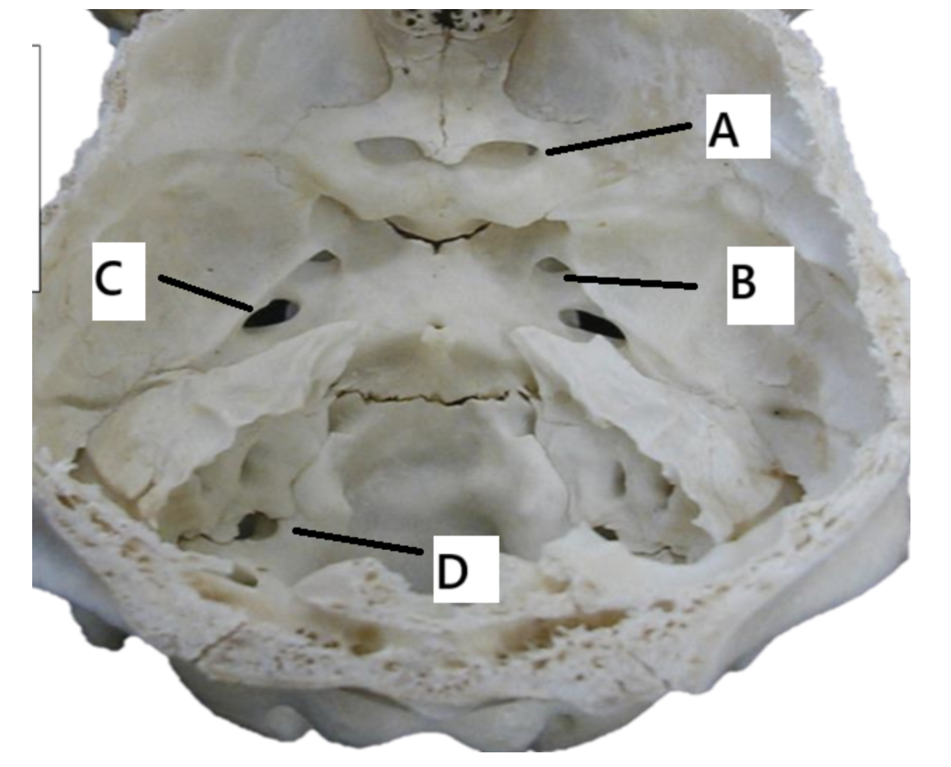
label the picture
A optic canal optic n. CNII
B round foraman trigeminal cn V
C oval foraman trigeminal cn V
D Jugular foramen accessory n. (XI)
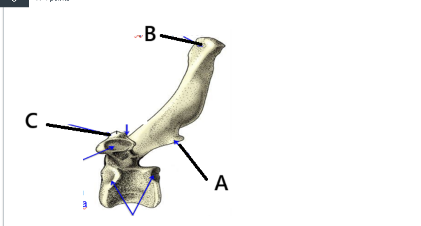
what bone is this?
What is A, B, C?
Bone: Thoracic vertebrae
caudal articular process
spinous process
mamillary process
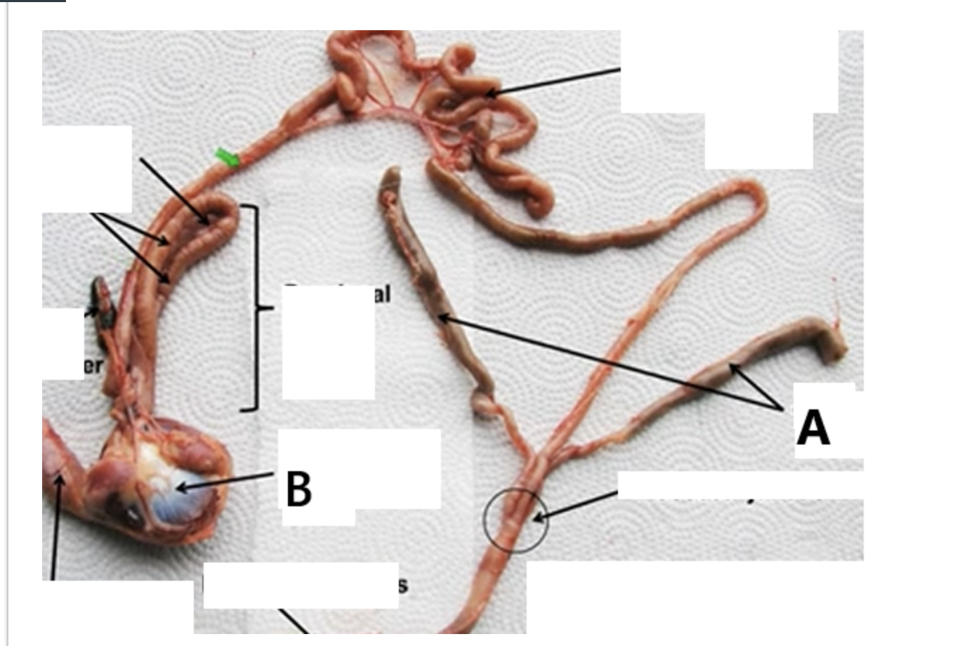
What is A & B
Ceca
Gizzard/ Ventricula
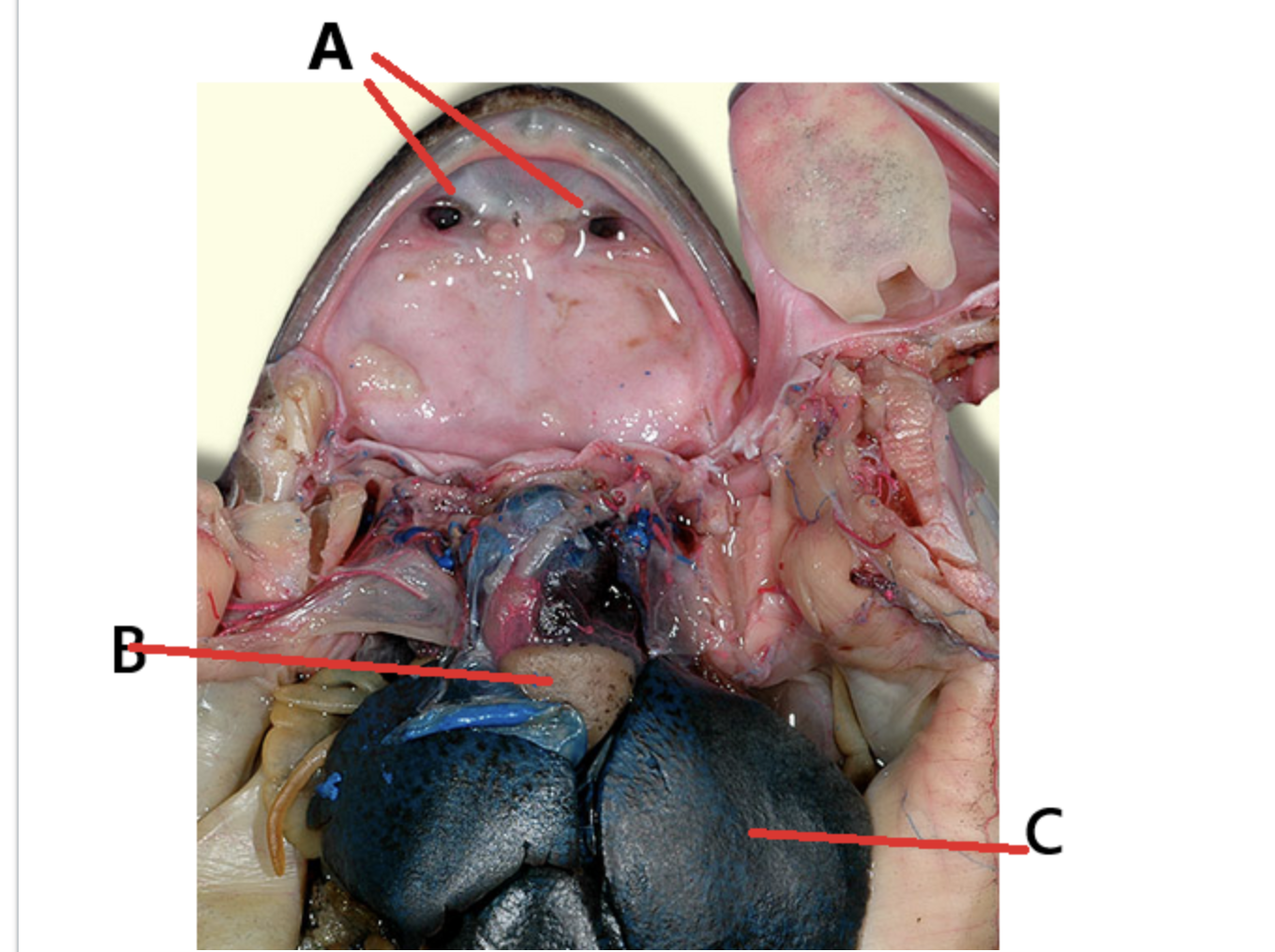
what is A, B, C
A: Internal nares
B: heart
C: liver
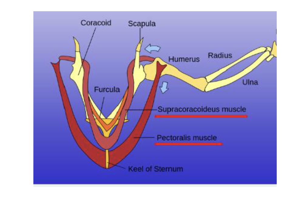
Name the two main muscles for flight and the bone structure that the 'upstroke' muscle passes through
Pectoralis m
Supracoracoid m
Triosseal canal bone structure
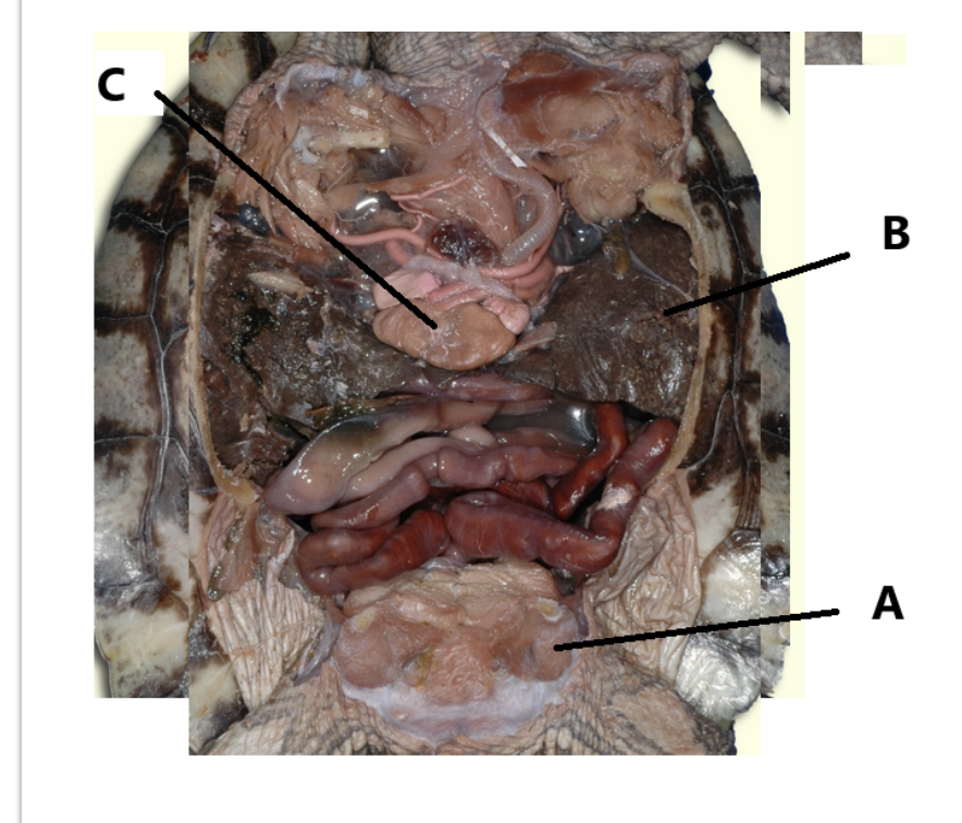
A,B, C
A: Bladder
B: Liver
C: Heart
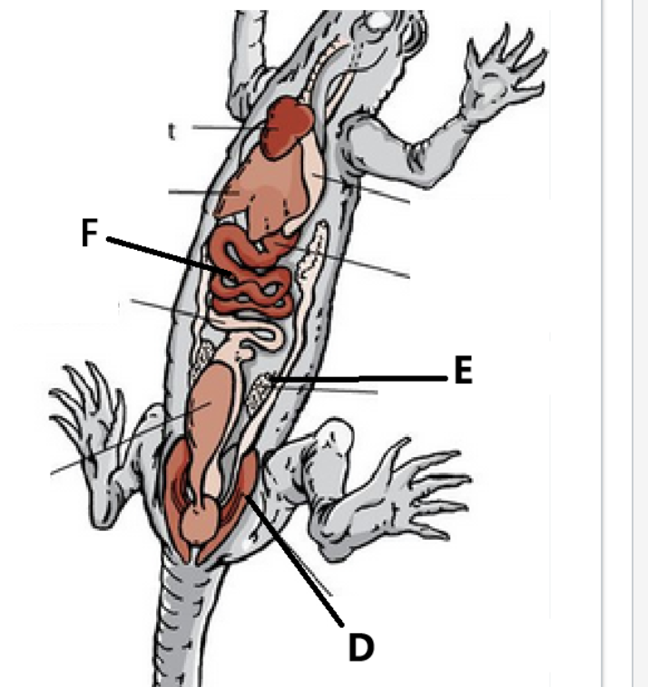
Label D,E,F
D: Kidney
E: Ovary
F: GI tract
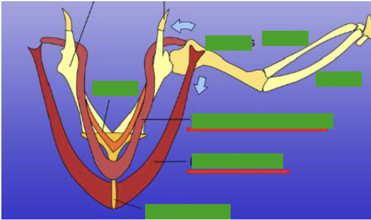
Label the scapula, coracoid and furcula (flying bones)
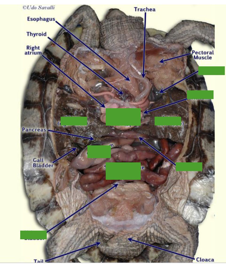
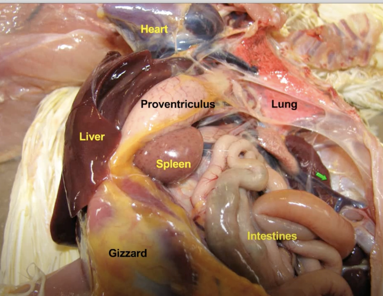
label the Stomach, liver, heart, lungs, bladder , GI tract
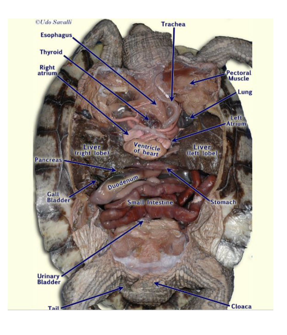
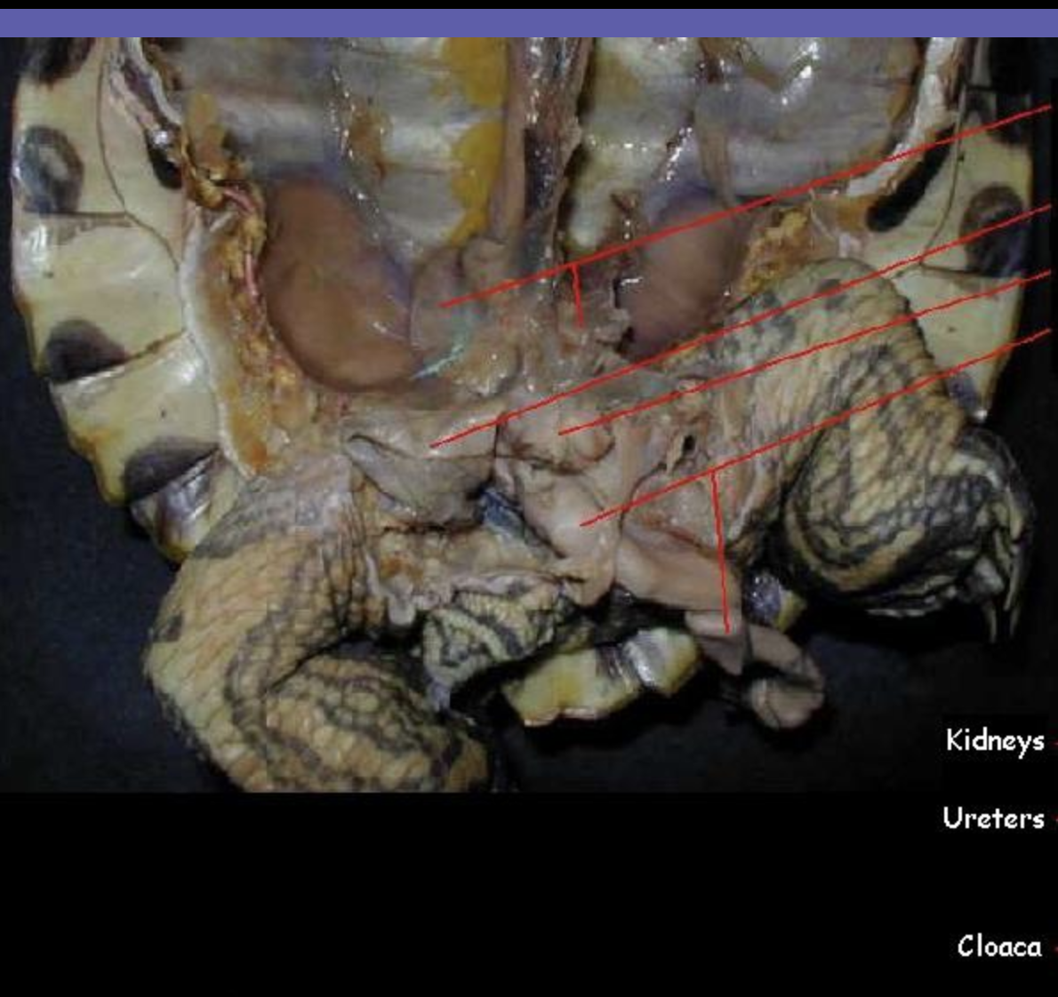
label the kidneys
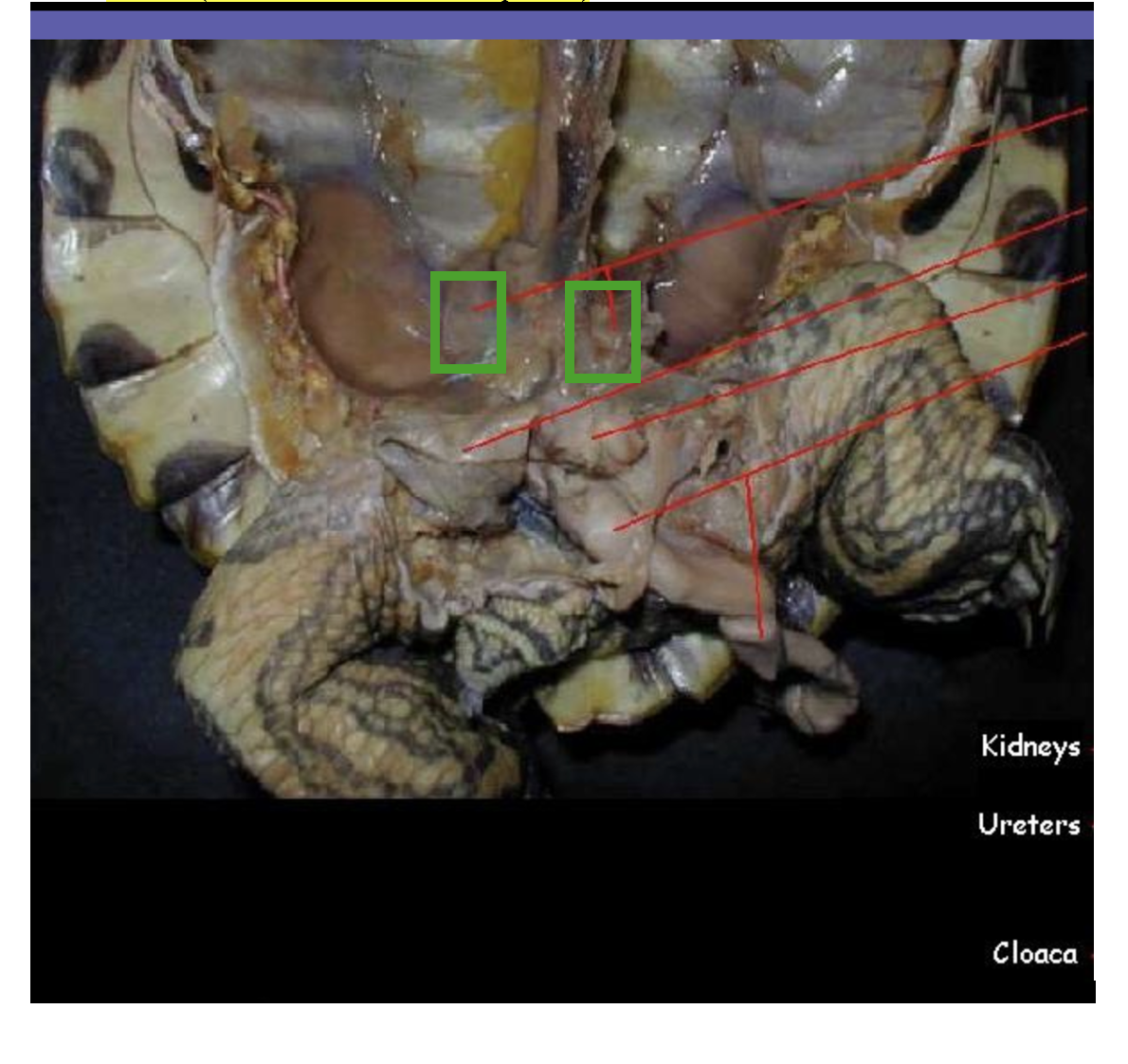
What is the green arrow pointing to
kidney
label this picture

what is this
corpus collosum