Chapter 14- The Brain and Cranial Nerves
1/25
There's no tags or description
Looks like no tags are added yet.
Name | Mastery | Learn | Test | Matching | Spaced |
|---|
No study sessions yet.
26 Terms
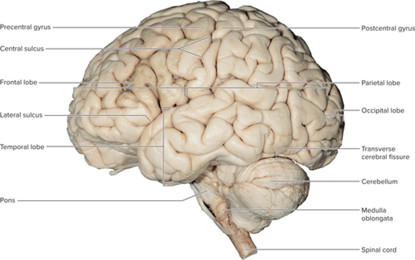
Describe the major subdivisions and anatomical landmarks of the brain.
Forebrain
cerebrum: 83% of brain volume
diencephalon: underlies cerebrum medially
cerebellum : occupies the posterior cranial fossa
brainstem
midbrain
pons
medulla oblongata
Describe the locations of its gray and white matter.
• Gray matter— exterior of brain
• White matter—inside brain
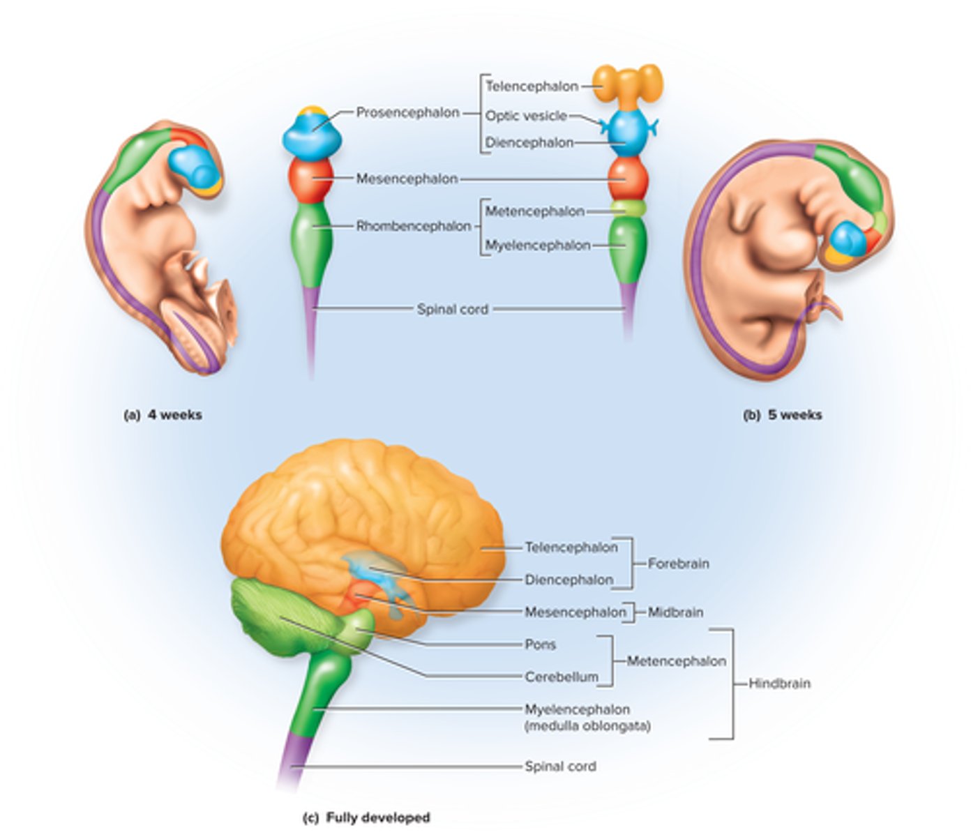
Describe the embryonic development of the CNS and relate this to adult brain anatomy.
Start: ectoderm
3 weeks: neurulation- forms neural plate that later makes neurtal fold and groove
Day 26: neural tube forms
4 weeks: forebrain, midbrain, hindbrain
5 weeks: Subdivides into 5 secondary vesicles
Forebrain: telencephalon, diencephalon
mid: mesencephalon
hindbrain: metencephalon, myelencephalon
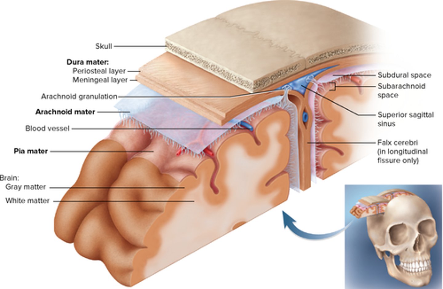
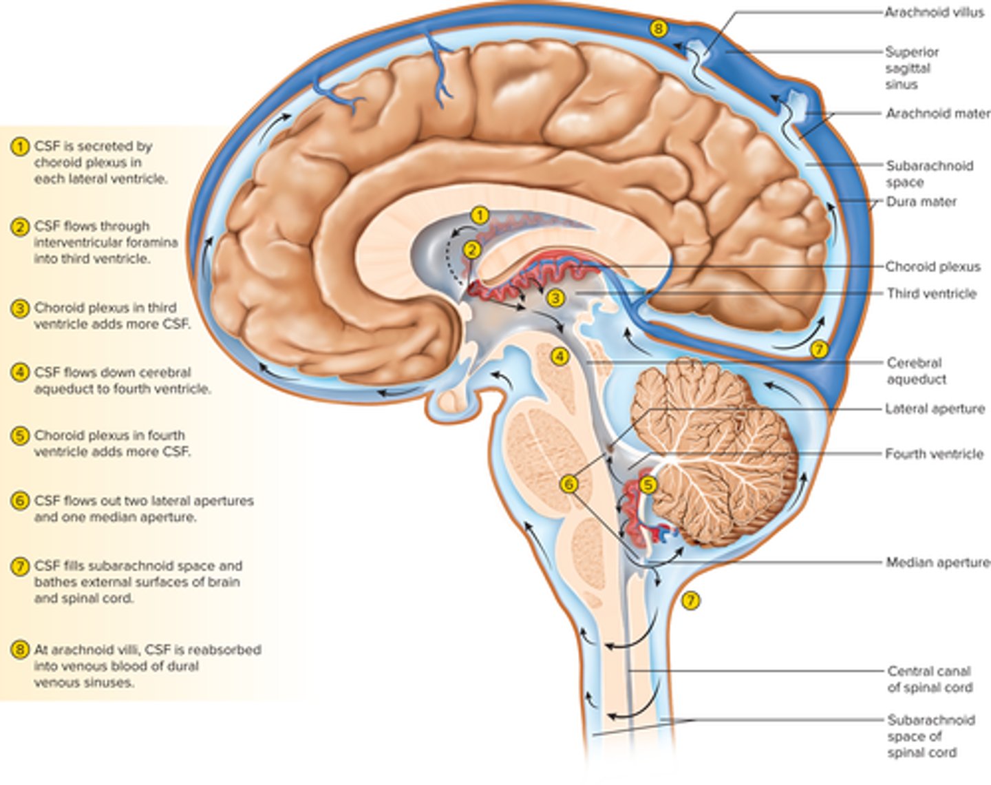
Describe the meninges of the brain.
Cranial dura mater- Dura mater is pressed closely against cranial bones
Outer periosteal—equivalent to periosteum of cranial bones
Inner meningeal—continues into vertebral canal and forms dural sheath around spinal cord
arachnoid mater: transparent membrane over the brain surface, deep to dura
pia mater: deep to arachnoid mater; thin, delicate membrane
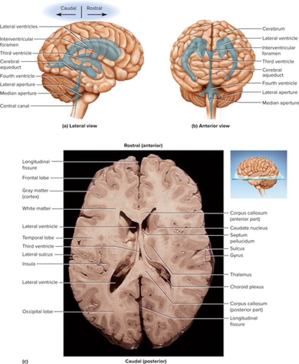
Describe the fluid-filled chamber within the brain.
Two lateral ventricles: one in each cerebral hemisphere
Third ventricle: narrow medial space beneath corpus callosum walled by diencephalon
Fourth ventricle: small triangular chamber between pons and cerebellum
Discuss the production, circulation, and function of the CSF that fills these chambers.
• Ependyma—Produces cerebrospinal fluid
• Cerebrospinal fluid (CSF)—clear, colorless liquid that fills the ventricles and canals of CNS and bathes external surface in subarachnoid space
Production: filtration of blood plasma through capillaries of the brain, so
CSF has more sodium and chloride than plasma, but less potassium, calcium, glucose, and very little protein
2)CSF flows through interventricular foramina into third ventricle.
3)Choroid plexus adds more CSF.
4)CSF flows down cerebral aqueduct to
fourth ventricle.
5)Choroid plexus in fourth ventricle adds more CSF.
6)CSF flows out two lateral apertures and one median aperture.
7)CSF fills subarachnoid space and bathes external surfaces of brain and spinal cord.
8)At arachnoid villi, CSF is reabsorbed into venous blood of dural venous sinuses
• Functions of CSF
Buoyancy: Allows brain to attain considerable size without being impaired by its own weight
Protection: Protects the brain from striking the cranium when the head is jolted
Chemical stability: rinses away metabolic wastes from nervous tissue and maintains homeostasis
Explain the significance of the brain barrier system.
• Brain barrier system—regulates what substances can get from bloodstream into tissue fluid of the brain
• Two points of entry must be guarded
Blood capillaries
Capillaries
Blood-brain barrier—protects blood capillaries throughout brain tissue
Induce the endothelial cells to form tight junctions that completely seal off gaps between them
forces things to leave through cells and not through blood
Blood-CSF barrier—protects brain at the choroid plexus
Forms tight junctions between the ependymal cells
Tight junctions are absent from ependymal cells elsewhere to allow exchange between brain tissue and CSF
• Brain barrier system is highly permeable to water, glucose, and lipid-soluble substances such as oxygen, carbon dioxide, alcohol, caffeine, nicotine, and anesthetics
• Only slightly permeable to sodium, potassium, and chloride
• Circumventricular organs (CVOs)—places in the third and fourth ventricles where the barrier is absent
• Blood has direct access to the brain
• Enables the brain to monitor and respond to fluctuations in blood glucose, pH, osmolarity, and other variables
List the components of the hindbrain and midbrain and their functions.
Hindbrain:
Medulla Oblongata
• All ascending and descending fibers connecting brain and spinal cord pass through medulla
• Pyramids—pair of ridges on anterior surface separated by anterior median fissure
- Contain corticospinal tracts
- Pyramidal decussation
• Medulla houses neurosomas of second order sensory neurons in gracile and cuneate nuclei
- Their axons decussate and form medial lemniscus
• Inferior olivary nucleus— relay center for signals to cerebellum
• Reticular formation— loose network of nuclei extending throughout the medulla, pons, and
midbrain
- Contains cardiac, vasomotor, and respiratory centers
• Four pairs of cranial nerves begin or end in medulla—VIII (in part), IX, X, and XII
• Pons—anterior bulge in brainstem, rostral to medulla
- Cerebellar peduncles—stalks on posterior pons that connect it (and the midbrain) to the cerebellum
• Ascending sensory tracts
• Descending motor tracts
• Pathways in and out of cerebellum
• Cranial nerves V, VI, VII, and VIII
- Sensory roles: hearing, equilibrium, taste, facial sensations
- Motor roles: eye movement, facial expressions, chewing, swallowing, urination, and secretion of saliva and tears
• Reticular formation in pons contains additional nuclei concerned with sleep, respiration, posture
Midbrain:
- Tectum: roof-like part of the midbrain posterior to cerebral aqueduct
• Exhibits four bulges, the corpora quadrigemina
- Upper pair, the superior colliculi, function in visual attention, tracking moving objects, and some visual reflexes
- Lower pair, the inferior colliculi, receives signals from the inner ear and control reflexive head turning in response to sound
- Cerebral peduncles: two anterior midbrain stalks
that anchor the cerebrum to the brainstem
• Each peduncle has three parts: tegmentum, substantia nigra, and cerebral crus
• Tegmentum
- Dominated by red nucleus
• Pink color due to high density of blood vessels
• Connections go to and from cerebellum for motor control
• Substantia nigra
- Black nucleus pigmented with melanin
- Motor center that relays inhibitory signals to thalamus and basal nuclei preventing unwanted body movement
- Degeneration of neurons leads to tremors of Parkinson disease
• Cerebral crus
- Bundle of nerve fibers that connect cerebrum to pons
- Carries corticospinal tracts
Describe the location and functions of the reticular formation.
-Reticular Formation: gray matter that runs vertically through all levels of the brainstem and has connections with many areas of cerebrum
• Functions:
1. Somatic motor control
• Adjust muscle tension to maintain tone,
balance, and posture, especially during body
movements
• Relay signals from eyes and ears to cerebellum
• Integrate visual, auditory, balance and motion
stimuli into motor coordination
• Gaze centers—allow eyes to track and fixate on
objects
• Central pattern generators—neural pools that
produce rhythmic signals to the muscles of
breathing and swallowing
2. Cardiovascular control
• Cardiac and vasomotor centers of medulla
oblongata
3. Pain modulation
• Some pain signals ascend through the reticular formation
• Some descending analgesic pathways begin in the reticular formation
- They end in the spinal cord where they block transmission of pain signals
4. Sleep and consciousness
• Reticular formation plays a central role in
consciousness, alertness and sleep
• Injury here can result in irreversible coma
5. Habituation
• Reticular activating system modulates activity in cerebral cortex so that it ignores repetitive, inconsequential stimuli
Cerebellum
• Consists of right and left cerebellar hemispheres connected by vermis
• Superficial cortex of gray matter with folds (folia), branching white matter (arbor vitae), and deep
nuclei
• Contains more than half of all brain neurons—about 100 billion
- Many small granule cells
- Large Purkinje cells have axons that synapse on deep nuclei
• Cerebellar peduncles—three pairs of stalks that connect brainstem and cerebellum (their fibers carry signals to and from cerebellum)
- Inferior peduncles: connected to medulla oblongata
• Most spinal input enters the cerebellum through inferior peduncle
- Middle peduncles: connected to pons
• Most input from rest of the brain enters through middle peduncle
- Superior peduncles: connected to the midbrain
• Carries cerebellar output
• Cerebellum has long been known to be important for motor coordination
• Receives input from regions of cerebral cortex that plan and initiate highly skilled movement
• Also receive input from sensory systems that monitor the course of movements
• Comparison of the two allows for reduction of "motor error"
- Correction occurs in real time and is stored as motor memory
- Error correction is mediated by fibers from the olivary nuclei that contact the Purkinje cells in the cortex
• Cerebellar disorders lead to ataxia
• Recent studies have revealed several other nonmotor functions
Name the three major components of the diencephalon and describe their locations and functions.
• Diencephalon has three parts: thalamus, hypothalamus, epithalamus
- Thalamus—ovoid mass on each side of the brain perched at the superior end of the brainstem beneath the cerebral hemispheres
• Two thalami are joined medially by a narrow intermediate mass
• Composed of at least 23 nuclei within five major functional groups
• "Gateway to the cerebral cortex": nearly all input to the cerebrum passes by way of synapses in the
thalamic nuclei, filters information on its way to cerebral cortex
• Plays key role in motor control by relaying signals from cerebellum to cerebrum and providing feedback loops between the cerebral cortex and the basal nuclei
• Involved in the memory and emotional functions of the limbic system: a complex of structures that include some cerebral cortex of the temporal and frontal lobes and some of the anterior thalamic
nuclei
-Hypothalamus—forms part of the walls and floor of the third ventricle
• Extends anteriorly to optic chiasm and posteriorly to mammillary bodies
• Each mammillary body contains three or four mammillary nuclei
- Relay signals from the limbic system to the thalamus to store memories
• Infundibulum—stalk attaching pituitary to hypothalamus
• Hypothalamus is a major control center of autonomic nervous system and endocrine system
- Plays essential role in homeostatic regulation of all body systems
• Functions of hypothalamic nuclei
- Hormone secretion
• Controls anterior pituitary, thereby regulating growth, metabolism, reproduction, and stress responses
• Produces posterior pituitary hormones for labor contractions, lactation, and water conservation
- Autonomic effects
• Major integrating center for autonomic nervous system
• Influences heart rate, blood pressure, gastrointestinal secretions, motility, etc.
• Hypothalamic functions include:
- Thermoregulation
• Hypothalamic thermostat monitors body temperature
- Food and water intake
• Regulates hunger and satiety; responds to hormones influencing hunger, energy expenditure, and long-term control of body mass
• Thirst center monitors osmolarity of blood and can stimulate production of antidiuretic hormone
- Sleep and circadian rhythms
• Suprachiasmatic nucleus sits above optic chiasm
- Memory
• Mammillary nuclei receive signals from hippocampus
- Emotional behavior and sexual response
• Anger, aggression, fear, pleasure, contentment, sexual drive
• Epithalamus—very small mass of tissue composed
of:
- Pineal gland: endocrine gland
- Habenula: relay from the limbic system to the midbrain
- Thin roof over the third ventricle
Identify the five lobes of the cerebrum and their functions.
Frontal lobe:
Abstract thought
Explicit memory
Mood
Motivation
Foresight and planning
Decision making
Emotional control
Social judgment
Voluntary motor control
Speech production
Insula:
Taste
Pain
Visceral sensation
Consciousness
Emotion and empathy
Cardiovascular homeostasis
Parietal lobe:
Taste
Somatic sensation
Sensory integration
Visual processing
Spatial perception
Language processing
Numerical awareness
Occipital lobe:
Visual awareness
Visual processing
Temporal lobe:
Hearing
Smell
Emotion
Learning
Language comprehension
Memory consolidation
Verbal memory
Visual and auditory memory
Language
Identify the three types of tracts in the cerebral white matter.
• Projection tracts
- Extend vertically between higher and lower brain and spinal cord centers
• Example: corticospinal tracts
• Commissural tracts
- Cross from one cerebral hemisphere to the other
allowing communication between two sides of cerebrum
• Largest example: corpus callusum
• Other crossing tracts: anterior and posterior commissures
• Association tracts
- Connect different regions within the same cerebral hemisphere
• Long fibers connect different lobes; short fibers connect gyri within a lobe
Describe the distinctive cell types and histological arrangement of the cerebral cortex.
Contains two principal types of neurons:
- Stellate cells
• Have spheroid neurosomas with dendrites projecting in all directions
• Receive sensory input and process information on a local level
- Pyramidal cells
• Tall, and conical, with apex toward the brain surface
• A thick dendrite with many branches with small, knobby dendritic spines
• Include the output neurons of the cerebrum
• Only neurons that leave the cortex and connect with other parts of the CNS.
Describe the location and functions of the basal nuclei and limbic system.
Limbic system—important center of emotion and learning
• Prominent components:
- Cingulate gyrus: arches over corpus callosum in frontal and parietal lobes
- Hippocampus: in medial temporal lobe (memory functions)
- Amygdala: immediately rostral to hippocampus (emotion functions)
• There is a limbic system in each cerebral hemisphere
• Limbic system components are connected through a loop of fiber tracts including the fornix
• Limbic system structures have centers for both gratification and aversion
- Gratification: sensations of pleasure or reward
- Aversion: sensations of fear or sorrow
Basal nuclei—masses of cerebral gray matter buried deep in the white matter, lateral to the thalamus
- Receive input from the substantia nigra of the midbrain and the motor
areas of the cortex
- Send signals back to both of these locations
- Involved in motor control
• At least three brain centers form the basal nuclei and are collectively called the corpus striatum
- Caudate nucleus
- Putamen
- Globus pallidus
• Lentiform nucleus—putamen and globus pallidus together
List the types of brain waves and discuss their relationship to mental states.
• Alpha waves 8 to 13 Hz
- Awake and resting with eyes closed and mind wandering
- Suppressed when eyes open or performing a mental task
• Beta waves 14 to 30 Hz
- Eyes open and performing mental tasks
- Accentuated during mental activity and sensory stimulation
• Theta waves 4 to 7 Hz
- Drowsy or sleeping adults
- If awake and under emotional stress
• Delta waves < 3.5 Hz
- Deep sleep
- Infants
Describe the stages of sleep, their relationship to the brain waves, and the neural mechanisms of sleep.
Four stages of sleep:
- Stage 1
• Drowsy, relaxed, eyes closed, drifting sensation, easily awakened
• Alpha waves dominate EEG
- Stage 2
• Light sleep
• EEG frequency decreases but amplitude increases with occasional sleep spindles
- Stage 3
• Moderate to deep sleep; muscles relax, vital signs fall
• Theta and delta EEG waves appear
- Stage 4
• Muscles very relaxed, vitals very low, difficult to
awaken
• EEG dominated by lowfrequency, high-amplitude
delta EEG waves (slow wave sleep)
• About five times a night, a sleeper backtracks from stage 3 or 4 to stage 2 and exhibits bouts of rapid eye movement (REM) sleep
- Eyes oscillate back and forth
- Also called paradoxical sleep, because EEG
resembles the waking state
- Sleeper is harder to arouse than during any other stage
- Vivid and long dreams
- Sleep paralysis is stronger, preventing dreams from being acted out
• Rhythm of sleep is controlled by complex interaction between cerebral cortex, thalamus,
hypothalamus, and reticular formation
• Suprachiasmatic nucleus (red)— important hypothalamic area located above optic chasm
- Input from eye allows SCN to synchronize multiple body rhythms with external rhythms of night and day.
• Hypothalamus produces orexins—neuropeptides that stimulate wakefulness
- Orexin levels are low in narcolepsy (excessive daytime sleepiness)
• Sleep has a restorative effect, and sleep deprivation can be fatal to experimental animals
- Sleep may be the time to replenish such energy sources as glycogen and ATP
- REM sleep may consolidate and strengthen memories by reinforcing some synapses, and eliminating others
Identify the brain regions concerned with consciousness and thought.
• Cognition—the range of mental processes by which we acquire and use knowledge
- Sensory perception, thought, reasoning, judgment, memory, imagination, and intuition
• Cognition is accomplished by distributed association areas of cerebral cortex
- Constitute about 75% of all brain tissue
• Researchers learn about cognition from studies of patients with brain lesions and from imaging studies using PET and fMRI
• Cognitive functions in association areas of cortex:
- Parietal lobe helps perceive and attend to stimuli
• Lesions can cause contralateral neglect syndrome—unaware of objects on opposite side of the body
- Temporal lobe helps identify stimuli
• Lesions can cause agnosia—inability to recognize, identify
familiar objects; example: prosopagnosia—cannot recognize faces
- Frontal lobe helps us think about the world, and plan and execute appropriate behaviors
• Lesions can cause personality disorders and socially inappropriate behaviors
Identify the brain regions concerned with memory.
• Information management entails:
- Learning: acquiring new information
- Memory: information storage and retrieval
- Forgetting: eliminating trivial information; as important as
remembering
• Amnesia—defects in declarative memory: inability to
describe past events
- Anterograde amnesia: unable to store new information
- Retrograde amnesia: person cannot recall things known
before the injury
• Procedural memory—ability to tie one's shoes
• Hippocampus—important limbic system area for memory
- Functions in memory consolidation: the process of "teaching the cerebral cortex" until a long-term memory is established in the cortex (e.g., memory of faces in temporal lobe cortex)
- Hippocampus organizes cognitive information into a unified long-term memory but does not hold the memory itself
Identify the brain regions concerned with emotion.
• Amygdala—emotional memory
• Amygdala receives input from sensory systems
- Role in fear, food intake, sexual behavior, and drawing attention to novel stimuli
- One output goes to hypothalamus, influencing somatic and visceral motor systems
• Heart races, blood pressure rises, hair stands on end, vomiting ensues
- Other output to prefrontal cortex important in controlling expression of emotions
• Ability to express love, control anger, or overcome fear
• Prefrontal cortex—seat of judgment, intent, and control over expression of emotions
• Feelings (e.g., fear) arise from hypothalamus and
amygdala
Identify the brain regions concerned with sensation.
• Primary sensory cortex—sites where sensory input is first received and one becomes conscious of the stimulus
• Association areas near primary sensory areas process and interpret that sensory information
- E.g., Primary visual cortex is bordered by visual association areas: make cognitive sense of visual stimuli
- Multimodal association areas: receive input from multiple senses and integrate this into an overall perception of our surroundings
• Vision
- Visual primary cortex in far posterior region of occipital lobe
- Visual association area: anterior, and occupies all the remaining occipital lobe
• Much of inferior temporal lobe deals with recognizing faces and familiar objects
• Hearing
- Primary auditory cortex in the superior region of the temporal lobe and insula
- Auditory association area: temporal lobe deep and inferior to primary auditory cortex
• Recognizes spoken words, a familiar piece of music, or a voice on the phone
• Equilibrium
- Signals for balance and sense of motion project mainly to the cerebellum and several brainstem
nuclei concerned with head and eye movements and visceral functions
- Association cortex in the roof of the lateral sulcus near the lower end of the central sulcus
• Seat of consciousness of our body movements and orientation in space
• Taste and smell
- Gustatory (taste) signals received by primary gustatory cortex in inferior end of the postcentral
gyrus of the parietal lobe and anterior region of insula
- Olfactory (smell) signals received by the primary olfactory cortex in the medial surface of the
temporal lobe and inferior surface of the frontal lobe
• General (somesthetic, somatosensory, or
somatic) senses—distributed over entire body
and employ simple receptors
- Include touch, pressure, stretch, movement, heat,
cold, and pain
• For the head, cranial nerves carry general
sensory information
• For the rest of the body, ascending tracts bring
general sensory information to the brain
- Thalamus processes the input from contralateral side
- Selectively relays signals to postcentral gyrus of
parietal lobe
• Cerebral fold that is immediately caudal to the
central sulcus
• Functionally known as the primary somesthetic
cortex
• Provides awareness of stimulus
- Somesthetic association area: caudal to the
postcentral gyrus and in the roof of the lateral sulcus
• Makes cognitive sense of stimulus
Identify the brain regions concerned with motor control.
• The intention to contract a muscle begins in motor association (premotor) area of frontal lobes
- Where we plan our behavior
- Where neurons compile a program for degree and sequence of muscle contraction required for an action
• Program transmitted to neurons of the precentral
gyrus (primary motor area)
- Most posterior gyrus of the frontal lobe
- These neurons send signals to the brainstem and spinal cord leading ultimately to muscle contractions
• Precentral gyrus also exhibits somatotopy
- Neurons for toe movement are deep in the longitudinal fissure of the medial side of the gyrus
- The summit of the gyrus controls the trunk, shoulder, and arm
- The inferolateral region controls the facial muscles
- Motor homunculus has a distorted look because the amount of cortex devoted to a given body region is proportional to the number of muscles and motor units in that body region (not body
region size)
• Pyramidal cells of the precentral gyrus are called upper motor neurons
- Their fibers project caudally
- About 19 million fibers ending in nuclei of the
brainstem
- About 1 million forming the corticospinal tracts
- Most fibers decussate in lower medulla
oblongata
- Form lateral corticospinal tracts on each side of
the spinal cord
• In brainstem or spinal cord, the fibers from
upper motor neurons synapse with lower motor neurons whose axons innervate skeletal muscles
Basal nuclei and cerebellum are also important in
muscle control:
• Basal nuclei
- Important motor functions include helping to control:
• Onset and cessation of intentional movements
• Repetitive hip and shoulder movements in walking
• Highly practiced, learned behaviors such as writing, typing, driving
a car
- Lie in a feedback circuit from the cerebrum, to the basal
nuclei, to the thalamus, and back to the cerebrum
- Dyskinesias: movement disorders caused by lesions in the
basal nuclei involving abnormal movement initiation
• Cerebellum
- Highly important in motor coordination
- Aids in learning motor skills
- Maintains muscle tone and posture
- Smooths muscle contraction
- Coordinates eye and body movements
- Coordinates motions of different joints with each
other
- Lesions can cause ataxia: clumsy, awkward gait
Identify the brain regions concerned with language.
• Language includes several abilities: reading, writing, speaking, and understanding words
• Wernicke area
- Posterior to lateral sulcus usually in left hemisphere
- Permits recognition of spoken and written language
- When we intend to speak, Wernicke area formulates phrases and transmits plan of
speech to Broca area
• Broca area
- Inferior prefrontal cortex usually in left hemisphere
- Generates motor program for the muscles of the larynx, tongue, cheeks, and lips for speaking and for hands when signing
- Transmits program to primary motor cortex for commands to the lower motor neurons that supply
relevant muscles
• Affective language area usually in right hemisphere
- Lesions produce aprosody—flat emotionless speech
Discuss the functional differences between the right and left cerebral hemispheres.
• Cerebral lateralization—the difference in the structure and function of the cerebral hemispheres
• Left hemisphere—usually the categorical hemisphere
- Specialized for spoken and written language
- Sequential and analytical reasoning (math and science)
- Breaks information into fragments and analyzes it
• Right hemisphere—usually the representational hemisphere
- Perceives information in a more integrated way
- Seat of imagination and insight
- Musical and artistic skill
- Perception of patterns and spatial relationships
- Comparison of sights, sounds, smells, and taste
define 1. rostral and 2.caudal
towards the nose/forehead/higher
towards the tail/spinal cord
define Gyri (folia) and Sulci
—thick folds
—shallow grooves