GB US
 Call Kai
 Learn
Learn Practice Test
 Spaced Repetition
 Match
Match Flashcards
 Knowt Play
1/23

There's no tags or description
Looks like no tags are added yet.
Name | Mastery | Learn | Test | Matching | Spaced | Call with Kai | Chat |
|---|
No analytics yet
Send a link to your students to track their progress
24 Terms
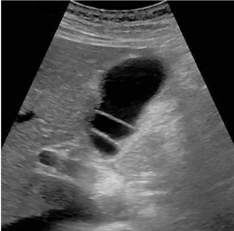
Describe this image of the GB
GB septations
Focal GB thickening can be caused by
Stones, polyps, masses or adenomyomatosis
Non-Biliary edema of GB Wall traits
thicker than that caused by Cholecystitis
Concentric thickening with a layered appearance with both linear hypoechoic (edema) and echogenic components
Often assoc w/ascites
What can simulate GB thickenining and is associated with Portal Vein Thrombosis?
GB varices, commonly associated with portal vein thrombosis, may simulate wall thickening but are distinguishable on color Doppler
Biliary Causes of GB thickening
Cholecystitis
Adenomyomatosis
Cancer
AIDS cholangiopathy
Sclerosing cholangitis
Cholangiopathy
any disease or damage affecting the bile ducts
Secondary causes of GB diffuse thickening
hepatitis
pancreatitis
heart failure
hypoprotenemia
Cirrhosis
Portal hypertension
Lymphatic obstruction
Mononucleosis

These picture are showing what?
Gallbladder (GB) wall thickening in different patients.
Longitudinal views of the GB show diffuse thickening (cursors) secondary to congestive heart failure (A), cirrhosis and portal hypertension (B), and acute hepatitis (C). A nodular liver surface is seen in the patient with cirrhosis (arrow). In the patient with hepatitis, the GB lumen is completely contracted and the coapted mucosal layers are seen as a thin echogenic line in the center of the GB.
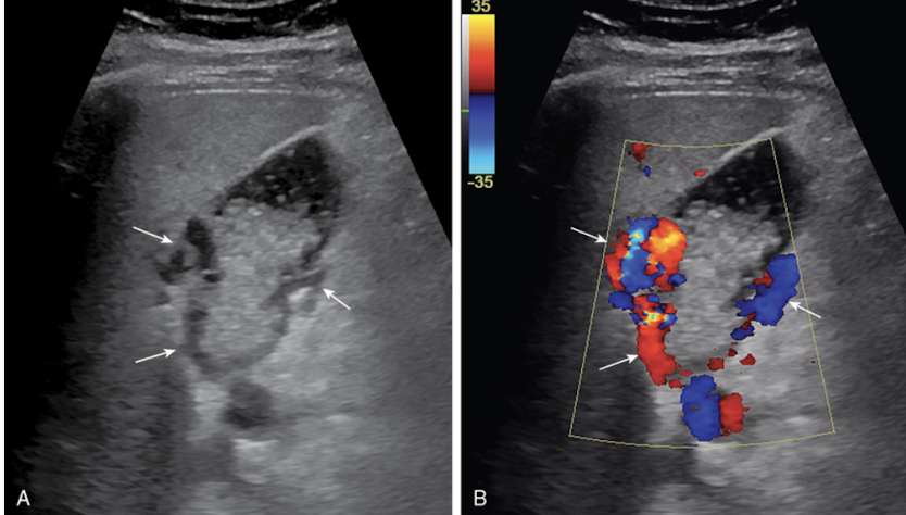
this a picture of what?
GB varices with sludge in the lumen
what chance to GB polyps have of turning cancerous
Risk of malignancy extremely small and related to size
US findings of GB polyp
Elevations or protuberances from the GB wall
Usually <1 cm; often multiple
Immobile; nonshadowing
Important to determine whether pedunculated or sessile
Doppler flow may differentiate true polyp from sludge ball; lack of Doppler flow does not exclude a polyp.
GB Carcinoma is associated ? percentage with Gallstones
95% associated with gallstones
Often appears as soft tissue mass centered in the GB fossa that completely or partially obliterates lumen
GB carcinoma
how big are gb carcinoma’s typically?
greater than 1cm (usually much larger) and sessile
GB metastases most often come from?
melanoma
if you see a Mass centered in gallbladder fossa with associated stones Eccentric, irregular wall thickening Bulky intraluminal polypoid mass…. what other areas should you check or look for?
invasion of liver
Obstruction of Bile ducts or vessels
Lymphadenopathy
nodal mets
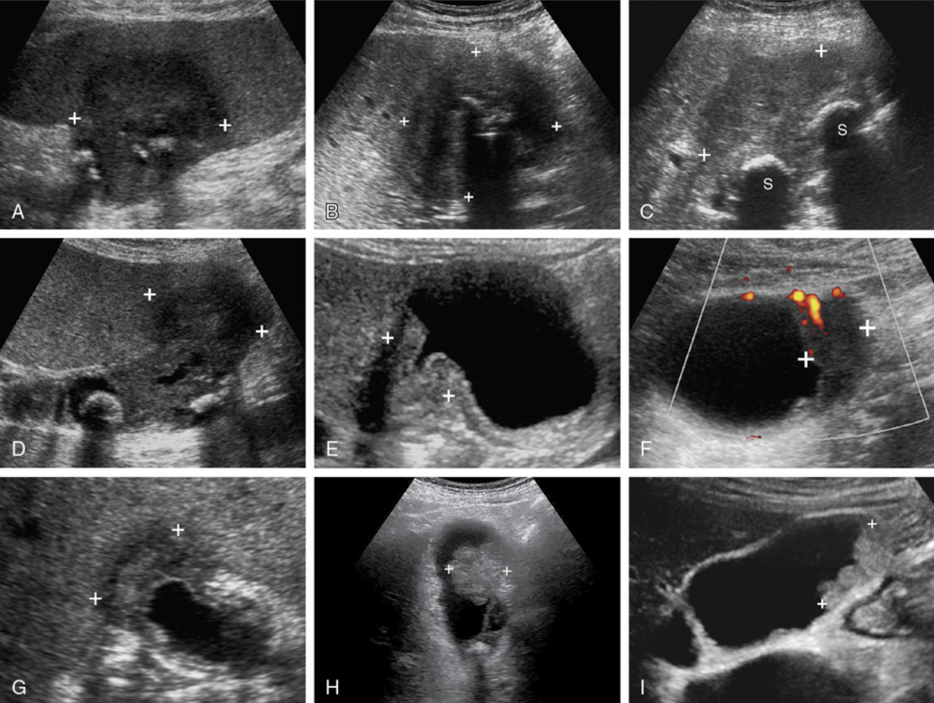
What are these pictures of?
GB carcinoma
These pictures represent
adenomyomatosis
This condition is char. by an acquired mucosal hyperplasia and thickening of the muscularis propria of the GB which causes Mucosal invaginations into the muscular wall form cystic spaces (Rokitansky-Aschoff sinuses)
adenomyomatosis
3 types of adenomyomatosis
Segmental annular thickening; often narrows lumen
Localized mass; most often fundal
Diffuse wall thickening
Classic sign of adenomyomatosis
Creates classic bright reflections with short comet-tail artifacts (most common finding)
Seen almost exclusively on near wall since comet tails only visible in background of anechoic bile within lumen
Characterized by calcification of GB wall and Associated with chronic GB inflammation
Porcelain GB
us findings of Porcelain GB
Echogenic arc with dense posterior shadowing
Less extensive calcification produces only partial shadowing; back wall of GB remains visible
In early cases, only segments of GB wall may be affected

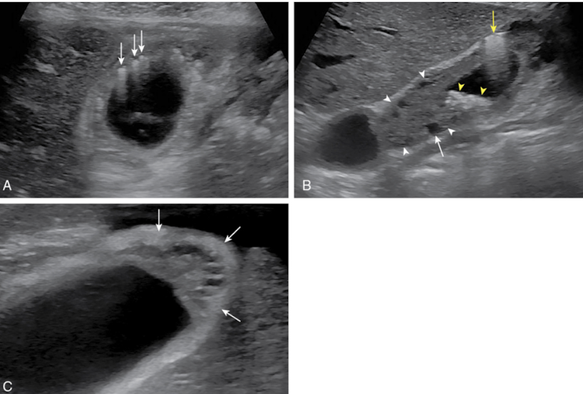
These pics are of what GB condition
porcelain gb
(A) Transverse view shows an echogenic superficial GB wall with complete shadowing of the remainder of the GB lumen and back wall.
(B) Longitudinal view of the GB shows an echogenic superficial wall with shadowing of the deep GB wall in the region of the fundus but sound penetration and visualization of the back wall in the body of the GB (arrows).
(C) Longitudinal view shows scattered areas of wall calcification (arrows) and a stone (S) in the GB neck.