The eye
1/367
Earn XP
Description and Tags
Week 9 NMSK
Name | Mastery | Learn | Test | Matching | Spaced |
|---|
No study sessions yet.
368 Terms
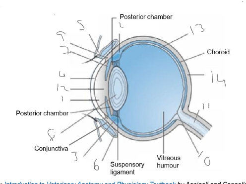
Label the parts of the eye in this image
Anterior chamber
ciliary body
ciliary muscles
cornea
eyelid
fornix
iris
lens
limbus
optic disk
optic nerve
pupil
retina
sclera

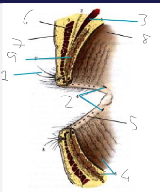
Label this diagram
cilia
lacrimal puncta
levator palpebrae superioris
meibomian glands
meibomian gland orifices
orbicularis oculi
outer skin
palpebral conjunctiva
tarsal plate

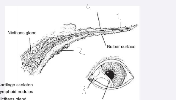
Label this diagram
cartilage skeleton
lymphoid nodules
nictitans gland
palpebral surface
Very bottom one is the T-shaped cartilage skeleton


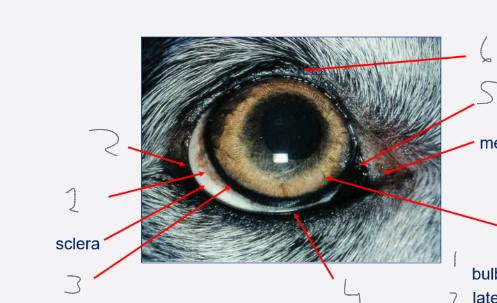
label this diagram
bulbar conjunctiva
lateral canthus
limbus
lower eyelid, no lashes
third eyelid
upper eyelid with lashes
On the right with no number
Top = medial canthus
Bottom = iris
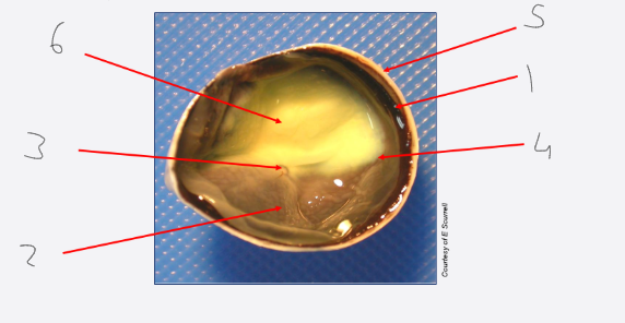
Label this diagram and what kind of segment of the eye is this?
calotte/cadaver: posterior segment (view of fundus)
choroid
non-tapetal fundus
optic nerve head/disc
retina
sclera
tapetal fundus

what does ‘calotte’ mean
half of the eye after sectioning (cadaver specimen)
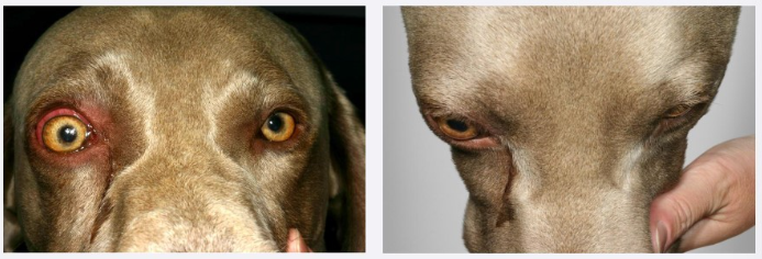
Define exophthalmos
abnormal position of the eye from the orbit
how can we recognise exophthalmos?
globe is the normal size
the globe position is pushed forward/protruding
the globe is normal

define enophthalmos
abnormal recession of the eye within the orbit
How can we recognise enophthalmos?
globe size is reduced
globe is sunken
globe appears normal

define hydrophthalmos
Enlargement of the globe
How can we recognise hydrophthalmos?
globe has an enlarged size
globe is protruding
globe appears abnormal

define microphthalmos
Congenitally abnormal (small) eye
How can we recognise microphthalmos?
globe is reduced in size
globe position is normal
globe appearance is abnormal
what is the orbit
a cavity within the skull that encloses the eye
what is the purpose of the orbit
protection
separation of the eye from the cranial cavity
why are there foramina in the walls of the orbit?
pathway for blood vessels and nerves to reach the eye
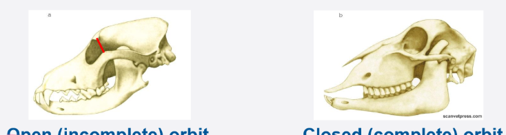
what are the 2 kinds of orbit
open/incomplete
closed/complete
Compare an open orbit to a closed orbit
Open: - lateral orbital ligament, facilitates access for a biopsy, ultrasound and surgery
Closed: fusion of zygomatic and frontal bones
What are the 2 kinds of visual fields?
monocular
binocular
What kind of vision do a)carnivores e.g. cats and b)herbivores e.g. horses have?
cats = binocular
monocular
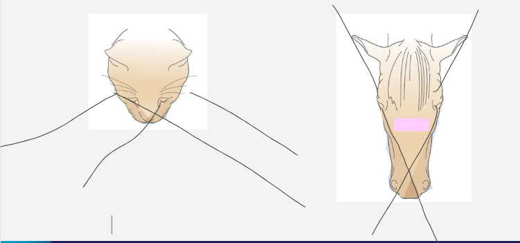
Compare:
a) orbit type
b) composition of later wall
c) degree of globe protection
d) visual field
e) degree of binocular vision
between herbivores and carnivores
a) Closed/complete Open/incomplete
b) fusion of frontal and zygomatic Lateral orbital ligament
c) greater protection less protection
d)wide field of vision narrower field of vision
e) more monocular more binocular
why do carnivores have an open/incomplete orbit
enables greater jaw movement
Provide:
A definition
depth of orbit
degree of protection offered
breed examples
of brachycephalic breeds
Skull is short and broad
shallow
reduced
pug, boston, boxer, french bulldog
Provide:
A definition
depth of orbit
degree of protection offered
breed examples
of mesocephalic breeds (mesaticephalic)
medium skull length, ‘normal’
normal
moderate
lab, german shepard, doberman, dalmation, beagle
Provide:
A definition
depth of orbit
degree of protection offered
breed examples
of dolicocephalic breeds
long skull
deep
increased
great dane, greyhound, daschund, irish wolfhound, saluki and poodle
what problems might be associated with having a brachycephalic conformation
these dogs have a greater risk of developmental disorders
some examples: shallow orbit, microphthalamia, anophthalamia and hydrocephalus with orbital malformation
Outline the anatomy of the orbit
5-7 bones (species dependent)
orbital rim: frontal, lacrimal and zygomatic bones
soft tissue structures
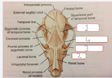
Label this image
frontal bone
maxilla
zygomatic bone
zygomatic process
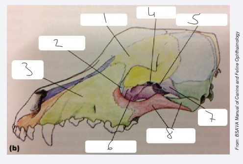
Label this image
frontal bone
lacrimal bone
maxilla
orbital canal
orbital fissure
palatine bone
sphenoid bone
zygomatic arch

outline the limits of the bony orbit
medial limit - frontal bone (separates the orbit from the nasal cavity), very thin
Dorsal limit - projection of the frontal bone, contains frontal sinus
Rostral and lateral limits: zygomatic, lacrimal and maxillary bones
caudal limit: sphenoid bone (contains the optic canal and orbital fissure)
Outline the soft tissue orbit
Dorsolateral limit - temporal muscle and the orbital ligament
rostral and lateral limits: masseter muscle (medial and ventral to zygomatic arch)
ventral floor: pterygoid muscle and neurovascular structures that transverse the orbital floor
How many foramina do we have in the orbit
8 but species variation
what are the 2 most important foramina in the orbit?
optical canal
orbital fissure
what nerves/vessels pass through the optical canal?
optic nerve
internal opthalmic artery
what nerves pass through the orbital fissure/foramen?
abducens nerve
oculomotor nerve
opthalmic nerve
trochlear nerve
what muscles does CNIII innervate around the eye
dorsal rectus
medial rectus
ventral rectus
ventral oblique
what muscles does CNVI innervate around the eye?
lateral rectus
retractor bulbi
what muscles does CNIV innervate?
dorsal oblique
where are the 2 places we can find soft tissue structures in the orbit?
intraconal
extraconal
how can we define the intraconal area?
4 rectus muscles and the periorbital fascial sheath
shaped like an ice cream cone
what does the intraconal space include
multiple nerves: optic nerve and nerves supplying extraocular muscles
vessels
smooth muscle
fat
orbital lacrimal gland
all other soft tissue orbital structures are within the extraconal space
what other important structures (not bone or soft tissue) are located within the orbit
tooth roots
zygomatic salivary glands (dogs)
paranasal sinuses
What other important structures (not bone or soft tissue) are found outside the orbit
nasolacrimal duct
base of the nictitating membrane
orbital fat cushion
where is the orbital fat cushion and what does it do
surrounds the eye and the muscles
protection
Outline the arterial blood supply to the eye
eye has a high metabolic activity; needs a rich blood supply
opthalmic artery - derived from the internal carotid artery
^ they supply the highly vascular uveal tract
retina has a dual blood supply
Outline the venous drainage of the eye and orbit
vortex veing and orbital venous plexus
smaller route via opthalmic vein
all goes to the external jugular vein
What is the clinical relevance of high ocular blood supply?
systemic hypertension → ocular damage
system diseases may cause uveitis (inflammation of the uveal tract)
what is the clinical relevance of venous damage?
When enucleating in rabbits, they have a very well formed orbital venous plexus - BE CAREFUL
careful when restraining around the neck
what is an example of a disease that affects the nasal cavity and paranasal sinuses
disease within the nasal cavity or paranasal sinus
neoplasia
what is an example of a disease that affects the caudal roots of the 4th premolar and 1st/2nd molar teeth
risk trauma during tooth extraction
tooth root abscess
what is an example of a disease that affects the brain
neoplastic or inflammatory CNS disease
what is an example of a disease that affects the temporal and masseter muscles
masticatory muscle myositis
what is an example of a disease that affects the zygomatic salivary glands
sialadenitis
define anisocoria
unequal size of the pupils
define miosis
excessive constriction of the pupil
define mydriasis
dilation of the pupil
define strabismus
cross eyed, they have an inability to align both eyes simultaneously
define nystagmus
involuntary eye movement, may result in limited or reduced vision
what is the ramus of the mandible
where the mandible moves between the zygomatic bone and cranium
it enables the jaw to be opened and closedwhat is an example of a disease that affects the
how does the ramus move
jaw opens - ramus moves towards the globe
jaw closed - aways from the globe
what is the clinical relevance of the ramus
orbital disease will often cause pain during:
eating
yawning
examination of the oral cavity
animal may yelp when trying to open the mouth
what is a useful clinical method to identify exophthalmos
looking at the animal’s head from above
How do we assess retropulsion
gently pushing on the globe through the upper eyelid
equal and non-painful protrusion of the globe
brachycephalic breeds - it’s limited because the orbit is normally shallow anyway
What should we look for whilst conducting an oral exam for indicators of eye problems?
pain on jaw movement
assess for dental disease
any history of reduced appetite, especially of dry food
What are the functions of the eye
vision
collects light from the environment
focuses light onto photoreceptors in the retina
converts light into a nerve impulse = PHOTOTRANSDUCTION
sensation of vision
Outline where light is refracted and what this is
refraction = bending of light rays
2 places:
cornea - major refraction 48/60 dioptres (man)
lens = aqueous-lens interface and the len-vitreous interface
why does major refraction take place at the cornea
air-cornea interface
what is a dioptre
unit of power of lens
recriprocal of focal length in metres
how do our eye and brain orientate images?
Eye - image is upside-down on the retina
Processing in the visual cortex - converts the image to being the correct way up
what 2 kinds of lenses do we have and what do they do to light rays
convex lens - converges light rays to a focal point
concave lens - diverge light rays
what kind of lenses are the cornea and lens
convex
What are 11 key points for vision in dogs
sensitive to motion
sensitive to flickering light
good day and night vision
visual perspective is closer to the ground
larger field of view
reduced depth perception
binocular vision
most are emmetropic (no visual error)
poor acuity
dichromatic
better at distinguishing shades of grey
what is the clinical relevance that dogs are emmetropic
short and long-sighted vision is uncommon
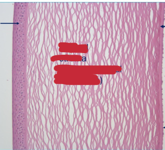
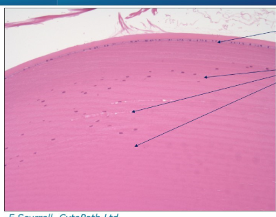
What structure is this and what can we identify?
It’s the cornea
corneal epithelium on the left (multiple layers)
corneal stroma (90% corneal thickness)
Endothelium layer on the right
Descemet’s membrane (basement membrane for endothelium)
What structure is this and what can we identify?
Lens
Anterior lens capsule (thick) on the left
Posterior lens capsule (thin) on the right
What structure is this and what can we identify
Lens equator
Lens epithelium on the top
lens fibres running through
what is the equator of the lens?
the marginal circumference where the anterior and posterior surfaces meet, playing a crucial role in the lens’s structure and function
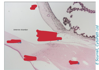
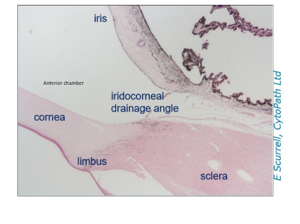
What are we looking at in this image?
Iris (top)
Iridocorneal drainage angle (middle)
Cornea (bottom left)
Limbus (bottom middle)
Sclera (bottom right)
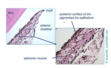
What are we looking at in this section?
Iris - uveal tract
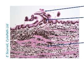
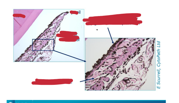
What are we looking at in this section and what can we identify
ciliary body (uveal tract)
lens zonule fragments (top)
ciliary processes
ciliary body stroma
What is the fundus?
interior surface at the back of the eye, comprising of essential structures like the retina, optic disc and blood vessels
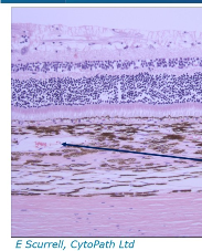
What are we looking at here and what structures can we identify?
vitreous (top)
Retina (middle)
Blood vessels in the choroid
sclera

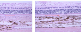
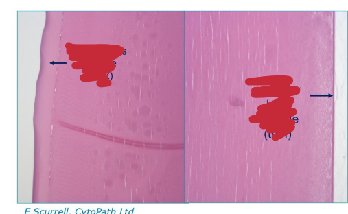
What are we looking at in this image and what is the difference between the 2?
these are images of the retina
On the left = tapetal retina
On the right = non-tapetal retina

what are the layers that are visible in these structures
Top to bottom:
retina
tapetum (more on left, as it’s tapetal retina and less on the right)
choroid
sclera
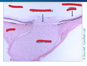
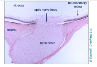
What are we looking at and what structures can we identify
Optic nerve
What setting do we want for an ocular ultrasound
B-mode US
what do we use ocular ultrasound for?
imaging inside teh globe if we can’t see inside due to cloudy cornea, cataract etc.
measure globe size
assess orbital disease
How do we prepare our patient for ocular ultrasound
conscious ± light sedation
topical anaesthetic eye drops to numb cornea
direct corneal contact, eyelids open
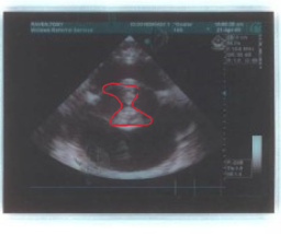
What do we see on the ocular ultrasound
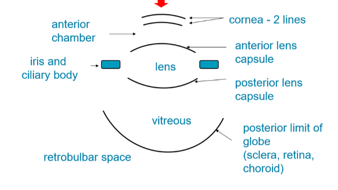
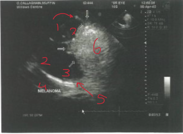
What structures are identified here?
anterior lens capsule
posterior lens capsule
vitreous
retrobellar space
posterior limit of the globe (retina, choroid)
melanoma (tumour)
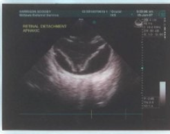
What problem can be identified in this ocular ultrasound
retinal detachment
What problem can be identified on this ocular ultrasound
cataract

What are some common indications we need a CT and MRI
orbital disease: FB, neoplasia, abscess
central blindness: brain tumour
investigation of tear duct disorders
What are 2 indicators may we get on an ocular ultrasound that we need a CT/MRI?
abnormal masses but can’t decipher what
swollen muscles visible
what fungus can cause orbital fungal infection
blastomyces dermatitidis
What diagnostic test can we use to assess optic neuritis
MRI scan
will show an abnormal optic nerve, swollen and retinal detachment
What diagnostic test may we need to use for a presentation of a swelling near the tear duct
skull radiograph with liquid contrast
CT

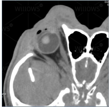
Is this a normal canine orbit? What diagnostic tool is used?
Yes
CT scan
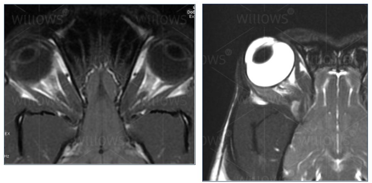
Are these normal canine orbits and what diagnostic imaging tool has been used?
MRI
yes