KIN 313 Lecture 2 - Motor units: Morphology
1/52
There's no tags or description
Looks like no tags are added yet.
Name | Mastery | Learn | Test | Matching | Spaced | Call with Kai |
|---|
No analytics yet
Send a link to your students to track their progress
53 Terms
afferent neuron direction
from periphery to CNS
who is the sexiest man of all time
Saad, duh
Large dicks have larger _____ ?
Motor units
efferent neurons direction
from CNS to periphery
motor units exit the spinal cord how?
from anterior horn via ventral root
these are efferent axons
how does sensory info enter the spinal cord?
comes through dorsal route ganglion, down the dorsal routeand into the spinal cord
it enters the grey matter and can either talk directly to motor neurons (via interneurons) or go to the brain via sensory axons
where are sensory axons in spinal cord
posterior aspect of grey matter
why is grey matter grey
packed with cell bodies
posterior aspect of spinal cord contains
mostly sensory axons going to the brain
ventral side of spinal cord contains
mostly motor pathways going down/out to periphery
morphological features/anatomy of a motor neuron
large dendritic tree (branches of collateral, spherical, going out in all directions like a dandelion)
soma (cell body)
axonal hillock (where APs are generated)
axon (heavily myelinated, APs travel down this to reach muscle)
presynaptic terminal
motor neuron pool
all the motor neurons innervating one particular muscle
every muscle has its own MN pool
MN pool exists in the brainstem and spinal cord
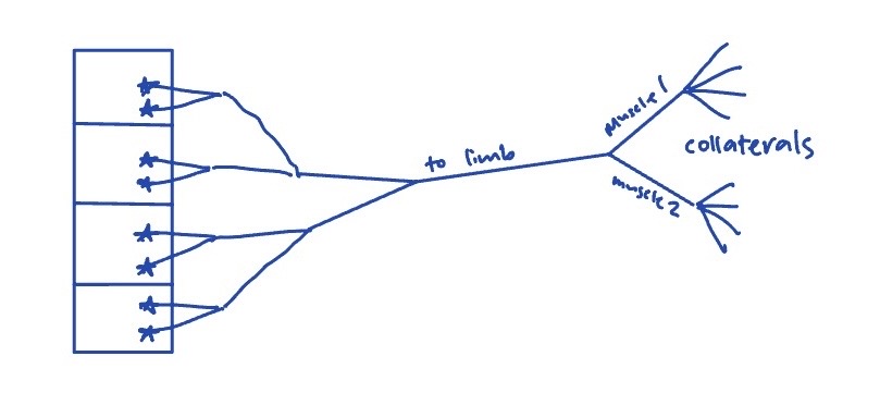
the axon of a motor neuron biforcates to form…
collaterals… other neurons can come synapse onto these collaterals to make connections
Neuromuscular synapse is modified specifically to…
release ACh onto the surface of the muscle over the zone of overlap of the fillaments
neuronal cell bodies for a muscle are found..
housed together in the spinal cord, but across several segments of the spinal cord
all motor neurons go out the respective ventral route of their segment to eventually reach their muscle
motor neuron pools crossing several segments: implications for spinal cord injuries
if you get an injury in a place where there are still some motor neurons for a particular muscle leaving from higher segments, then you will still have some form of functionality of the muscle
spinal cord enlargement based on location?
the fact that the grey matter of the spinal cord is bigger around the arms (cervical) and legs (lumbar) because there are a lot of muscles in these areas that need to be controlled
mediolateral distribution of spinal cord
location of motor neurons withing the spinal cord is indicative of…
the muscle they are innervating
motor neurons for proximal muscles are found i nthe medial portion of the spinal cord
motor neurons for distal muscles are found in the lateral portion of the spinal cord
the motor unit
the cell body and dendrites of a motor neuron, the multiple branches of its axon (collaterals), and the muscle fibres that it innervates
muscle unit
all the muscle fibres belonging to a motor unitm
motor unit vs muscle unit
motor unit includes all parts of a motor neuron and the muscle fibres that it innervates.
muscle unit only refers to the muscle fibres that belong to a motor unit
how many motor neurons can innervate a muscle fibre
only one motor neuron can speak to a muscle fibre. there is not competition
path of an AP from motor neuron in spinal cord to the muscle
MotN sends AP, which goes down axon, down each collateral, until it reaches the end plates, leading to a contraction of the muscle
implications of one motor neuron innervating a lot of muscle fibres
one AP causes ALL those muscle fibres to contract at the same time
the muscle unit contracts together
path of motor neurons going from spinal cord to the muscle
they leave the spinal cord through the ventral route of whichever segment they are in, then join together as the nerve leaving that segment, then join with the motor neurons from the other segments to form plexi before eventually spliting off to form the nerves for specific muscles and then splitting off further to innervate their respective muscle fibres
distribution of a muscle unit in a muscle
why?
the muscle fibers are not all packed together, but spread out throughout a compartment of a muscle
dispersing reduces twitchy movements
innervation ratio
the number of muscle fibres innervated by a single motor neuron
relationship between force production and innervation ratio
high innervation ratio = high force production (gastroc = 1:1900)
lower innervation ratios = lower force production (eye muscles = 1:15)
relationship between innervation ratio and control/precision
aka implications of innervation ratio for motor control
high innervation = high force production but low control/precision
low innervation = low force production but high control/precision
gastroc = high = strong = low precision
FDI = medium
eye = low = weak = high precision
innervation ratio and aging
Innervation ratio increases
# of MotUs decreases, so other MotUs have to pick up the slack
nervous system sends collaterals from healthy neurons to connect with the muscle fibres that are no longer receiving signals so they can work again
this results in an increased innervation ratio
Amyotrophic Lateral Sclerosis (ALS)
death of motor neurons
healthy neurons send out collaterals to try to pick up slack
death outpaces help until there are no MotUs left in that muscle
Signs and Symptoms: muscle fibres that are abandoned contract out of sync = squirming sensation
Progression: 2-5 years
three types of skeletal muscel fibres
slow oxidative (SO)
fast oxidative-glycolytic (FOG)
fast glycolitic (FG)
Characteristics of Slow MotorNeurons
low innervation ratio
low CSA
conducts signals slower than others
high input resistance = easier to excite (takes less current to trigger AP) → low rheobase
long afterhyperpolarization period (long refractory period, longer time between APs being fired)
Innervates Slow Oxidative muscles
high vascularity and mitochondria → can make lots of ATP → slow to fatigue
Characteristics of Fatigue Resistant Motor Neurons
Hybrid
medium innervation ratio
medium CSA
medium conduction speed
medium input resistance = medium current needed to trigger AP
medium rheobase
medium refractory period
Innervate FOG muscles
medium vascularity and mitochondria → makes some ATP → fatigue resistant
more blood supply than FG, more stored pyruvate than SO
Characteristics of Fast Fatiguable Motor Neurons
high innervation ratio
high CSA
fast conduction speed
Low input resistance (large current needed to trigger AP) → high rheobase
low refractory period (can fire another AP quickly after the last one)
innervates fast glycolytic muscle fibres
low vascularity, low mitochondria → fatiguable
high pyruvate storage → fast ATP making but runs out fast
high force production with contraction but very fatiguable
how motor neurons can be classified
who they connect to (fibre type)
muscle they innervate
innervation ratio
relationship between input resistance and size
large motor neuron = low input resistance (harder to excite)
small motor neuron = high input resistance (easier to excite)
small have high resistance because after putting in a little bit of current, its already hard to put in more. they are easier to excite because then they are already full and at threshold
opposite applies to large motor neurons
equation for Voltage (needed to fire AP)
Voltage = Current x Resistance
if you have a large input resistance, how much current do you need to reach voltage required to trigger an AP
small current
Rheobase
how much current is required to excite the motor neuron enough to trigger an action potential
low rheobase level in small Motor Neurons because their resistance is so high
absolute refractory period
where the polarization of a neuron goes super negative, too negative to be able to fire another AP
what is a twitch and how to calculate
a force time response of a motor unit to a single input/stimulation
twitch = change in force divided by time
contraction time
time from start of force build up to peak force
temporal summation
one motor neuron firing repeatedly so that the motor unit doesnt get to fully reset between twitches
unfused tetanus
oscillating force from muscle fibres due to summation of impulses
fused tetanus
APs firing so fast there is no oscilation in force production
physiologically doesnt happen naturally bc the muscle would burn out
would happen if you get shocked by electricity
Half relaxation time (HRT)
time for a motor unit to go from peak force to 50% force
in order for temporal summation to work, must hit the unit again before it hits HRT
peak, contraction time, and HRT of a slow contracting fatigue resistant motor unit
low peak, long CT, long HRT
peak, contraction time, and HRT of a fast contracting fatigue resistant motor unit
medium peak, meadium CT, medium HRT
peak, contraction time, and HRT of a fast contracting fast fatiguing motor unit
big peak, short CT, short HRT
why is it hard to fatigue a slow oxidative muscle
high vascularity
the whole metabolic system is there to take waste out and bring atp in, so it can generate force much longer
why fast glycolytic muscles fatigue so fast
low vascularity, rely on the pyruvates already in muscle
use this up quickly, then theres none left = fatigued = cant keep generating significant force even though it is stimulated
why the best powerlifter cant become the best marathon runner no matter the training
born with what you have
training can change muscle fibre size, but cannot change innervation ratio, vascularity
behaviour of muscle fibres can change, but not dramatically
you cant change their physiology