Fetal Environment and Maternal Complications
 Call Kai
 Learn
Learn Practice Test
 Spaced Repetition
 Match
Match Flashcards
 Knowt Play
1/130

There's no tags or description
Looks like no tags are added yet.
Name | Mastery | Learn | Test | Matching | Spaced | Call with Kai | Chat |
|---|
No analytics yet
Send a link to your students to track their progress
131 Terms
what is the maternal contribution of the placenta
decidua basalis, the endometrium beneath the developing placenta
what is the fetal contribution to the placenta
The chorion frondosum, the portion derived from the blastocyst and containing the chorionic villi
The placenta produces human chorionic gonadotropin, which maintains the
corpus luteum of the ovary.
In later pregnancy, the placenta also produces
estrogen and progesterone, taking over that function from the corpus luteum.
. One major function of the placenta is to
act as an excretory organ for the fetus, performing imperative exchanges of waste products and gases with valuable nutrients and oxygen from the mother
A definitive placenta may not be identified sonographically until after
after 10 to 12 weeks.
The placenta consists of three parts:
the chorionic plate: element of the placenta closest to the fetus.
the placental substance: the functional parts of the placenta and is located btw chorionic plate and basal layer
the basal layer or basal plate: area adjacent to the uterus.

what are Venous lakes, also referred to as maternal lakes or placental lakes
pools of maternal blood within the placental substance. Anechoic or hypoechoic and may contain swirling blood. Little clinical significance

A bilobed placenta consists of
two separate discs of equal size connected by an isthmus of placental tissue

what is an accessory lobe or a succenturiate lobe of the placenta,
additional smaller lobes located separate from the main segment of the placenta
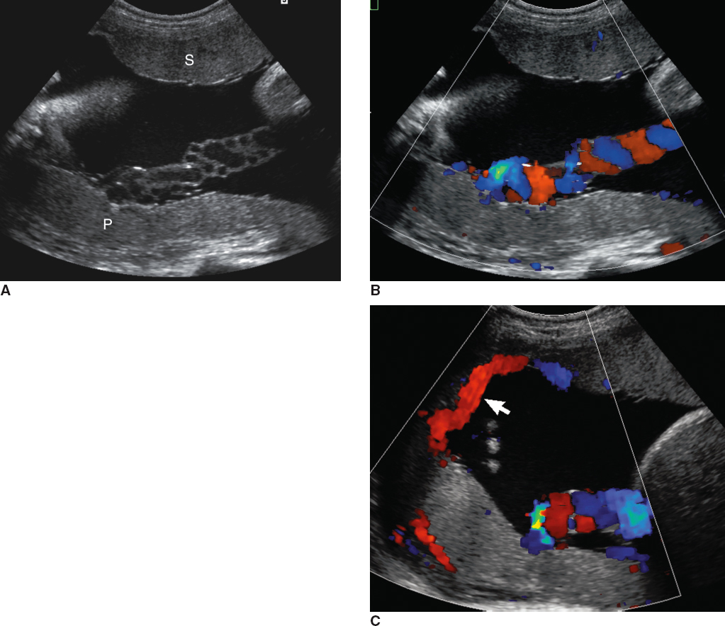
what is a circumvallate placenta
an abnormally shaped placenta caused by the membranes inserting inward from the edge of the placenta, producing a curled-up placental contour

A circumvallate placenta may lead to
vaginal bleeding and placental abruption, among other complications.
with advancing gestation,
Calcifications may be noted within the placenta, and indentations may be seen within the basal and chorionic plates
Classic placental grading with associated sonographic findings
Grade 0 - Uninterrupted chorionic plate and homogeneous placental substance
Grade I - Subtle indentations on the chorionic plate, with some small calcifications within the placental substance
Grade II - Moderate indentations in the chorionic plate with “comma-like” calcification in the placental substance
Grade III - Prominent indentation in the chorionic plate that extends to the basal layer with diffuse echogenic and anechoic areas noted within the placental substance

The thickness of the placenta should be evaluated with sonography. It should not exceed
4 cm
a thick (placentomegaly) and thin placenta are both associated with
maternal and/or fetal abnormalities

Possible causes of a thick placenta (6 causes)
Diabetes mellitus
Maternal anemia
Infection
Fetal hydrops
Rh isoimmunization
Multiple gestations
Possible causes of a thin placenta (6 causes)
Diabetes mellitus (long standing)
IUGR
Placental insufficiency
Polyhydramnios
Preeclampsia
Small-for-dates fetus
what is placenta previa
placenta covers the internal os of the cervix.
Placenta previa is a common cause of
painless vaginal bleeding in the second and third trimesters
placenta previa is more commonly found in mothers who have
Multiparous
AMA
Prior abortion
Prior c-section
Term Associated with Placenta Previa
Complete (total) previa - Placenta covers the internal os completely
Partial previa - Placenta partially covers the internal os
Marginal previa - Placenta lies at the edge of the internal os
Low-lying previa - placenta edge is 2cm away from internal os

The placenta should be evaluated for placenta previa after
20 weeks with an empty maternal bladder using a transabdominal approach
a cause of false-positive placenta previa is
painless myometrial contractions that occur in the lower uterine segment.
Clinical Findings of Placenta Previa
Previous C-section or uterine surgery
Painless vaginal bleeding
Possibly asymptomatic
what is vasa previa
fetal vessels resting over the internal os of the cervix. May lead to exsanguination of the fetus
Vasa previa is often associated with
velamentous cord insertion and, possibly, a succenturiate lobe
Sonographic Findings of Vasa Previa
Identification of vessels over the internal os of the cervix with the use of color Doppler
Velamentous cord insertion

Placental abruption, also referred to as abruptio placentae, is the
premature separation of the placenta from the uterine wall before the birth of the fetus, thus causing hemorrhage
Placental abruption can be further described as
complete abruption -- (most severe) retroplacental hematoma between uterus and myometrium
partial abruption - few cm of separation
marginal abruption - subchorionic hemorrhage (edge of placenta)

Maternal conditions that are linked to the development of placental abruption include (6 conditions)
hypertension
preeclampsia
cocaine use
cigarette smoking
poor nutrition
trauma.
Clinical Findings of Placental Abruption
Abdominal pain (often sudden onset)
Possible vaginal bleeding
Uterine contraction
Uterine tenderness
Sonographic Findings of Placental Abruption
Hematoma located either at the edge of the placenta or between the placenta and the myometrium

Placenta accreta is frequently used as a universal term to describe the condition that is defined as the
abnormal adherence of the placenta to the myometrium in an area where the decidua is either absent or minimal. The placenta will not detach after birth
The placenta may attach to a uterine scar following a previous C-section and/or after uterine surgery. This explains the association between
anterior placenta previa and placenta accreta.
There are three different terms associated with this abnormality:
placenta accreta - adhesion and loss of clear zone
placenta increta - invasion of the placenta into the myometrium
placenta percreta - progression to urinary bladder or other organs, thus causing urinary complications

Depending upon the amount of invasion or penetration of the placenta into the myometrium, the patient could suffer from heavy bleeding at delivery and, possibly, uterine rupture. Therefore, an
emergency hysterectomy may be warranted
Clinical Findings of Placenta Accreta, Placenta Increta, and Placenta Percreta
Previous C-section or uterine surgery
Painless vaginal bleeding if placenta previa is present
Possibly asymptomatic
Sonographic Findings of Placenta Accreta, Placenta Increta, and Placenta Percreta
Placenta previa (frequent associated finding)
Loss of the normal hypoechoic interface between the placenta and the myometrium

what is the retroplacental clear zone?
normal hypoechoic interface between the placenta and the myometrium

what is the most common placental tumor
Chorioangioma
The most common location of Chorioangioma
is adjacent to the umbilical cord insertion site at the placenta
They typically do not carry any risks to the fetus or mother, although larger chorioangiomas have been associated with
polyhydramnios
intrauterine growth restriction (IUGR)
fetal hydrops
Clinical Findings of a Chorioangioma
Possible elevation in maternal serum alpha-fetoprotein
Sonographic Findings of a Chorioangioma
Solid hypoechoic or hyperechoic mass within the placenta (common area is adjacent to chord insertion)
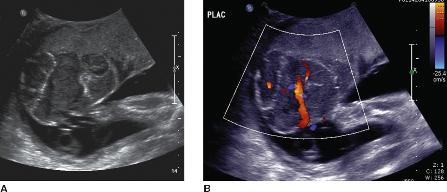
Uterine synechiae, also referred to as amniotic sheets, are
linear bands of scar tissue within the uterus.
Although uterine synechiae are typically isolated, uterine synechiae have been associated with
premature rupture of membranes (PROM), premature delivery, and placental abruption.
Sonographic Findings of Uterine Synechia(E)
Linear, echogenic band of tissue traversing the uterine cavity
The band does not involve fetal parts, and the normal fetus appears to move freely

The umbilical cord, which normally inserts into the middle of the placenta, has
two arteries and one vein, surrounded by a gelatinous material called Wharton jelly
The cord develops from the fusion of the
yolk stalk and the vitelline duct (omphalomesenteric duct) early in gestation.
The 3 Vessel View

what is the most common abnormality of the umbilical cord
A single umbilical artery, or two-vessel cord (2VC); assoc. with abnormalities of all major organ systems and IUGR
Fetuses with a 2VC have an approximate
20% chance of having additional abnormalities, a thorough examination of the fetus for other findings is warranted.
The umbilical cord normally inserts into the central portion of the placenta. Abnormal cord insertion sites are described as either
Marginal cord insertion (battledore placenta) - insertion at edge of the placenta
Velamentous cord insertion - insertion in membranes beyond the placental edge

abnormal insertion is often seen in association with
vasa previa
Occasionally, the umbilical cord may be seen encircling the fetal neck. This is termed
nuchal cord

what is An allantoic cyst
a mass that may be noted in the umbilical cord adjacent to the umbilical vessels (NOT VASCULAR)
Sonographic Findings of Allantoic Cysts
Cystic mass within the umbilical cord
Most often noted close to the fetal abdomen

Umbilical cord cysts are most often found near the fetal abdomen and have been seen in connection with
omphalocele and aneuploidy, especially if noted in the second or third trimester
what is an umbilical vein varix
the focal dilatation of the abdominal portion of the umbilical vein

umbilical vein varix has been associated with
fetal aneuploidy,
IUGR,
hydrops,
demise.
What is the most common tumor of the umbilical cord
hemangioma
Sonographic Findings of Hemangiomas of the Umbilical Cord
Solid hyperechoic mass within the umbilical cord
Most often noted close to the cord insertion into the placenta

Fetal well-being can be evaluated using pulsed Doppler of the umbilical cord by measuring the systolic-to-diastolic ratio (S/D ratio). The S/D ratio assesses the vascular resistance in the placenta by taking a sample of the umbilical artery. It can be performed anywhere along the length of the cord, although a free loop of cord will tend to offer the most accurate measurement. Normally, the S/D ratio will
decrease with advancing gestation
Therefore, an elevated S/D ratio with absence or reversal of diastolic flow in the umbilical artery is associated with
increased placental resistance and an increase in the risk of perinatal mortality and morbidity i.e : IUGR and oligohydramnios

. Echogenic debris in the amniotic fluid may be
vernix or meconium, with meconium being the least likely to be observed
The fluid that is seen in early gestation is thought to arise from water and various other materials passing freely through the membranes surrounding the embryo. In the second half of the pregnancy, the
fetal kidneys and lungs produce the majority of amniotic fluid, with urine being the greatest contributor
The maximum vertical pocket, also referred to as the deepest vertical pocket, can be used to asses volume of amniotic fluid. This pocket should contain no fetal parts or umbilical cord and measure at least
2 cm, with a normal range between 2 and 8 cm
The most widely accepted means of evaluating the volume of amniotic fluid is the amniotic fluid index (AFI). The AFI is measured using the
anteroposterior dimensions obtained from the four quadrants of the amniotic sac and adding them together. measurements should not include fetal parts or umbilical cord.
Fetal malformation and complications associated with oligohydramnios (GENITOURINARY)
Bilateral multicystic dysplastic kidney disease
Bilateral renal agenesis
Infantile polycystic kidney disease (ARPKD)
IUGR
Posterior urethral valves
PROM
Fetal malformations and complications associated with polyhydramnios
Cardiac and/or chest abnormalities
Chromosomal abnormalities
Duodenal atresia
Esophageal atresia
Gastroschisis
Omphalocele
Neural tube defects
Rh incompatibility
TTS
TORCH, an acronym that stands for
toxoplasmosis, other infections, rubella, cytomegalovirus, and herpes simplex virus, is a group of infections that can cross the placenta and influence the development of the fetus.
what is the most common congenital infection
cytomegalovirus
Sonographic Findings of Torch
Intracranial calcifications
Microcephaly
Microphthalmia
Ventriculomegaly
Hepatosplenomegaly

what is the most common cause of intellectual disability in the United States
Fetal alcohol syndrome (FAS)
Sonographic Findings of Fetal Alcohol Syndrome
Microcephaly
Dysgenesis of the corpus callosum
Microphthalmia
Malformed ears
Long round philtrum
Cleft palate
Heart defects such as ventricular septal defects

what is Intrauterine Growth Restriction
an estimated fetal weight (EFW) that is below the 10th percentile at a given gestational age.
IUGR typically results from the
inadequate transfer of nutrients from the mother to the fetus and thus is the dysfunction of the placenta
The fetus is at risk of IUGR if the mother suffers from
chronic disease,
drinks alcohol,
smokes cigarettes,
has poor nutrition,
is younger than 17 or older than 35 years,
or has a history of previous pregnancies that were considered growth restricted
The measurement that should be scrutinized closely in fetuses that are at risk for growth abnormalities is the
abdominal circumference (AC), because it carries a sensitivity of greater than 95% for the diagnosis of IUGR.
The fetus with IUGR can be monitored with sonography by evaluating the flow within the umbilical artery with the S/D ratio . The S/D ratio evaluates the sufficiency of the placenta by pulsed Doppler interrogation of the umbilical artery. An abnormally high S/D ratio, resulting from a reversal or absence of diastolic flow within the umbilical artery, is associated with a
poor outcome
Suspected causes of symmetric intrauterine growth restriction
Genetic disorders
Fetal infections
Congenital malformations
Syndromes
Suspected causes of asymmetric intrauterine growth restriction
Placental dysfunction
Nutritional deficiency
Oxygen deficiency
When a fetus presents with measurements that lead to suspected small for gestational age, sonography can further assess the pregnancy for signs of IUGR by performing the biophysical profile

Doppler assessment of the middle cerebral artery has also been shown effective at evaluating for
hypoxia in a fetus that is measuring small for dates
the pulsatility index of the middle cerebral artery varies with gestational age but normally decreases as the pregnancy progresses toward term. When comparing the two Doppler signals, the middle cerebral artery should generate a
higher resistance flow pattern than the umbilical artery

Maternal uterine artery Doppler may be useful at anticipating the progression of IUGR in high-risk pregnancies. In the first trimester, the uterine artery is analyzed with spectral Doppler before it enters the uterus at the level cervix. the portion of the main uterine artery as it crosses over the external iliac artery may be utilized for this sample in the second/third trimester. The normal flow pattern is said to be
low resistance (high resistance is abnormal)

Abnormal flow may also be a forewarning of
preeclampsia and preterm delivery.
In the neonatal period, macrosomia is technically defined as the
neonate that measures more than 4,500 g in nondiabetic mothers and 4,000 g in diabetic mothers (90th precentile)
Mothers who are prone to have a macrosomic fetus are those who suffer from
diabetes, whether pregestational or gestational.
A macrosomic fetus is predisposed to
shoulder dystocia secondary to fetal size and has an increased risk of hypoglycemia and lifelong struggles with obesity. Therefore, they will most often be delivered by means of C-section.
occasionally, during the second half of pregnancy, the cervix can be difficult to image with a transabdominal approach. Translabial scanning, also referred to as transperineal scanning, can offer a useful, noninvasive glimpse at the cervix
ccasionally, during the second half of pregnancy, the cervix can be difficult to image with a transabdominal approach. Translabial scanning, also referred to as transperineal scanning, can offer a useful, noninvasive glimpse at the cervix
Cervical incompetence, or an incompetent cervix, is the
painless dilation of the cervix in the second or early third trimester.
what is an early sign of an incompetent cervix
Funneling of the cervix is a result of the premature opening of the internal os and the subsequent bulging of the membranes into the dilated cervix.
Funneling width can be measured
May take a “U” or “V” shape

Patients who are at risk for cervical incompetence include those with
uterine malformations
previous pregnancy loss in the second trimester
Intrauterine exposure to diethylstilbestrol
A sonographic assessment of the cervix can be performed with transvaginal or translabial imaging as mentioned earlier. There are two measurements of the cervix that can be obtained utilizing these techniques. The most often employed sonographic measurement for the assessment of cervical incompetence is the length measurement taken from the
internal os to the external os,
The cervical length should measure at least 3 cm.
Therefore, the shorter the cervical length, the more likely the patient will suffer from preterm delivery
The treatment of an incompetent cervix is a
cerclage (Shirodkar and the McDonald.)
Clinical Findings of Cervical Incompetence
Painless dilation of the cervix
PROM
Vaginal bleeding
Sonographic Findings of Cervical Incompetence
Cervical length of less than 3 cm
Funneling of the cervix (can produce a “U” or “V” shape)

Fetal hydrops can be categorized as either immune or nonimmune
Immune hydrops is caused by the absence of a detectable circulating fetal antibody against the red blood cells in the mother. This results in incompatibility between the fetal and maternal red blood cells, a condition known as erythroblastosis fetalis