microbio test 1
 Call Kai
 Learn
Learn Practice Test
 Spaced Repetition
 Match
Match Flashcards
 Knowt Play
1/124

There's no tags or description
Looks like no tags are added yet.
Name | Mastery | Learn | Test | Matching | Spaced | Call with Kai | Chat |
|---|
No analytics yet
Send a link to your students to track their progress
125 Terms
hippocrates
“father of western medicine”
thucydides
father of scientific history
early concept of immunity
marcus terentius varro
proposed that things we cannot see could cause disease
al-Razi (rhazes)
experimental methods to test aspects of medicine
meat rotting & hospital location
efforts to identify disease cause, not just focus on symptoms
ibn sina (avicenna)
canon of medicine
described contagion, infection, and transmission
illness isolation & quarantine
leeuwenhoek
used magnifying lens to observe “animalcules” in rain drop
father of microbiology
Louis Pasteur
Fermentation and spoilage caused by microbes
Pasteurization
Vaccines (e.g. rabies)
Robert Koch
Established method for connecting pathogen with specific disease
Koch’s postulates
Anthrax, cholera, tuberculosis

Phylogenetic analysis
Molecular techniques were implemented by Woese & Fox
Strains
Closely related genetic variants within a species
Sublevels: kingdom, phylum, class, order, family, genus, species, strain
Naming organisms
Binomial nomenclature – two-word system that names organisms based on genus and species
Names generally have Latin or Greek roots
Written out in italics
Genus is capitalized; species is lower case
Genus can be abbreviated after initial use
Cellular Microbes
Prokaryotes
- Bacteria
- Archaea
Eukaryotes
- Protists
- Fungi
- Helminths
Acellular microbes
Viruses
Prions
Archaea
Different evolutionary history, genetics, metabolism, physiology from bacteria
Pseudopeptidoglycan
Extreme environments
Protists
algae, protozoa, water molds & slime molds
Algae
plant-like; photosynthetic and important in ecosystems and consumer products
Protozoa
animal-like: very diverse and backbone of many food webs
Water molds
fungal-like; (oomycetes) often parasites or decomposers
Slime molds
fungal-like; “intelligent” and have both single-and multicellular life stages
Fungi
Multicellular and unicellular (yeasts)
Chitin cell walls
MOLDS: good & bad
Helminths
Multicellular parasitic worms
Microscopic eggs and larvae
Viruses and Prions
Acellular
Require a host to reproduce
Prion: proteinaceous disease-causing agent
chapter 2 starts here : )
>>>>
Properties of light
Wavelength = length between peaks
Amplitude = height of peaks
Frequency = rate of peaks in time
Lower wavelengths
lower frequency
Light interactions
Reflection = wave bounces off material
Absorbance = wave is captured
Transmission = wave travels through
Interference = interacts w/ another wave
Diffraction = bent or scattered by object opening
Refraction = change direction and/or speed
Refraction
Refractive index – degree of change in transmission speed
Higher refractive index causes more bending light
refractive index = speed of light in vacuum / speed of light through material
Convex lens refraction occurs on a curved boundary to meet a focal point (microscope)
Concave lens refracts light away from a focal point (flash lights)
Electromagnetic radiation spectrum
Higher frequency waves have higher energy (photons move faster)
Certain materials can refract non-visible into visible (e.g fluorescence)
Magnification
ability of a lens to enlarge the image of an object (microscopes refract light to magnify images)
Contrast
visible differences between parts of a specimen through brightness or intensity; enhanced by staining
Resolution
ability to tell that two separate points/objects are separate
wavelengths
SHORTER wavelengths = HIGHER resolution
LONGER wavelengths = LOWER resolution
Antonie van Leeuwenhoek
utilized 1st simple microscope
Galileo Galilei
used a compound microscope
Robert Hooke
first to describe “cells” using compound microscope and observing cork cells
Darkfield microscopy
Brightfield with modified condenser with an opaque disk (light stop)
Light that is viewed through objective lens is reflected/refracted from specimen
Good for viewing live specimen and/or ones that cannot be stained
Phase contrast microscope
Increases contrast w/o stain
Good for viewing live specimen and organelles
Features are highlighted based on refractive index
Differential interference contrast microscope
Similar to phase contrast
Two different wavelengths are passed thru specimen and combined for differential effects
Fluorescent microscope
Fluorochromes are used to absorb & refract into visible light
Direct & indirect (immunofluorescent)
Confocal microscope
Scans multiple z-planes
Thick specimen (e.g. biofilms)
Modified fluorescence to avoid “bleaching”
Two-photon microscope
Good for viewing thicker materials (brain slices, embryos, organs, etc)
Modified confocal scope near with near infrared light
Minimizes light scattering through tissue
Electron microscopy
Recall: resolution is limited by wavelength
Electron microscopes use electrons and have short wavelengths (no use of light)
Two types
Transmission electron microscope (TEM)
Scanning electron microscope (SEM)
Transmission electron microscope
Forms image based on varying opacity of specimen\
Specimen are cut very thin
View internal structures
Scanning electron microscope
Electrons are bounced off specimen w/ coating
Surfaces are easily observed
TEM
Samples analyzed must have very thin sections (ultramicrotome)
Specimen embedded in plastic resin and dehydrated
Stained with electron dense heavy metals
SEM
More dehydrated – critical point drying with liquid CO2
Sputter-coated with metal
Staining specimen
Wet mount – good for viewing live specimens
Fixed mount (smear) -- good for staining
Basic stain
positively charged ions
Basic fuchsin, crystal violet, malachite green, safranin, methylene blue
Acidic stain
negatively charged ions
Eosin, rose Bengal, acid fuchsin
Positive stain
Dye/stain is absorbed into cells
Negative stain
Dye/stain absorbed into background
simple stains
emphasizes structure
single stain
differential stains
differentiates organisms based on stain interactions
2+ stains
gram stain
distinguishes diff cell wall components
important clinical diagnostic tool
gram stain (STEPS)
primary stain (crystal violet)
mordant (iodine)
decolorizer (alcohol)
counter stain (safranin)
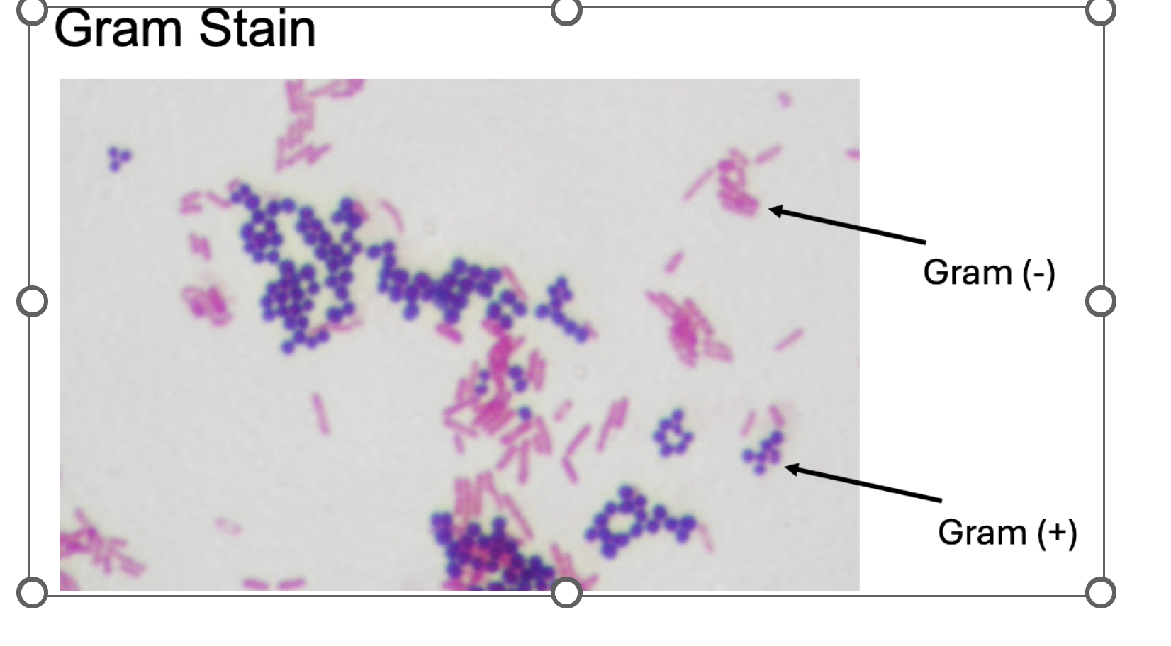
pink = negative
purple/violet = positive
acid fast stain
diagnostic tool for detection of mycolic and Mycobacterium spp
Ziehl-Neelson method (w/ heat)
Kinyoun method (w/o heat)
acid fast stain (STEPS)
heat fix smear
primary stain (carbolfuschin)
decolorizer
counter stain (methylene blue)
endospore stain
identification of endospore formers
Schaeffer Fulton method
endospore stain (STEPS)
heat fix smear
primary stain (malachite green)
decolorzier (water)
counter stain (safranin)
capsule stain
diagnostic tool for detection of protective coating
dyes do not penetrate capsule
capsule stain (STEPS)
no heat smear
primary stain (india ink)
flagella stain
identification of flagella appendages
bacteria, archaea, eukaryotes
flagella stain (STEPS)
no heat smear
primary stain (specialized)
decolorizer (water)
counter stain (carbol fuschin)
chapter 3 starts here : )
>>>>>
cell theory
robert hooke - cork tissue “cells” (1665)
matthias schleiden - plant tissue (1838)
theodor schwann - compared plant and animal cells (1839)
robert remak - cells come from other cells (1852) cell division
rudolf virchow - published “cellular pathology” (1855) “all cells arise from cells”
endosymbiotic theory
lynn margulis (1967) - mitochondria and chloroplasts of prokaryotic origin
^^ 1981 - symbiosis and cell evolution
DNA, ribosomes, binary fission
**** mitochondria and chloroplasts were orginally prokaryotic cells thar established a symbiotic relationship within a eukaryotic host
germ theory of disease
Diseases may result from microbial infection
• Girolamo Fracastoro (1546)
• ”spores” can be transferred between individuals
• Ignaz Semmelweis (1847)
• “Contaminated” physicians transferred causative agent to patients
• Promoted handwashing as solution
• John Snow (1848)
• cholera outbreaks in London traced to sewage in drinking water
louis pasteur (1856)
organisms could spoil food and therefore people
joseph lister (1857)
handwashing + carbolic acid in surgery for disinfection
robert koch (1884)
koch’s postulates - a specific microbe can cause a specific disease
the prokaryotic cell
nucleoid
inclusions
plasmids
pili
fimbriae
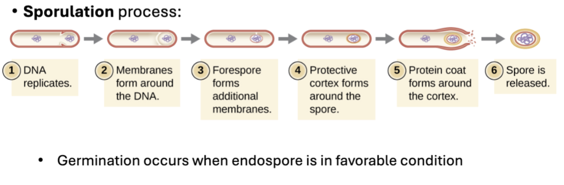
endospore
nucleiod (prokaryotic cell)
Central region of cell w/ DNA & DNA associated proteins
Chromosomes(s) are circular and haploid
Plasmids – circular, non-essential DNA
Prokaryotic ribosomes
Sites of protein synthesis
Prokaryotic – 70S size (50S + 30S)
inclusions (prokaryotic cell)
Structures used for storage of excess nutrients, movement, or specialized metabolism
Storing nutrients in a polymerized form aids in reducing osmotic pressure
endospores (prokaryotic cell)
Structure formed in dormant state to protect genome
Most resilient state of bacteria and primary goal/target for sterilization processes
Vegetative cells are present in normal favorable conditions
Cell (plasma) membrane (prokaryotic cell)
Present in all organism
Has selective permeability to move molecules in and out
Bilayer of phospholipids linked with esters and embedded with proteins and carbs
membrane transport mechanisms
passive transport - diffusion of molecules
facilitated transport - carrier proteins that ferry larger molecules across w/o ATP
active transport - membrane proteins that move molecules with ATP
group translocation - molecule chemically modified as it enters a cell against unfavorable concentration gradient
cell wall (prokaryotic cell)
Found outside the cell membrane
Function is to protect cell from adverse conditions
Different for bacteria, archaea, and eukaryotes
Peptidoglycan (bacteria) is a major component and provides shape
Components include alternating chains of N-acetylglucosamine & N-acetylmuramic acid with different linkages
Cell Wall: Archaea
Pseudopeptidoglycan
Similar to some bacterial species
A few archaea do not have cell walls
Glycocalyces
Glycocalyx = sugar coat outside cell wall that allows cells to adhrere to surfaces, protect against desiccation and/or antibiotics and disinfectants
Capsules = organized; polysaccharides or proteins
Slime layer = loosely attached and made of polysaccharides, glycoproteins, or glycolipids
S-layer
- composed of structural proteins and found outside cell wall
- provides rigidness and helps against osmotic pressure changes
Filamentous Appendages
Fimbriae = short bristly proteins that aid in attachment
Pili = longer appendages aid in attachment OR transfer of DNA
Prokaryotic Flagella
Structures used to move in aqueous environments towards an enviromental signal
Phototaxis = light
Chemotaxis = chemicals
Magnetotaxis = magnetic fiels
Aerotaxis = oxygen
Basic structures includes; basal body, hook, & filament
Different bacteria have different arrangements
Movement and direction occurs by changing rotation of the flagella
Nucleus (eukaryotic cell)
DNA material is surrounded by a membrane
^^ chromosomes are linear
Nucleolus (eukaryotic cell)
dense region in nucleus where ribosomal RNA (rRNA) is synthesized
ribosomes (eukaryotic cell)
80s (40s small subunit + 60s large subunit)
^^ chloroplasts & mitochondria have smaller (70s) ribosomes (recall prokaryotes)
Endomembrane system (eukaryotic cell)
Function = create and move components around within the cell
Included organelles:
- Endoplasmic reticulum (smooth & rough)
-Golgi apparatus
-Lysosomes
-Vesicles
Endoplasmic reticulum (eukaryotic cell)
Group of tubules & flat sacs
Function = synthesis of various molecules
RER – ribosomes are present & synthesizes proteins
SER – NO ribosomes and synthesizes lipids, metabolizes
Golgi apparatus (eukaryotic cell)
Membranous disks stacked together
Function = modifies lipids & proteins that arrive from ER
Vesicles carry modified molecules to other parts of the cell
Lysosomes (eukaryotic cell)
Membrane bound organelles that contain digestive enzymes
Function = to break down food, damaged organelles or cellular debris
Peroxisomes (NOT part of endomembrane system)
Membrane bound organelles that produce hydrogen peroxide
Function = contribute to lipid synthesis as well as degradation of molecules
Some can be socialized and modified
Cytoskeleton
Filaments inside the cell that provide structure and network for transport
Microfilaments (cytoskeleton)
Involved in cell motility, cytoplasmic streaming, cell division, muscle movement
Amoeba locomotion
Pseudopodia – temporary extensions of membrane that fill with microfilaments
Intermediate filaments (cytoskeleton)
^^^ diverse group of monomers that act as cables within the cell
Mid-size component of cytoskeleton
Anchor nucleus and other organelles, and form nuclear lamina