pupil
1/23
There's no tags or description
Looks like no tags are added yet.
Name | Mastery | Learn | Test | Matching | Spaced | Call with Kai |
|---|
No analytics yet
Send a link to your students to track their progress
24 Terms
what controls the pupil
A: The pupil is controlled by both sympathetic and parasympathetic nervous fibers.
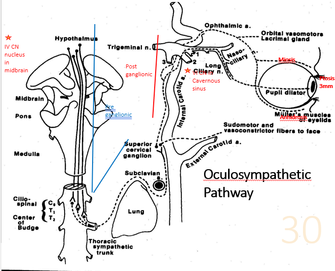
How is pupillary dilation mediated?
Pupillary dilation - 3-neuron sympathetic pathway
1st order central neuron
Starts: hypothalamus & ↓ cervical spinal cord (C8-T2), (ciliospinal center of Budge)
2nd (preganglionic)
neuron exits spinal cord & travels → cervical sympathetic chain → synapses superior cervical ganglion
3rd (postganglionic)
neuron enters the cranium via internal carotid artery → orbit & innervates iris dilator muscles & Müller's muscle in eyelids.
Pupillary dilation - three-neuron sympathetic pathway
1st order central neuron
Starts: hypothalamus & ↓ cervical spinal cord (C8-T2), (ciliospinal center of Budge)
A: 2nd (preganglionic) neuron exits spinal cord & travels → cervical sympathetic chain → passes over the pulmonary apex & synapses superior cervical ganglion near mandible & carotid artery bifurcation.
A: 3rd (postganglionic) neuron enters the cranium via internal carotid artery → trigeminal nerve, → orbit & innervates iris dilator muscles & Müller's muscle in eyelids.
Pupillary Constriction
A: Pupillary constriction is controlled by parasympathetic fibers traveling with the oculomotor nerve (cranial nerve III).
What is spasmus nutans and when does it occur?
A: Spasmus nutans is an acquired form of nystagmus seen in children, usually within the first 2 years of life.
: The triad of spasmus nutans includes:
1) Nystagmus (involuntary eye movement),
2) Head bobbing,
3) Torticollis (twisting of the neck).
: Head bobbing and torticollis are thought to be compensatory mechanisms to improve vision by reducing the nystagmus' frequency and asymmetry.
Ocular Motor Apraxia (OMA)
A: OMA is a condition where there is a defect or absence of voluntary eye movement control. Children with OMA struggle to move their eyes in a specific direction, especially horizontally.
A: To compensate, they use a head thrust to track objects, as they cannot initiate horizontal eye movements. Vertical eye movements typically remain unaffected.
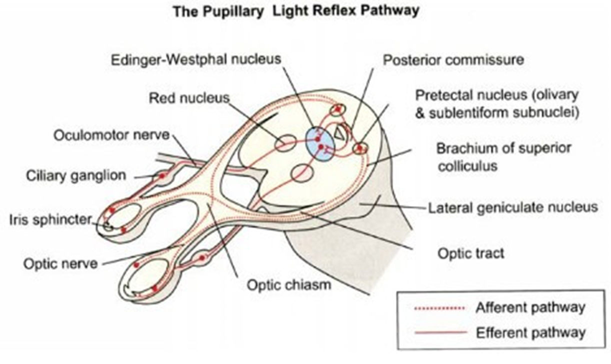
Q: Describe the pupillary light reflex pathway.
A: 1) Light hits the retina, initiating signals in retinal ganglion cells.
2) Signals travel through the visual pathway.
3) Some axons diverge to the pretectal nucleus, relaying to the Edinger-Westphal nucleus (EWN)
4) EWN sends signals via the third cranial nerve, through the ciliary ganglion to the sphincter muscle of the iris, causing pupil constriction.
Describe the sympathetic pathway for pupil dilation.
A: 1) Originates in the hypothalamus.
2) First-order neuron descends to the ciliospinal center of Budge (T1-T2).
3) Second-order neuron exits spinal cord, enters sympathetic chain, terminates in superior cervical ganglion.
4) Third-order neuron travels along internal carotid artery, joins ophthalmic nerve, enters orbit to innervate dilator muscle of the iris.

Oculocardiac Reflex (OCR)
OCR is triggered by stimulation of the vagus nerve due to traction on the extraocular muscles (EOMs), often seen in pediatric fractures, causing a reduction in heart rate (bradycardia)
Q: Describe the oculocardiac reflex (OCR) pathway.
A: 1) Afferent limb: Trigeminal nerve (CN V) carries signals from stretch receptors via ciliary nerves to ciliary ganglion, then to trigeminal nucleus.
2) Efferent limb: CNS processes information, communicates with vagus nerve (CN X) motor nucleus.
3) Vagal impulses travel to sinoatrial node, causing bradycardia.
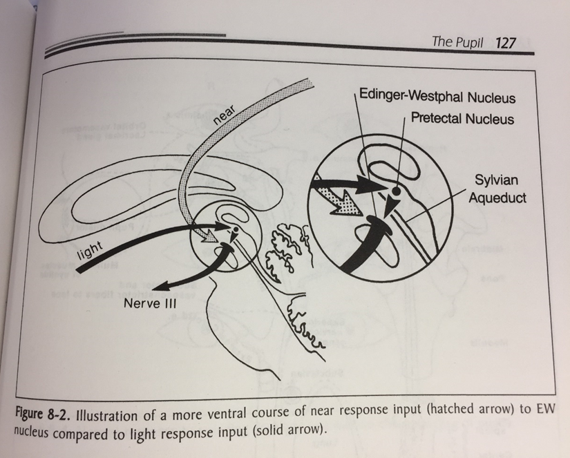
what is the Near reflex
The near reflex (or near response) occurs when focusing on a close object.
Accommodation:
Convergence:
Pupillary Constriction:
The neurons carry on to the visual cortex from LGB, optic radiations = area 19 - visual cortex
area 19 - interprets near object.
Signals → via descending fibers → thru internal capsule.
fibers synapse in the EW nucleus of midbrain.
Parasympathetic fibers (via CN III and ciliary ganglion) cause:
Accommodation (lens thickens).
Pupillary constriction.
Signals to MR muscles of both eyes = convergence.
pupil size depends on
Age
Hippus
Light intensity
Accommodation
Drugs
Pharmaceutical
Recreational
Psychosensory
Attraction
Fear
How do you asses pupil
Observation
Shape
Size (look for anisocoria)
Direct light reflex
Consensual light reflex
Swinging flashlight test
Accommodative reflex
Look for other abnormalities
Eyelids
Eye position
what are the main pupil abnormalities
Anisocoria
Mydriatic pupil
Miosed pupil(s)
Irregular pupils
Trauma
Iris tumours
Coloboma
Posterior adhesions to the lens (Synechiae)
Causes of a mydriatic pupil
Failure to constrict……..?
IIIrd N palsy
Holmes-Adie Tonic pupil
Dorsal midbrain syndrome
Acute glaucoma
Trauma
Pharmacological accident
Hutchinson pupil (coma)
IIIrd N palsy
Compressive
Aneurysm: Junction of Posterior communicating artery (PCA) and internal carotid artery
Associated with other EOM defects
Accommodation affected
Aberrant regeneration
Holmes-Adie Tonic Pupil
Lesion in the ciliary ganglion
Bacterial/ viral infection
Mostly affects women (30s-40s)
No response to direct/consensual light reflex
Accommodative response impaired - N reduced
Due to denervation and supersensitivity
an enlarged pupil that constricts slowly in bright light:
WILL REACT TO 0.125% PILOCARPINE
or METACHOLINE CHLORIDE (2.5%)
Possible reduction in knee jerk reflex
Dorsal midbrain syndrome - bilateral light near dissociation
Pupillary light-near dissociation, lid retraction, convergence-retraction nystagmus, reduced upgaze, eye misalignment
Causes
Tumors, strokes, hydrocephalus, head trauma, multiple sclerosis, A/V malformation
pharmacological dilation
causes of miosis
Failure to dilate…….?
Uniocular
Horner’s syndrome
Anterior segment inflammation
Binocular
Argyll Robertson
Convergence/Accommodative spasm
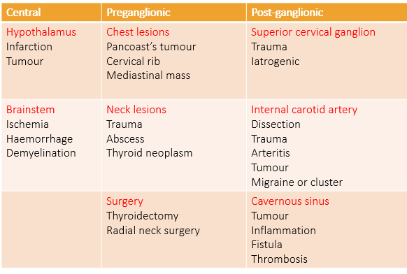
Horners syndrome
Characteristics: - PAREDINE - order neurone TEST & COCAINE
PTOSIS, MIOSIS, ANHYDROSIS
Additional characteristics
Heterochromia (congenital cases)
Apparent enophthalmos
Can be associated with contralateral IVth N (nuclear/fascicular) or VIth N (cavernous sinus)
Anisocoria increses in dim light
Cocaine (2-4%) dilates a nomal pupil but not a Horner’s !
1st (central), 2nd (Preganglionic) or 3rd (Post-ganglionic)order neuron lesion differential diagnosis
Differential diagnosis test: PAREDRINE (1% Hydroxyamphetamine) drops will fully dilate the pupil if 1st or 2nd order neuron lesion, but subnormal dilation if 3rd neuron.
Second Order neuron lesion- Pancoast’s tumour
can lead to horners
Argyll Robertson
Usually bilateral miotic pupils
May be asymmetrical
Poor dilation in the dark and to mydriatics
Light-Near dissociation
NO response to light
Responds to near target
Hallmark of neurosyphilis
Site of lesion: Region of the Sylvian aqueduct in the rostral midbrain
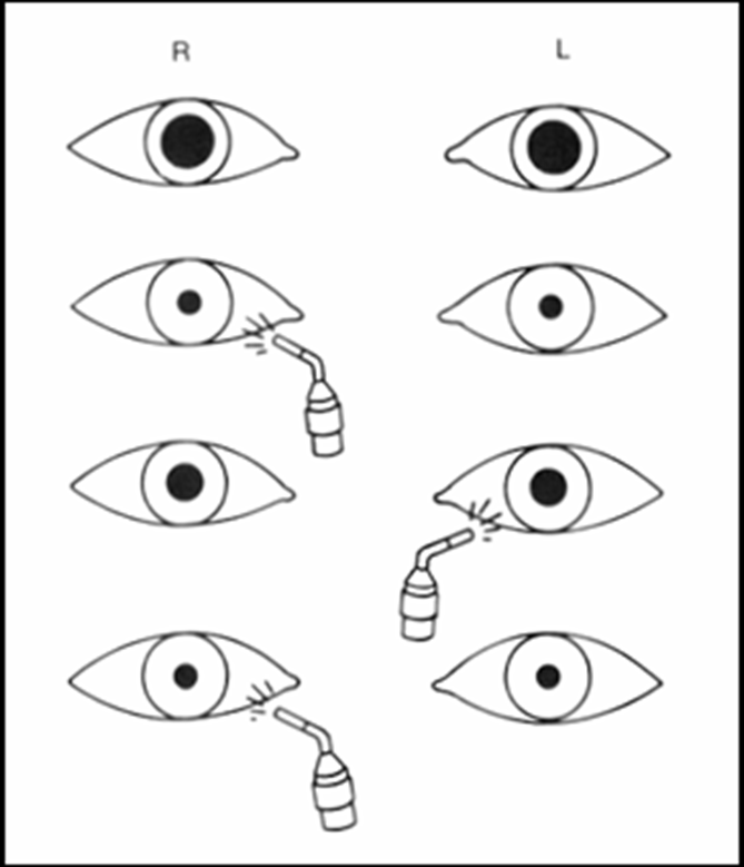
Relative Afferent Pupillary Defect (RAPD) (Marcus Gunn Pupil)
Lesion - afferent pathway
Retina, optic nerve or anterior visual pathway
Diagnosed by the swinging flashlight test
The affected pupil will dilate instead of constrict when the light is transferred from the normal eye to the abnormal eye.
Direct response<consensual response
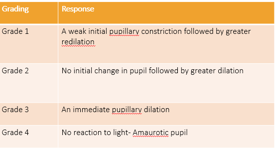
Afferent Pupillary Defect (Marcus Gunn Pupil)
Some of the conditions that exhibit RAPD are:
Optic Neuropathy
Extensive retinal damage (Central retinal artery/vein occlusion, marked retinal detachment)
TED – optic nerve compression
Amaurotic pupil- “Blind Eye”
Mild RAPD- amblyopia
Light Near Dissociation
Better pupillary responses to a “near”accommodative target than to a “light”
Conditions include:
Optic neuropathy
Adies Tonic Pupil
Argyll Robertson
Parinaud’s syndrome (dorsal midbrain syndrome)
Rare Pupil disorders
Springing pupil-pupil dilation associated with migraines
Tadpole pupils-Sectoral pupil dilation
Midbrain corectopia- eccentric or oval pupil associated with rostral midbrain disease
parasympathetic pathway - constriction
Parasympathetic Pathway
Retinal Ganglion Cells: Afferent signals originate here.
Synapse in Pretectal Nucleus: Axons diverge from the optic tract to the superior colliculus.
Edinger-Westphal Nucleus: Bilateral relay of information.
Third Nerve Pathway: Preganglionic fibers synapse in the ciliary ganglion.
Iris Sphincter Activation: Postganglionic fibers constrict the pupil.