BMS2052 - W2: Bacterial Pathogenesis
 Call Kai
 Learn
Learn Practice Test
 Spaced Repetition
 Match
Match Flashcards
 Knowt Play
1/24

There's no tags or description
Looks like no tags are added yet.
Name | Mastery | Learn | Test | Matching | Spaced | Call with Kai | Chat |
|---|
No analytics yet
Send a link to your students to track their progress
25 Terms
Define colonisation, infection and disease
Colonisation: presence of a microorganism on/in host, with multiplication of the organism but not necessarily exerting an impact on the host.
Infection: colonisation by an infectious agent. This has the potential to develop into a disease
Disease: clinical signs and symptoms, pathology
Factors that influence infection and disease
pathogen factors (e.g. virulence factors)
host factors (e.g. immune function, pregnancy)
environmental factors (e.g. water quality)
Name the types of microbes from good to bad
commensals, opportunist, pathogens
Pathogens - what are virulence factors?
Virulence factors: Substances, products, appendages or strategies that are essential for survival, infection and pathogenicity
What are the 5 steps that most pathogens must do to cause disease?
enter the body (transmission)
colonise the host
evade hose defenses
multiply and disseminate
cause damage to the host
How can virulence factors be acquiared?
mutation
horizontal gene transfer
Horizontal gene transfer
Horizontal gene transfer helps bacteria adapt
Recipient bacteria becomes a transformant, transductant or transconjugant
Transformation
uptake of foreign DNA
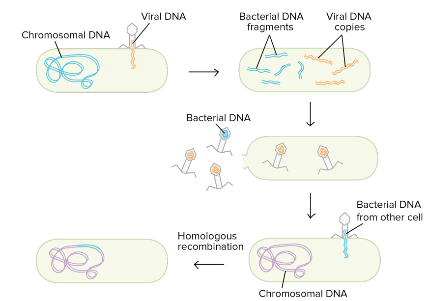
Transduction
bacteriophage can inject its genetic info into bacteria which can be incorporated in bacterial chromosome
bacteriophage can take up genetic info from a bacteria and inject into another bacteria (transconjugant)
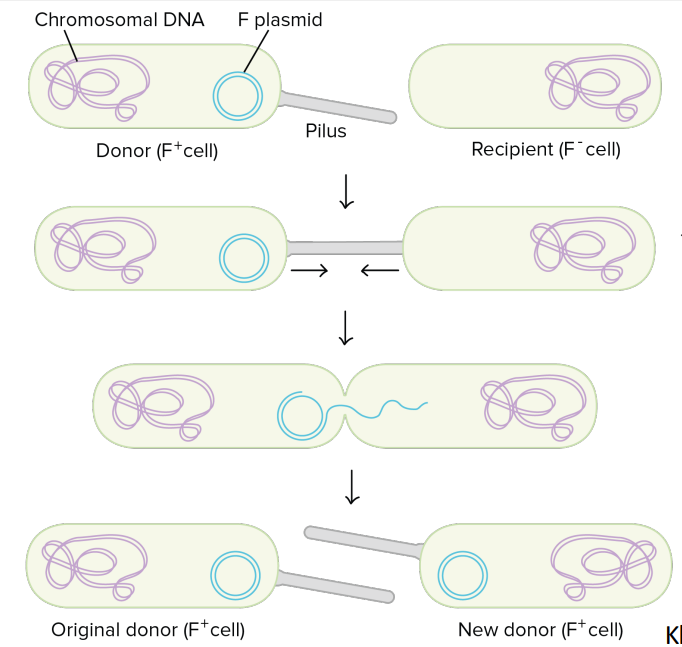
Conjugation
plasmids can transfer from one bacteria to another
What are plasmids
Bacteria usually have bacterial DNA and one or more plasmids
Plasmid:
Extrachromosomal double stranded circular DNA
Self-replicating
many plasmids are associated with virulence as virulence factors can be encoded on these plasmids
can be transferred between bacteria (conjugation)
Conjugation
Transduction
Lysogenic Phage: Integrates into the bacterial genome (prophage)
Lytic phage: Infects and rapidly kills the bacterium through it’s replication
basically turns the bacteria into a replication machine to create more bacteriophages
bacterial DNA can accidentally get incorporated into some of the bacteriophages produced - can inject bacterial DNA into another bacteria (transduction)
Transposons
Fragment of DNA that can move around, in and out of a bacterial chromosome, plasmids and bacteriophages
Can transfer antibiotic resistance genes, toxin encoding genes and other virulence factors
Pathogenicity islands
Pathogenicity islands are elements that have been horizontally acquired, are stable (now are just part of the chromosomes)
on a bacterial chromosome that contains genes encoding for virulence factors
pathogens can have more 1 pathogenicity island
permanently integrated
Can be detected by the different G+C ratio of the DNA = different origin to the rest of the genome
Genes from mobile elements can accumulate in gene clusters. These clusters of genes on chromosomes can work together to mediate virulence = pathogenicity islands
Regulation of virulence
Virulence factors are not being made all the time (waste of energy) as they are only required at specific times and in specific locations
bacteria detect external signals to determine when to express virulence factors
essential for energy conservation
can be switched on/off (e.g. flagella, capsule production)
What factors influence successful infection?
Portal of entry (need to be at the correct niche)
host specificity (some bacteria can infect many animal types, while others cannot)
bacterial load (number of bacteria entering the body)
colonisation
Pathogens (or normal microbiota) establish themselves in a host
must first overcome physical and innate immune barriers
must outcompete normal flora
Adhesion - needed for colonisation
contact with host cell (non specific electrostatic attraction)
binding to host cell (surface interactions with host molecules, mediated by virulence factors termed adhesins)
Adhesins:
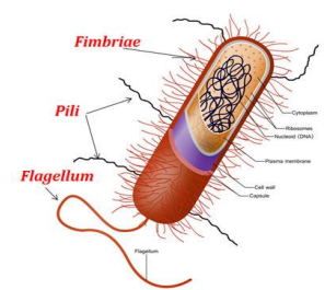
fimbrial adhesins (long extensions that interest with host cell surface)
functions: attachment to surfaces, motility, DNA transfer, biofilm formation
fimbriae - thinner and shorter fibers than pili
adhesion via tip of pilus
afimbrial adhesins (membrane proteins that interact with components on surface of host cell)
establishes very close and strong adhesion with host cell
capsule
polymeric structure surrounding the cell wall
composed of polysaccharides
protects bacterial cell, facilitates adhesion (to each other and other surfaces)
different adhesin types (which respond to different molecules) contributes to pathogen’s ability to colonise different niches (e.i. bladder vs kidney) and animals.
Invasion: What is it?
What does invasion mean?
to disseminate to deeper tissues or invade a previously uncolosnised site/ tissue
to invade a particular cell
Advantages:
avoid immune recognition
Disadvantages:
cell autonomous defense
Obligate intracellular bacteria: Needs that condition to survive
Facultative intracellular bacteria: does not necessarily need the condition to survive
Invasion mechanisms: How do bacterial pathogens get inside our cells?
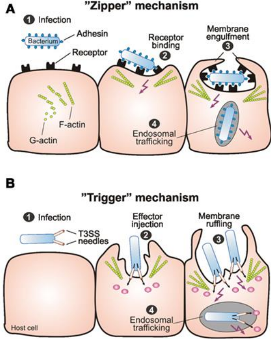
Zipper:
exploits host cell pathways normally used for adhesion
induces receptor immobilisation
bacteria is engulfed (similarly to endocytosis)
very close interaction with host cell surface
e.g. Listeria monocytogenes
gram +ve, food-borne pathogen
internalisation requires the actin cytoskeleton and bacterial protein Internalin A (an invasin) → interacts with E-cadherin which normally forms tight junctions, but Internalin A causes clustering of these E-cadherins to facilitate entry
Trigger:
via secretion system, pathogens inject effector proteins into cells to manipulate cell cytoskeleton → ruffling
e.g. Salmonella
mediated by a type II secretion system. Needle like structure injects into the host cell
How can pathogens overcome the innate immune system?
evade phagocytosis
resist killing
Phagocytosis:
Phagocytes destroy microbes through ‘cellular eating’
can recognise pathogens through pattern recognition receptors (recognise elements on the bacteria), C3b receptor (component of compliment), Fc receptor (arm of an antibody sticking out on the bacterium)
Strategies to evade phagocytosis: Extracellular pathogens
Direct evasion of phagocytosis
kill phagocytes that come in contact
bacterial cell scan produce proteins that interact with specific components on phagocyte surface → create pore in the phagocyte surface → destruction of phagocyte
Capsules
can resemble host components (things that are usually found in human body) → not recognised by immune system
can mask underlying structures (hides the components like PAMPS which are recognised by immune cells) → avoid complement binding, recognition → prevent phagocytosis
Interfering with opsonins (substances that bind to microorganisms to facilitate phagocytosis e.g. antibodies, complement, mannose-binding protein/lectin) - bacteria can prevent themselves from being coated with opsonins
bacterial surface proteins can bind to C4BP or FH, leading to degradation of complement components
bacterial proteins can inactivate C3
secreted bacterial proteins can degrade complement proteins
Bacterial surface proteins that bind to the Fc region of antibodies (causing the antibody to bind in the wrong direction than it’s supposed to) can prevent complement binding = block phagocytosis
Resist Killing: intracellular pathogens
Resist killing by phagocytes
Once inside a phagocyte/ epithelial cells they can:
get out of the phagsome/ vacuole into the cytosol
prevent trafficking of vacuole to lysosomal niche (redirect to diff environment)
able to survive in lysosome niche
Overcoming adaptive immunity
direct immunosuppression
expression of weak antigens
antigen modification
antigen diversity
Multiplication and dissemination:
What do bacteria need to do to be able to multiply and disseminate?
What do bacteria need to do to be able to multiple and disseminate?
overcome immune system
outcompete normal microbiota
nutrients for cell division, growth, dissemination
Example of nutrient acquisition:
Iron acquisition strategies:
siderophores
excreted into extracellular environment
can interact with iron at very high affinity, forming complex
complex interacts with receptor on surface of bacteria → internalised
express receptors for host iron capture proteins (capture proteins which capture iron for host)
express high affinity iron binding receptor for haemoglobin (capture haemoglobin)
express toxins to release host iron in local environment which can be taken up by bacteria
Tissue damage:
Direct toxicity - mediated by the bacterial toxins
Induction of cytokines (immune mediated)
Induction of immunopathology (immune mediated)
Bacterial toxins
Directly mediate damage to humans
2 main categories:
Exotoxins - secreted out of the bacteria or released by bacterial lysis
Endotoxins - within the bacteria, usually cell wall components
Endotoxins:
within the bacteria, usually cell wall components
can be released into local environment when a bacterial cell dies and cell wall breaks down
can be produced by gram positive and gram negative bacteria
e.g. LPS, PG, LTA
Exotoxins:
secreted out of the bacteria or released by bacterial lysis
can be produced by gram positive and gram negative bacteria
usually produced by extracellular pathogens rather than intracellular pathogens (as bacterial do not want to kill its host)
Can be classified according to:
site of action (where the toxin acts)
intracellular
extracellular (toxin has to be taken up into the cell)
Tissue specificity
Enterotoxin (gut)
neurotoxin (nerve tissue)
cardiotoxin (heart)
Can be further classified as:
Heat-liable toxins (proteins) - can be denatured (inactived) by heat
Type I - superantigens (act to overstimulate our immune system)
causes overactivation of nonspecific T-cell production which can send the body into shock and lead to organ failure (VERY TOXIC!!)
Type II - membrane disrupting toxins (act on cell membrane)
damage cell membrane causing cell lysis
can create pores (loss of nutrients, etc. → cell death), cleave phospholipid bilayer → lysis/ cell death
Type II - A-B toxins (have an active component + binding component that binds to a receptor on host cell for interalisation)
gets internalised into host cells
usually made up on an A and B subunit (can have more subunits)
causes internal cellular function being disrupted, often leading to cell death
heat-stable toxins (non-proteins) - cannot be denatured (inactivated) by heat