Clinical Microbiology Refresher
 Call Kai
 Learn
Learn Practice Test
 Spaced Repetition
 Match
Match Flashcards
 Knowt Play
1/67

There's no tags or description
Looks like no tags are added yet.
Name | Mastery | Learn | Test | Matching | Spaced | Call with Kai | Chat |
|---|
No analytics yet
Send a link to your students to track their progress
68 Terms
Microbes That Cause Infectious Diseases
prokarya→bacteria
eukarya:
fungi (yeast+molds)
protozoa
helminths (worms)
-multicellular
viruses:
no membrane+cytoplasm
can’t make own energy
unable to synthesize proteins
Important Features of Cellular Microbes
structure:
cells: nucleus/nucleoid containing DNA
surrounded by cytoplasm
proteins synthesized
energy generated
viruses: inner core of genetic material (DNA/RNA)
no cytoplasm
depend on host cells for energy
method of replication:
cells:
binary fission
mitosis
viruses:
must replicate within host cells
disassemble→produce copies of nucleic acids+proteins→reassemble into viruses
nature of nucleic acid:
cells: both DNA and RNA
viruses: either DNA or RNA but not both
Eukaryotes vs Prokaryotes
eukaryotes:
DNA in nucleus within nuclear membrane
multiple chromosomes
histones
organelles
larger ribosome 80s
flexible plasma membranes (fungi→chitin cell walls)
cell membrane contains sterols
divide by mitosis (fungi→budding)
prokaryotes:
DNA in nucleoid region
no nuclear membrane
single circular chromosome
no histones
no organelles
smaller ribosome 70s
rigid peptidogycan cell walls
no sterols in cell membrane (exc: mycoplasma)
divide by binary fission
General Terminology/Nomenclature
bacteria/fungi/protozoa/helminths: genus+species
ex: escherichia (genus) coli (species)
virus: name-virus
ex: poliovirus
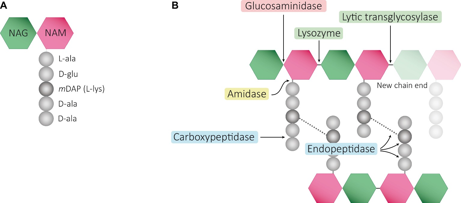
Peptidoglycan
a complex interwoven macromolecule network
surrounds entire cell
composed of a single covalently linked macromolecule
polymer of a prepeating disaccharide in rows:
n-acetylglucosamine (NAG)
n-acetylmuramic acid (NAM)
NAM-NAG complex
linked by polypeptides
found only in bacterial cell walls
rigid support for cell
maintains shape of cell
gives cell resistance to low osmotic pressure
Microscopy Staining
gram stain:
bacteria
fungi
acid-fast stain:
mycobacteria
calcofluor white stain:
fungi
gomori methenamine silver (GMS) stain:
fungi
Cell Wall
gram-positive: thicker cell wall
gram-negative: thinner cell wall
acid-fast: glycolic walls
atypical: PCR-based testing needed
Gram Positive+Aerobic: Staphylococci
all catalase positive
coagulase-positive→staph aureus
coagulase-negative:
staph lugdunensis
staph epidermis
staph saprophyticus
staph haemolyticus
Gram Positive+Aerobic: Streptococci
all catalase-negative
beta-hemolysis:
strep pyogenes (group A strep)
strep agalactiae (group B strep)
alpha-hemolysis:
strep pneumoniae
viridians group strep
non-hemolysis:
enterococcus faecalis
enterococcus faecium (harder to treat)
Gram Positive+Aerobic: Bacilli
listeria monocytogenes
bacillus anthracis
corynebacterium diptheria
Gram Negative+Aerobic: Cocci
neisseria meningitidis
neisseria gonorrhoeae
haemophillus influenzae
moraxella catarrhalis
Gram Negative+Aerobic: Spirochetes
trepodema pallidium (syphillis)
borrelia burgdorferi (lyme disease)
Gram Negative+Aerobic: Bacilli
lactose-fermenting:
oxidase-negative:
enterobactales
escherichia coli
klebsiella
enterobacter cloacae
citrobacter
serratia spp
providencia spp
proteus spp
oxidase-positive:
aeormonas spp
pasteurella spp
vibrio spp
non-lactose-fermenting:
oxidase-negative:
acinetobacter baumanii
sternotrophomonas spp
burkholderia spp
oxidase-positive:
pseudomonas aeurginosa (nosocomial)
Anaerobes: Cocci
gram positive: peptostreptococcus spp
gram-negative: veillonella spp
Anaerobes: Bacilli
gram-positive:
clostridioides/clostridium difficile (c-diff)
clostridium perfringens
clostridium tetani
clostridium botulinum
gram-negative:
bacteroides spp
prevotella spp
fusobacterium spp
Atypical Bacteria
chlamydia pneumonia
chlamydia trachomatis
mycoplasma pneumoniae
legionella pneumophila
Acid-Fast Bacteria
myocbacterium tuberculosis
mycobacterium avium complex
nocardia spp
actinomyces
Classification of Medically Important Bacteria
based on nature of cell wall:
rigid:
-free-living
-non-free-living
flexible→spirochetes
absent→mycoplasmas
used under genera+species
Gram-Positive Medically Important Bacteria
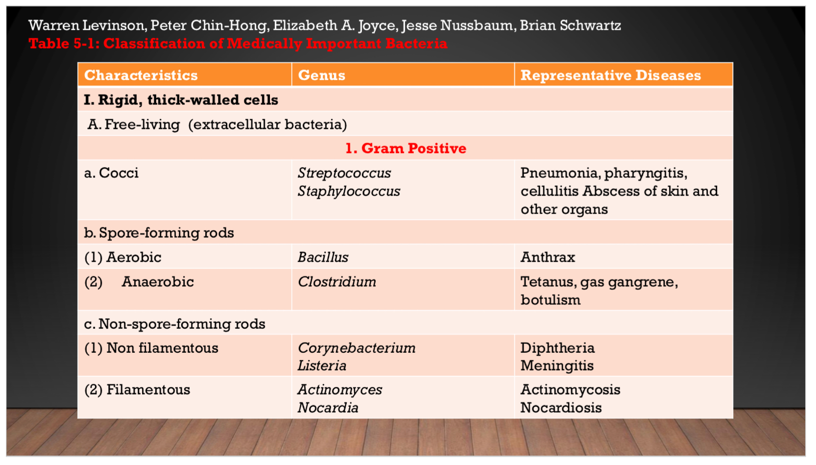
Gram-Negative Medically Important Bacteria
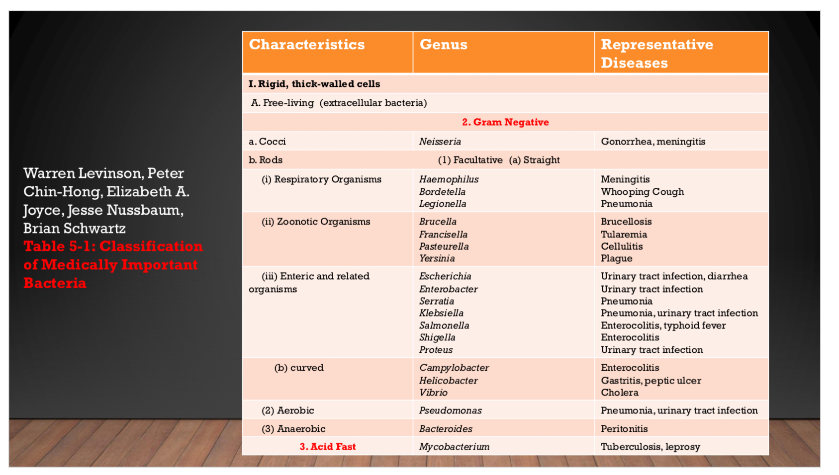
Atypical Medically Important Bacteria
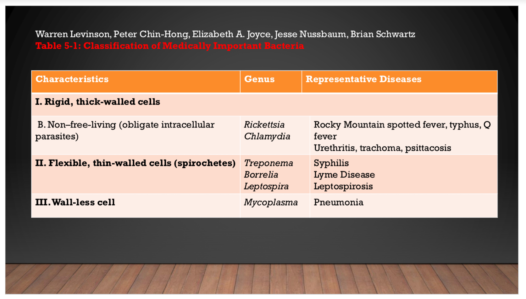
Staphylococcus Aureus
PP: gram-positive cocci
grape-like clusters
R: human→nose
E: abscesses
various pyogenic infections:
-endocarditis
-septic arthritis
-osteomyelitis)
food poisoning
scalded skin syndrome
toxic shock syndrome
CM: post-surgical wound infections
pneumonia:
empyema
lung abscess
conjunctivitis:
unilateral burning eye pain
hyperemia of conjunctiva
purulent discharge
abscesses
scalded skin syndrome
impetigo
carbuncle
osteomyelitis
septic arthritis
P: pneumonia
septicemia
surgical-wound infections
skin+soft tissue infections (SSTIs):
-folliculitis
-cellulitis
-impetigo
bacterial conjunctivitis
Streptococcus Pyogenes
PP: gram-positive cocci
chains or pairs
CM:
pyogenic dx:
pharyngitis
cellulitis
toxigenic:
scarlet fever
toxic shock syndrome
immunologic dx:
rheumatic fever
acute glomerulonephritis
group A:
cellulitis
erysipelas
necrotizing facsiitis
impetigo
P: group A→pharyngitis
cellulitis
group-B→neonatal sepsis
E. Faecalis: UTI
endocarditis
S. Viridians→endocarditis (rare)
Clostridium Spp.
PP: gram-positive rods
anaerobic
spore-forming
E/P:
clostridium perfringens: gas gangrene
food poisoning
clostridium difficile
Mycobacteria
mycobacterium tuberculosis→tuberculosis
mycobacterium leprae→leprosy
mycobacteria avium-intracellulare complex (MAI, MAC)→tuberculosis-like dx
mycobacterium kansasii→tuberculosis-like dx
mycobacterium chelonae→immunocompromised/implant amputee dx
Spirochetes
treponema: syphillis
non-veneral treponematoses
yaws
pinta
E: direct contact
borrelia: lyme disease
relapsing fever
E: infected tick
leptospira: leptospirosis
E: animals
animal urine contaminated water
CM: 7-13 day asx incubation period→CM
fever
chills
headache
conjunctival suffusion
myalgia
Viruses (General)
not considered cells
not capable of independent replication→must reproduce within cells (obligate intracellular parasites)
can’t make own energy+proteins
too small to be seen with microscope
particles composed of internal core with DNA/RNA (not both) covered by protein coat
±outer lipoprotein membrane→envelope
don’t have: nucleus
cytoplasm
mitochondria
ribosomes
Pathogenesis of Viruses+Stages of Viral Infection
pathogenesis:
transmission of virus→entry into host
replication of virus→damage to cells
spread of virus to other cells+organs
innate+adaptive immune responses activate
potential persistence of virus
stages of infection:
incubation period→asx
prodromal period→nonspecific sx
specific-illness period→CM sx
recovery period→dx wanes→pt normal health
Herpesvirus
PP: replicate in nucleus→form intranuclear inclusions
obtain envelope from nuclear membrane
E:
HSV-1: saliva
face infections
HSV-2: sexual contact
genital infections
CM:
HSV-1: above the waist
acute gingivostomatitis
recurrent herpes labialis (cold sores)
keratoconjunctivitis (keratitis)
encephalitis
erythema multiforme
HSV-2: below the waist
herpes genitalis (genital herpes)
neonatal encephalitis
neonatal herpes
aseptic meningitis
erythema multiforme
Respiratory Viruses
PP: infect respiratory tract→infect mucosal cells of respiratory tract→cause sx in respiratory tract+CM found outside respiratory area
most have RNA (one has DNA)
most enveloped (exc: rhinovirus+adenovirus)
E: measles
mumps
rubella
varicella zoster
orthomyxoviruses
paramyxoviruses
coronaviruses
CM: found outside respiratory tract
DX: severe→PCR-based assay from respiratory tract secretions
Human Immunodeficiency Virus (HIV)
PP: infects+kills CD4/helper T lymphocytes→suppression+loss of cell-mediated immunity (destroys immune system)→opportunistic infections
E: retrovirus
HIV-1→worldwide
HIV-2→west africa
caused by acquired immunodeficiency syndrome (AIDS)
humans+certain primates
non-endogenous to humans (no sequences found in normal human cell DNA)
CM:
early/acute
middle/latent
late/immunodeficiency
P: opportunistic infections
Fungi Structure
eukaryotic organisms
different from prokaryotic bacteria
2 medically important fungal structures:
fungal cell wall: made of chitin (not peptidoglycan)
insensitive to certain cell-wall synthesis-inhibiting abx (penicillins/cephalosporins)
contain other polysaccharides: beta-glucan (long polymer of D-glucose+site of action for caspofungin)
chitin: polysaccharide
composed of long chains of N-aceytlglucosamine
fungal cell membrane: contains ergosterol (not cholesterol)
Fungi Growth
yeasts:
unicellular
budding→daughter cells unequal in size
molds:
multicellular
contain hyphae/long filaments of cells
cell division→daughter cells equal in size
sexual reproduction spores:
classified by sexual spores
zygospores→single large+thick walls
ascospores→formed in ascus sac
basidiospores→formed externally on tip of pedestal called basidium
dimorphism:
exist as yeasts or molds depending on temperature
mold→room temperature
yeast→body temperature
Parasite Classification
protozoa:
single-celled
classified on most important site of infection:
-giardia→intestines
-trichomonas→urogenital
-plasmodium→blood
-tissue→toxoplasma
metazoa:
multicellular
helminths/worms
two phyla:
platyhelminthes: flatworms
cestoda→tapeworms
trematoda→flukes
nemathelminthses: roundworms
nematodes
Protozoa Forms+Transmission
trophozoite:
motile/feeding/reproducing form of protozoan
flexible cell membrane
cyst:
non metabolizing/nonreproducing form of a protozoan
thick wall
transmission:
intestinal protozoa→ingestion of cysts
blood+tissue protozoa→insect vectors
exc: toxoplasma→ingestion of cysts from cat feces/placenta transfer
Helminths Transmission
life cycle: egg→larva→adult
transmission:
ingestion of eggs
penetration of skin by larvae
insect bite
DX: stool→ova+parasites
Microbiology DX Process
pt infected
1-3 hrs→specimens collected+sent to lab
automated culture system/agar plate
no growth: incubate for over 5 days
growth: rapid dx test in 1-3 hr
gram stain T-24h in 1 hr→24-48 hr→identified microorganism
Minimum Inhibitory Concentration (MIC)
lowest concentration of an abx that inhibits visible growth
Dx: broth microdilution (BMD)
disk diffusion/kirby-bauer
epsilometer strip/e-test
automated: vitek2
microscan walkaway plus system
Interpretation of Phenotypic Susceptibility Results
breakpoints/interpretive criteria: Breakpoints at which successful eradication of an organism has been confirmed in practice, as established by the Clinical and Laboratory Standards Institute (CLSI M100)
criteria:
susceptible (S)
susceptible dose dependent (S-DD)
intermediate (I)
resistant (R)
Contaminant
organism present in a culture specimen that is deemed to not be the primary cause of infection
occurs by chance+improper technique/cross-contamination
interpreting contaminants as pathogens could expose pt to unnecessary abx
Colonizer
an organism present at a body site but doesn’t normally cause sx
often not pathogenic
may be symbiotic
Ex: healthy GI flora is essential for healthy GI tract
Gut Microbiota
the microorganism and viruses that live within the colon
Gut Microbiome
the genome of the gut microbiota which is used for identification through sequencing
Prebiotics
fiber that can pass through the upper GI undigested
Probiotics
live microorganisms that can be ingested for health benefits
Pathogen
any organism/microorganism that can cause disease in its host
Opportunistic Pathogen
a microorganism
in healthy individuals→typically does not cause disease
in weakened/compromised immune system→causes disease
Virulence
the capacity of a microorganism to cause disease
Infectious Dose
the minimum number of pathogenic organisms/pathogens required to establish an infection/disease in a host
Gram Positive vs Gram Negative Bacteria+Gram Stain
gram positive
thick peptidoglycan layer
retain purple stain
gram negative
thin peptidoglycan layer+outer membrane
turn pink after stain
gram stain
determines difference between gram positive/gram negative bacteria
stain order: gram positive→gram negative
Role of Cell Wall With Physical Signs and Medications
Bacteria
possess a cell wall→made of peptidoglycan
exception→Mycoplasma
Gram-negative bacteria
have a thin peptidoglycan layer
covered by an outer lipid membrane→contain endotoxin→a key inducer of septic shock
Gram-positive bacteria
have a thick peptidoglycan layer
no outer membrane
periplasmic space
in gram-negative bacteria
between inner+outer membranes
houses β-lactamases→enzymes→degrade β-lactam antibiotics
Peptidoglycan (unique to bacteria)
forms a network providing shape
composed of:
-sugar backbone
-peptide side chains
-(both) cross-linked by transpeptidase
Mycobacteria
higher lipid content in cell wall→resistant to Gram staining
susceptible to acid-fast staining
Lysozymes
breaks down peptidoglycan's glycan backbone→destroys bacteria
cytoplasmic membrane
regulates nutrient transport
regulates toxin secretion
Fungi Types
yeasts
single cells
reproduce asexually→budding
molds
long filaments→hyphae→form a mat→mycelium
form transverse walls→septate hyphae
don’t form transverse walls→nonseptate hyphae
-multinucleated→coenocytic
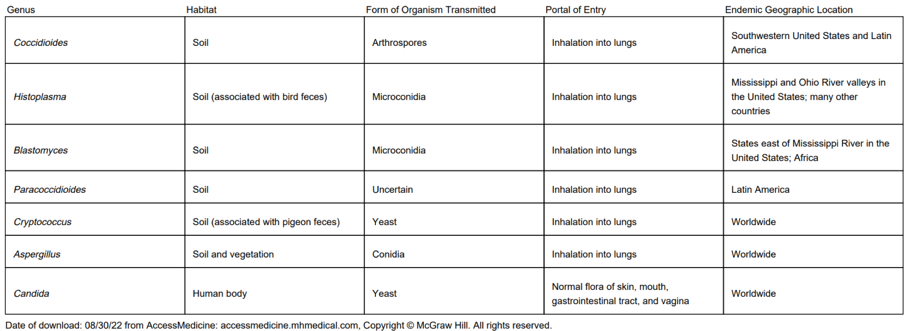
Fungi Route of Transmission+Habitat of Medically-Important Fungi
TABLE 47–2Transmission and Geographic Location of Some Important Fungi
Genus | Habitat | Form of Organism Transmitted | Portal of Entry | Endemic Geographic Location |
|---|---|---|---|---|
Coccidioides | Soil | Arthrospores | Inhalation into lungs | Southwestern United States and Latin America |
Histoplasma | Soil (associated with bird feces) | Microconidia | Inhalation into lungs | Mississippi and Ohio River valleys in the United States; many other countries |
Blastomyces | Soil | Microconidia | Inhalation into lungs | States east of Mississippi River in the United States; Africa |
Paracoccidioides | Soil | Uncertain | Inhalation into lungs | Latin America |
Cryptococcus | Soil (associated with pigeon feces) | Yeast | Inhalation into lungs | Worldwide |
Aspergillus | Soil and vegetation | Conidia | Inhalation into lungs | Worldwide |
Candida | Human body | Yeast | Normal flora of skin, mouth, gastrointestinal tract, and vagina | Worldwide |
Laboratory Diagnosis of Fungi
Direct Microscopic Examination
Specimens: sputum, lung biopsies, skin scrapings.
Looks for hyphae, yeasts, and asexual spores.
10% KOH dissolves tissue while leaving fungi intact.
Important findings:
Coccidioides immitis → spherules
Cryptococcus neoformans → wide capsule seen with India ink
Calcofluor white binds fungal cell walls and fluoresces, making fungi easier to see.
Methenamine silver stain can also highlight fungi in tissue.
Culture
Usually performed on Sabouraud agar.
Its low pH and antibiotics inhibit bacterial growth and favor fungi.
Fungi can be identified by:
Mycelium appearance
Asexual spore characteristics
PCR
Uses DNA probes to identify fungal organisms.
Can identify fungi faster than waiting for colonies to become visually recognizable.
Available for organisms such as:
Coccidioides
Histoplasma
Blastomyces
Cryptococcus
Serology
Detects antibodies in serum or spinal fluid
Particularly useful for systemic mycoses
Diagnosis→significant rise in antibody titer
Complement fixation can be used for:
Coccidioidomycosis
Histoplasmosis
Blastomycosis
Cryptococcal meningitis: detects C. neoformans capsular polysaccharide antigen in CSF using latex agglutination.
-known as the CRAG (cryptococcal antigen) test
Medical Mycoses
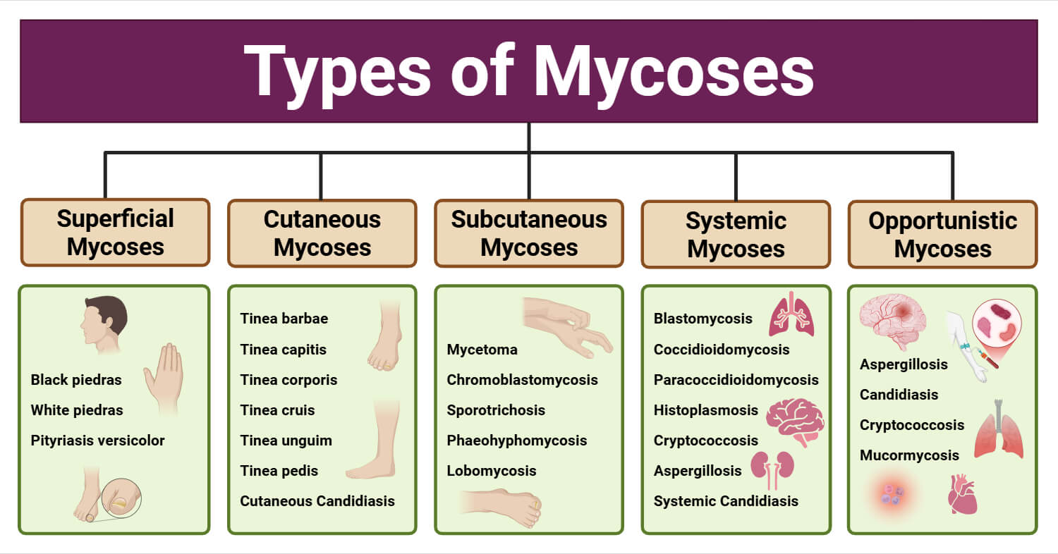
don’t need to know opportunistic mycoses
Bacteria vs Fungi (Table)
TABLE 47–1Comparison of Fungi and Bacteria
Feature | Fungi | Bacteria |
|---|---|---|
Diameter | Approximately 4 μm (Candida) | Approximately 1 μm (Staphylococcus) |
Nucleus | Eukaryotic | Prokaryotic |
Cytoplasm | Mitochondria and endoplasmic reticulum present | Mitochondria and endoplasmic reticulum are absent |
Cell membrane | Sterols present | Sterols absent (except Mycoplasma) |
Cell wall content | Chitin | Peptidoglycan |
Spores | Sexual and asexual spores for reproduction | Endospores for survival, not for reproduction |
Thermal dimorphism | Yes (some) | No |
Metabolism | Require organic carbon; no obligate anaerobes | Many do not require organic carbon; many obligate anaerobes |
Opportunistic Fungi+5 Genera of Medically Important Fungi
immunocompetent→don’t cause disease
immunocompromised/impaired host defenses→cause disease
table→only need to know genus
TABLE 50–1Important Features of Opportunistic Fungal Diseases
Genus | Form in Tissue Seen by Microscopy | Geographic Location | Important Clinical Findings | Laboratory Diagnosis |
|---|---|---|---|---|
Candida | Yeast forms pseudohyphae (also hyphae) | Worldwide | Thrush in mouth and vagina; endocarditis in intravenous drug users | Gram-positive; culture grows yeast colonies; Candida albicans forms germ tubes; polymerase chain reaction (PCR) assay; MALDI-TOF assay |
Cryptococcus | Yeast with large capsule | Worldwide | Meningitis | India ink stain shows yeast with large capsule; culture grows very mucoid colonies; PCR assay |
Aspergillus | Mold with septate hyphae | Worldwide | Fungus ball in lung; wound and burn infections; indwelling catheter infections; sinusitis | Culture grows mold with green spores; conidia in radiating chains |
Mucor and Rhizopus | Mold with nonseptate hyphae | Worldwide | Necrotic lesion formed when mold invades blood vessels; predisposing factors are diabetic ketoacidosis, renal acidosis, and cancer | Culture grows mold with black spores; conidia enclosed in a sac called a sporangium |
Dimorphic Fungi Properties+Pathogenesis+Important Clinical Findings
Clinical Features of Respiratory Viruses (Table)
Virus | Important Disease | Number of Serotypes | Causes Worldwide Epidemics (Pandemics) | Main Clinical Findings | Vaccine Available | Treatment |
Influenza virus | Influenza | Many | Yes | Sudden-onset headache, shaking chill, sore throat, cough, and myalgias | Yes | Oseltamivir, zanamivir, baloxavir |
Parainfluenza virus | Croup | Four | No | Barking cough | No | None |
Respiratory syncytial virus | Bronchiolitis in infants | Two | No | Cough, dyspnea, retractions, wheezing | No | Ribavirin |
Human metapneumovirus | Common cold, bronchiolitis, pneumonia | Two | No | Various (coryza, wheezing, cough) | No | None |
Coronaviruses, especially SARS-CoV-2 | Common cold, SARS,1 MERS,2 COVID-193 | Seven (4 common cold serotypes; 3 pneumonia serotypes) | Yes | Various (coryza, cough, severe pneumonia) | Yes, for SARS-CoV-2 | Remdesivir; Emergency Use Authorization for several drugs and monoclonal antibodies (see Text) |
Rhinovirus | Common cold | Many | No | Runny nose (coryza), sneezing, no fever | No | None |
Adenovirus | Pharyngitis, pneumonia, conjunctivitis | Many | No | Sore throat, cough, pneumonia, “pink eye” | Yes (for military only) | None |
Clinical Features of Important Childhood Viruses (Table)
Virus | Important Disease | Number of Serotypes | Causes Worldwide Epidemics (Pandemics) | Main Clinical Findings | Vaccine Available | Treatment |
Influenza virus | Influenza | Many | Yes | Sudden-onset headache, shaking chill, sore throat, cough, and myalgias | Yes | Oseltamivir, zanamivir, baloxavir |
Parainfluenza virus | Croup | Four | No | Barking cough | No | None |
Respiratory syncytial virus | Bronchiolitis in infants | Two | No | Cough, dyspnea, retractions, wheezing | No | Ribavirin |
Human metapneumovirus | Common cold, bronchiolitis, pneumonia | Two | No | Various (coryza, wheezing, cough) | No | None |
Coronaviruses, especially SARS-CoV-2 | Common cold, SARS,1 MERS,2 COVID-193 | Seven (4 common cold serotypes; 3 pneumonia serotypes) | Yes | Various (coryza, cough, severe pneumonia) | Yes, for SARS-CoV-2 | Remdesivir; Emergency Use Authorization for several drugs and monoclonal antibodies (see Text) |
Rhinovirus | Common cold | Many | No | Runny nose (coryza), sneezing, no fever | No | None |
Adenovirus | Pharyngitis, pneumonia, conjunctivitis | Many | No | Sore throat, cough, pneumonia, “pink eye” | Yes (for military only) | None |
Properties of Viruses Commonly Infecting the Intestinal Tract (Table)
Property | Norovirus | Rotavirus | Poliovirus | Coxsackie Virus | Echovirus |
Virus family | Caliciviruses | Reoviruses | Picornavirus | Picornavirus | Picornavirus |
Genome | Single-stranded RNA; positive polarity | Double-stranded RNA; 11 segments | Single-stranded RNA; positive polarity | Single-stranded RNA; positive polarity | Single-stranded RNA; positive polarity |
Virion RNA polymerase | No | Yes | No | No | No |
Nucleocapsid | Icosahedral | Icosahedral | Icosahedral | Icosahedral | Icosahedral |
Envelope | No | No | No | No | No |
Number of serotypes | Two or more | At least six | Three | Many | Many |
Clinical Features of Viruses Commonly Infecting the Intestinal Tract (Table)
Property | Norovirus | Rotavirus | Poliovirus | Coxsackie Virus | Echovirus |
Virus family | Caliciviruses | Reoviruses | Picornavirus | Picornavirus | Picornavirus |
Genome | Single-stranded RNA; positive polarity | Double-stranded RNA; 11 segments | Single-stranded RNA; positive polarity | Single-stranded RNA; positive polarity | Single-stranded RNA; positive polarity |
Virion RNA polymerase | No | Yes | No | No | No |
Nucleocapsid | Icosahedral | Icosahedral | Icosahedral | Icosahedral | Icosahedral |
Envelope | No | No | No | No | No |
Number of serotypes | Two or more | At least six | Three | Many | Many |
DNA Virus Classification

RNA Virus Classification

4 Main Effects of Virus Infection on the Cell
4 main outcomes
cell death
viruses→stop protein production of cell→trigger apoptosis→cell deathcell fusion
infected cells→fuse together→form multinucleated giant cells
ex:
-herpesvirus
-paramyxovirusesmalignant transformation
viruses→cause uncontrolled cell growth→contribute to cancerno obvious changes
virus→replicates without causing noticeable changes/damage to cell
Viral Infections on Different Types of Cells+Affect on Disease Symptoms+Severity
Viruses infect specific cells by binding viral surface proteins to specific receptors on host cells.
Example: HIV gp120 binds to the CD4 receptor on T cells.
The type of cell infected determines the symptoms and disease:
Rabies → neurons → encephalitis
Hepatitis B → hepatocytes → hepatitis
Viruses use normal cell receptors to enter cells and replicate.
Cell Death-Caused Viral Disease Signs+Symptoms
Cell death: Viruses often inhibit host protein synthesis while allowing viral proteins to be made. Apoptosis can also cause cell death through caspase activation.
Inclusion bodies: Viral proteins/particles can form visible inclusions inside cells.
Negri bodies → rabies
Owl’s eye inclusions → CMV
Cell fusion: Some viruses cause infected cells to fuse, forming multinucleated giant cells, especially herpesviruses and paramyxoviruses.
Cytopathic effect (CPE): Infected cells change appearance, typically becoming round and dark, followed by cell lysis or giant-cell formation. CPE helps detect and measure viruses in the laboratory.
Malignant transformation: Some viruses cause uncontrolled cell growth and prolonged survival, contributing to cancer.
No visible effect: Some viruses can replicate without causing obvious changes, allowing the cell to survive and continue functioning.
Virus Host Defense Evasion Methods
Viruses use immune evasion to avoid being destroyed by the immune system.
Block immune signals: Viruses make cytokine decoys that bind and block immune mediators like IL-1 and TNF.
Reduce MHC I: This makes infected cells harder for cytotoxic T cells (CTLs) to recognize and kill.
Block interferon: Viruses can prevent interferon production or interfere with its antiviral effects.
Avoid NK cells: Some viruses prevent infected cells from being recognized by natural killer (NK) cells.
Glycan shield: Surface sugars can hide viral antigens from antibodies.
Multiple serotypes: Some viruses have many antigenic types, so immunity to one type may not protect against another.
Rhinovirus: >100 serotypes → contributes to frequent common colds.
Influenza, HIV, HCV, SARS-CoV-2: multiple antigenic types → makes vaccination and long-term immunity more difficult.
TABLE 32–4Important Mechanisms by Which Viruses Evade Host Defenses
Host Defense Affected | Mechanism of Evasion | Virus That Employs the Mechanism |
|---|---|---|
Cytotoxic T cells | Reduces MHC class I proteins, thereby decreasing killing by cytotoxic T cells | HIV, HSV, CMV, adenovirus |
Helper (Th-1) T cells | Blocks IL-12, which reduces formation of Th-1 cells, thereby decreasing cell-mediated immunity | Measles virus |
Interferon | Blocks synthesis of interferon by virus-infected cells | EBV, HCV, SARS-CoV-2 |
Interferon | Blocks synthesis of kinase that phosphorylates initiation factor-2 | HIV, influenza, and HSV |
Interferon | NS1 protein blocks action of protein kinase and ribonuclease that inhibit viral replication | Influenza |
Interferon | Blocks action of protein kinase | Hepatitis C virus |
Interleukins | Encodes receptors for immune mediators; receptors are secreted by infected cells, binds mediators, and inactivates them | Vaccinia virus encodes receptor for IL-1 |
Chemokines | Encodes chemokine receptor; this blocks action of chemokine, thereby inhibiting migration of inflammatory cells to site of infection | Vaccinia virus, CMV |
Complement | Encodes protein that binds to complement protein C3b; this blocks opsonizing action of C3b as well as its ability to participate in forming the membrane attack complex | HSV |
Antibody | Glycans, especially oligomannose, coat envelope proteins decreasing antibody synthesis and binding | HIV, SARS-CoV-2, HCV |