Forelimb- bones and joints (blz. 54 the dog anatomy workbook)
1/18
There's no tags or description
Looks like no tags are added yet.
Name | Mastery | Learn | Test | Matching | Spaced |
|---|
No study sessions yet.
19 Terms

1
Scapula

2
Humerus (dog)
The humerus is the long bone located in the upper arm or forelimb. It runs from the shoulder to the elbow and is the largest bone in the arm. The humerus consists of a head, neck, and shaft, with an array of important features including the greater and lesser tubercles that serve as attachment points for muscles, as well as the medial and lateral epicondyles that provide attachment sites for ligaments and tendons. The humerus plays a crucial role in the movement of the arm and functions in various activities such as lifting and throwing.

3
Radius (dog)
The radius is one of the two long bones in the forearm, located on the lateral side (thumb side) when in the standard anatomical position. It extends from the elbow to the wrist and works in conjunction with the ulna, the other forearm bone, to facilitate the motion of the arm. The radius has several key features: the head, which is a circular-shaped surface that articulates with the humerus at the elbow joint; the neck, which is a narrowing just below the head; and the shaft, which tapers as it approaches the wrist. The distal end of the radius acts as a pivot point for the wrist and articulates with the carpal bones. The radius is crucial for forearm rotation, allowing movements such as supination (turning the palm up) and pronation (turning the palm down), and it provides attachment points for muscles that enable gripping and other hand movements.

4
Ulna (dog)
The ulna is one of the two long bones in the forearm, located on the medial side (pinky side) of the forearm in standard anatomical position. It is slightly longer than the radius and extends from the elbow to the wrist. The ulna has a distinct structure, including three key parts: the olecranon, which forms the bony prominence of the elbow; the coronoid process, which helps stabilize the joint; and the trochlear notch, which articulates with the humerus, allowing for hinge-like movement at the elbow. The ulna also features the ulnar head near the wrist, where it articulates with the carpal bones and is involved in the wrist joint. Important for forearm movement, the ulna provides attachment points for muscles associated with flexing and extending the elbow, as well as movements of the wrist and hand. Additionally, the interosseous membrane between the ulna and radius serves as a site for muscles and helps stabilize the two bones during movement.

5
Carpal Bones
The carpal bones are a group of eight small bones in the wrist that articulate with the radius and ulna to form the wrist joint. They are arranged in two rows: the proximal row and the distal row. The proximal row consists of the scaphoid, lunate, triquetrum, and pisiform bones. The scaphoid is the largest bone in the proximal row and is crucial for wrist mobility; it articulates with the radius. The lunate is crescent-shaped and also articulates with the radius, providing stability to the wrist. The triquetrum is pyramidal and articulates with the articular disc of the ulnocarpal joint, while the pisiform is a small bone that sits on top of the triquetrum and is often considered a sesamoid bone due to its embedded position within the tendons of the flexor carpi ulnaris. The distal row consists of the trapezium, trapezoid, capitate, and hamate bones, which facilitate the movements of the hand. The trapezium allows for the opposition of the thumb, which is critical for grasping. The capitate is the largest carpal bone, central to wrist function, while the hamate features a hook-like projection that provides attachment for the flexor retinaculum. Together, the carpal bones provide structural support to the wrist, enable a wide range of hand movements, and serve as attachment points for various ligaments and tendons.

6
Accessory carpal bone
The accessory carpal bone in dogs is a small, extra bone located on the ulnar side of the wrist (carpus) that is significant in veterinary anatomy and functional movement. It sits at the proximal end of the carpal bones and is found just medial to the pisiform bone. This accessory bone serves several important functions: it acts as a site for the attachment of ligaments, including the flexor carpi ulnaris and flexor retinaculum, helping to stabilize the carpal joint during movement. Additionally, the accessory carpal bone assists in the facilitation of wrist motion, especially during flexion, and impacts the overall biomechanics of the forelimb as the dog runs, jumps, or engages in other activities. Its presence and morphology can vary among breeds, with some dogs displaying a more prominent accessory carpal bone than others, which can have implications for certain athletic performances and potential injuries in the foreleg. Regular examination of this bone can also be important in diagnosing conditions related to wrist injuries or arthritis in older dogs.

7
Metacarpal bones (Dog)
The accessory carpal bone in dogs is a small, extra bone located on the ulnar side of the wrist (carpus) that plays a crucial role in the anatomy and function of the canine forelimb. It is situated at the proximal end of the carpal bones, just medial to the pisiform bone, and is positioned above the carpal joint. This accessory bone serves multiple vital functions:
Ligament Attachment: The accessory carpal bone is a key attachment site for several ligaments, including the flexor carpi ulnaris and the flexor retinaculum. These ligaments are essential for stabilizing the carpal joint during various activities, preventing excessive movement that could lead to injuries.
Wrist Motion Facilitation: It aids in the facilitation of wrist motion, particularly during flexion and extension. This is essential for actions such as running, jumping, and digging, allowing the dog to perform complex movements efficiently.
Biomechanical Support: The accessory carpal bone plays a significant role in the overall biomechanics of the forelimb. Its presence can affect the leverage and movement dynamics of the wrist, impacting a dog's athletic performance and agility during physical activities.
Morphological Variation: The size and shape of the accessory carpal bone can vary among different dog breeds, with some breeds exhibiting a larger and more prominent accessory carpal bone. This variability may influence their physical capabilities and susceptibility to injuries, particularly in athletic dogs.
Clinical Relevance: Regular examination of the accessory carpal bone is important in veterinary practice. It can be a site of injury or degeneration, especially in older dogs, making it critical for diagnosing conditions related to wrist injuries, arthritis, or tendon pathologies. Anomalies or changes in this bone can be indicative of underlying issues and may warrant further investigation.

A
Scapulohumeral (shoulder) joint (Dog)
The scapulohumeral joint, commonly known as the shoulder joint, is a crucial anatomical structure in dogs that connects the scapula (shoulder blade) to the humerus (the bone of the upper arm). This joint is classified as a ball-and-socket joint, which allows for a wide range of movement, including flexion, extension, abduction, adduction, and rotation. The head of the humerus articulates with the shallow glenoid cavity of the scapula, providing mobility while offering some stability through the arrangement of surrounding muscles and ligaments.
Key Components of the Scapulohumeral Joint:
Bones: The main bones involved are the scapula and the humerus. The scapula's glenoid fossa enhances movement but provides less inherent stability compared to deeper socket joints.
Ligaments: The joint is supported by several ligaments, including the glenohumeral ligaments that help maintain integrity and stability during movement, as well as the joint capsule that encases the joint.
Muscles: The rotator cuff muscles surround the shoulder joint, including the supraspinatus, infraspinatus, teres minor, and subscapularis. These muscles play a vital role in stabilizing the joint and enabling a wide range of motions critical for activities such as running, jumping, and playing.
Functional Importance: The scapulohumeral joint is essential for the dog's ability to perform daily activities. It facilitates the mobility of the forelimb, which is crucial for hunting, climbing, and manipulating objects in the environment. Strong shoulder muscles allow for powerful movements and the ability to withstand weight-bearing forces during tasks like running and jumping.
Veterinary Considerations: Injuries or conditions affecting the scapulohumeral joint can lead to pain and restricted movement, significantly impacting a dog's quality of life. Common issues include shoulder dislocation, arthritis, and injuries associated with overexertion in active breeds. Regular assessment and prompt treatment of shoulder injuries are essential to ensure optimal joint function and overall health in dogs.

B
Humeroulnar (elbow) joint (dog)
The humeroulnar joint, commonly referred to as the elbow joint, is a synovial hinge joint that connects the humerus (the upper arm bone) to the ulna (the inner bone of the forearm) in dogs. This joint is essential for the mobility and function of the forelimb, allowing for a range of movements such as flexion and extension.
Key Components of the Humeroulnar Joint:
Bones Involved: The primary bones involved in the humeroulnar joint are the humerus and the ulna. The trochlea of the humerus is shaped like a spool, which fits into the trochlear notch of the ulna, allowing the elbow to flex and extend smoothly.
Articular Cartilage: The surfaces of the joint are covered by articular cartilage, which reduces friction and absorbs shock during movement. This cartilage helps protect the underlying bone and facilitates smooth joint function.
Joint Capsule: The elbow joint is surrounded by a fibrous joint capsule that encloses the joint and contains synovial fluid. This fluid lubricates the joint surfaces, reducing friction and providing nutrients to the cartilage.
Ligaments: Several key ligaments stabilize the humeroulnar joint, including the medial (ulnar) collateral ligament and lateral (radial) collateral ligament. These ligaments provide support and help maintain the integrity of the joint during weight-bearing and dynamic activities.
Muscles: Muscle tendons crossing the elbow joint, including those of the biceps brachii, triceps brachii, and brachialis, play vital roles in executing movements at the joint. The biceps primarily facilitates flexion, while the triceps serves to extend the elbow.
Functional Importance: The humeroulnar joint is crucial for a dog's ability to perform a variety of movements, such as running, jumping, and digging. Its hinge-like structure allows for efficient lever action which is essential for rapid and powerful limb movements.
Veterinary Considerations: Conditions affecting the humeroulnar joint can significantly impact a dog’s mobility and quality of life. Common issues include elbow dysplasia, arthritis, fractures, and soft tissue injuries. Diagnostic imaging (like radiography or MRI) is often necessary for assessing joint integrity, while treatment may involve medical management, physical therapy, or surgical intervention for severe cases.
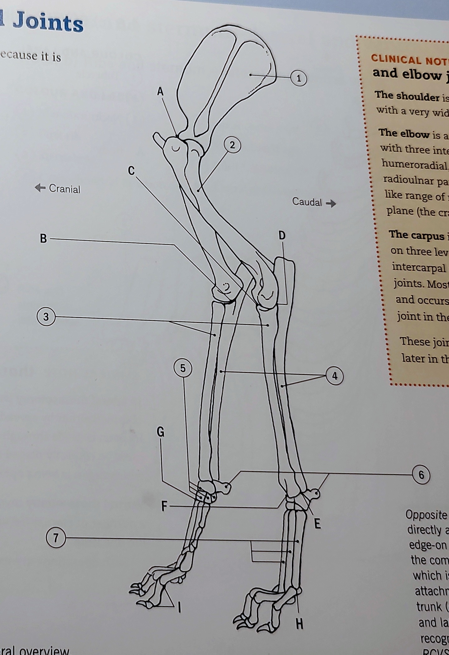
C
Humeroradial (elbow) joint (Dog)

D
Proximal Radioulnar (elbow) joint (dog)
The proximal radioulnar joint is a critical synovial joint located at the elbow region in dogs, connecting the radius and ulna, the two long bones of the forearm. This joint allows for rotational movements of the forearm, which are essential for various canine activities such as digging, turning, and grasping.
Key Components of the Proximal Radioulnar Joint:
Bones Involved:
The joint primarily involves the head of the radius and the radial notch of the ulna. The head of the radius is cylindrical and fits into the radial notch, allowing for smooth rotation during pronation and supination of the forearm.
Articular Cartilage:
The surfaces of the joint are covered with articular cartilage that reduces friction between the bones during movement, protecting them from wear and tear over time.
Joint Capsule:
Like other synovial joints, the proximal radioulnar joint is encased in a fibrous joint capsule that contains synovial fluid. This fluid lubricates the joint and provides nutrients to the cartilage, facilitating smooth movements and absorbing shocks.
Ligaments:
The joint is stabilized by several ligaments, including the annular ligament, which encircles the head of the radius and retains it in the radial notch of the ulna. This ligament is crucial for preventing dislocation of the radius during arm movements.
Muscles:
The flexor and extensor muscles of the forelimb play vital roles in the motion at the proximal radioulnar joint. Muscles such as the biceps brachii assist in flexion and enable supination, while muscles like the pronator teres facilitate pronation.
Functional Importance:
The proximal radioulnar joint is integral to a dog's forelimb function, allowing for versatile movements required during running, jumping, and various activities of daily living. Its ability to permit rotation of the forearm enhances the dog's capability to manipulate objects and perform athletic movements effectively.
Veterinary Considerations:
Injuries or conditions affecting the proximal radioulnar joint can result in significant mobility issues for dogs. Common problems include elbow luxation, fractures around the joint, arthritis, and conditions such as elbow dysplasia. Veterinary assessment through physical examination and imaging (x-rays or ultrasound) is essential for diagnosis and treatment planning. Treatments may range from conservative management, including rest and anti-inflammatory medications, to surgical intervention in more severe cases.

E
Distal radioulnar (carpus, wrist) joint (Dog)
The distal radioulnar joint is a critical synovial joint in dogs located near the wrist region, specifically at the distal ends of the radius and ulna. This joint plays a significant role in allowing forearm movement and enhancing the functionality of the wrist. Its primary function is to facilitate the rotational motion of the forelimb, which is essential for actions such as turning, digging, and grasping.
Key Components of the Distal Radioulnar Joint:
Bones Involved:The distal radioulnar joint consists of the distal end of the radius and the ulnar head. The ulnar notch on the radius allows for the articulation with the ulnar head, enabling movement between these two bones during forelimb rotation.
Articular Cartilage:The joint surfaces are covered with articular cartilage, which reduces friction during movement and protects the underlying bones from wear. This cartilage is crucial for maintaining the joint's health and function over time.
Joint Capsule:Like most synovial joints, the distal radioulnar joint is surrounded by a fibrous joint capsule. This capsule encloses the joint space and contains the synovial fluid, which lubricates the joint, reducing friction and providing nutrients to the cartilage.
Ligaments:The joint is stabilized by ligaments, including the triangular fibrocartilage complex (TFCC), which acts as a stabilizer for the distal radioulnar joint and provides support during dynamic motions. Ligaments play an essential role in preventing dislocation and maintaining the alignment of the joint during movement.
Muscular Interactions:The muscles surrounding the distal radioulnar joint, such as the pronator teres and supinator muscles, are important for facilitating pronation and supination of the forearm. These movements enable the dog to efficiently rotate its forelimb for a range of activities from running to playing.
Functional Importance:The distal radioulnar joint is crucial for a dog's overall mobility and functionality. It allows for twisting and rotating motions of the forelimb, which are vital for tasks such as climbing, digging, and interacting with the environment. Functional integrity of this joint affects a dog's ability to perform daily activities efficiently.
Veterinary Considerations:Injuries or conditions affecting the distal radioulnar joint, such as fractures, dislocations, or degenerative joint diseases, can lead to decreased mobility and pain. Common issues include distal radiculopathy and conditions affecting the TFCC, which may require diagnostic imaging (like x-rays or MRI) for proper evaluation. Treatment options may involve conservative management such as rest and anti-inflammatory medications or surgical intervention in severe cases. Regular veterinary check-ups are essential to monitor joint health, especially in active or aging dogs.

F
Antebrachiocarpal (carpus, wrist) joint (dog)
The antebrachiocarpal joint is the synovial joint located between the distal ends of the radius and ulna and the proximal row of carpal bones. It plays a key role in allowing flexion and extension movements of the wrist in dogs.

G
Carpometacarpal (carpus, wrist) joint (dog)
The carpometacarpal joint is a synovial joint located between the distal row of carpal bones and the proximal ends of the metacarpal bones. This joint facilitates limited movement, primarily allowing for flexion and extension of the digits, contributing to the overall dexterity of the dog's forelimb.

H
Metacarpophalangeal (digits) joint (dog)
The metacarpophalangeal joint is the synovial joint located between the metacarpal bones and the proximal phalanges of the digits. It allows for flexion and extension of the toes, playing a vital role in the dog's ability to walk, run, and perform various activities.

I
Proximal and distal Interphalangeal (digits) joints (dog)
These joints are crucial for grasping and manipulating objects. The proximal and distal interphalangeal joints are synovial joints located between the phalanges of the digits. They enable flexion and extension, allowing for fine motor control and movement of the dog's toes.
Voorbeen
Thoracic limb
omdat het aan de thorax vast zit door de spieren.
= another term for the forelimb of a dog, consisting of the shoulder, upper arm, forearm, carpus, and digits.
Wat is een spheroidal gewricht? (Kogelgewricht)
A spheroidal joint, also known as a ball-and-socket joint, allows for multi-directional movement and rotation. This type of joint is exemplified by the shoulder and hip joints in dogs. It consists of a spherical head fitting into a cup-like socket, enabling a wide range of motion.
wat is een composite gewricht?
A composite joint, also known as a compound joint, is formed by the union of two or more different types of joints, allowing for complex movement. In dogs, this can involve the interaction of multiple bones and joints, providing stability and flexibility. elbow, the carpus and stifle