Inflammatory and benign bone tumors
1/7
There's no tags or description
Looks like no tags are added yet.
Name | Mastery | Learn | Test | Matching | Spaced |
|---|
No study sessions yet.
8 Terms
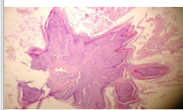
Organ: skin
Comment: skin polyp showing a pink, branching, vascular core covered by hyperplastic stratified squamous epithelium. The basal layer of the epithelium is formed of tall cuboidal basophilic cells with acanthosis where there is hyperplasia of the prickle layer appearing multiple layers of polyhedral cells and hyperkeratosis. The core contains inflammatory cells infiltrate.
Diagnosis: squamous cell papilloma
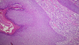
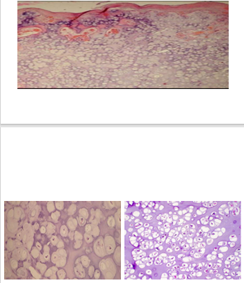
Organ: bony growth (the epiphyseal cartilage of a growing long bone)
Comment: the section shows a neoplastic bony growth (stalk with head) of mature bone trabeculae. It is covered by a cartilaginous cap formed of benign hyaline cartilage. The cartilage undergoes ossification to produce new bone formation. The cartilaginous matrix is eroded by dilated, thin-walled blood vessels. Beneath the stalk is a medullary cavity containing fat cells and congested capillaries.
Diagnosis: Osteochondroma
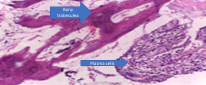
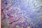
Organ: bone
Comment: it shows bony trabeculae with infiltration of chronic inflammatory cells which are plasma cells, lymphocytes and histiocytes, with granulation tissue formation.
Diagnosis: chronic osteomyelitis
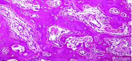
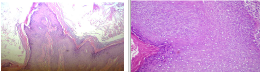
Organ: Bony growth
Comment: the section shows neoplastic bony growth of benign mature osteoblasts with eosinophilic cytoplasm and monochromatic nucleus admixed at the periphery of bone trabeculae. The background shows few osteoid matrix
Diagnosis: Osteoma
Organ: Bony growth
Comment: Section is lobulated and encapsulated showing benign faint blue hyaline matrix with benign chondrocytes. The chondrocytes reside in lacunae singly or in groups. They have a clear or vacuolated cytoplasm as glycolipids dissolves during processing and central dark nuclei
Diagnosis: Chondroma
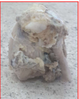
Orgab: Part of broken rib
Comment: Sows a broken rib with exophytic growth of whitish-grayish color, partially encapsulated, well-circumscribed, containing blue foci of chondroid matrix.
Diagnosis: Osteochondroma, rib