HMI204 - Spinal Cord, Spinal Nerves and Peripheral nerves
 Call Kai
 Learn
Learn Practice Test
 Spaced Repetition
 Match
Match Flashcards
 Knowt Play
1/103

There's no tags or description
Looks like no tags are added yet.
Name | Mastery | Learn | Test | Matching | Spaced | Call with Kai | Chat |
|---|
No analytics yet
Send a link to your students to track their progress
104 Terms
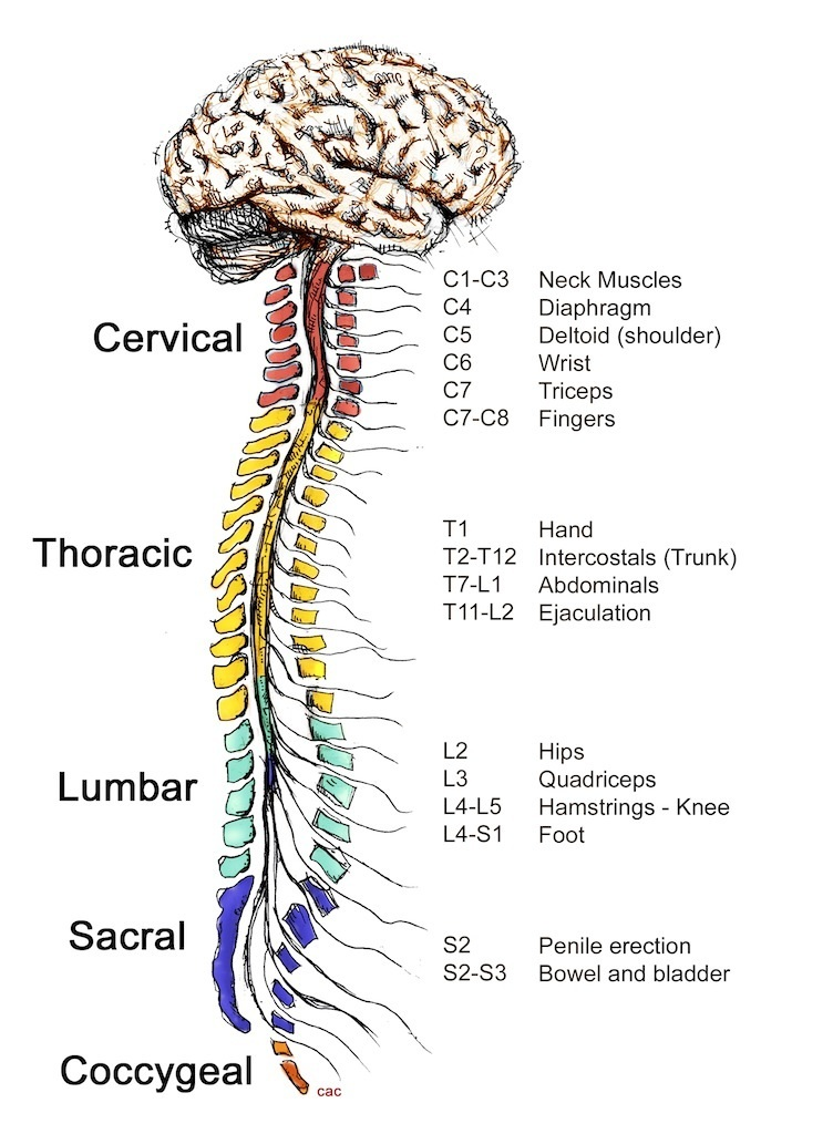
How many segments of the spinal cords are there?
31
8 segments in the cervical
12 thoracic
5 lumbar
5 sacral
1 coccygeal
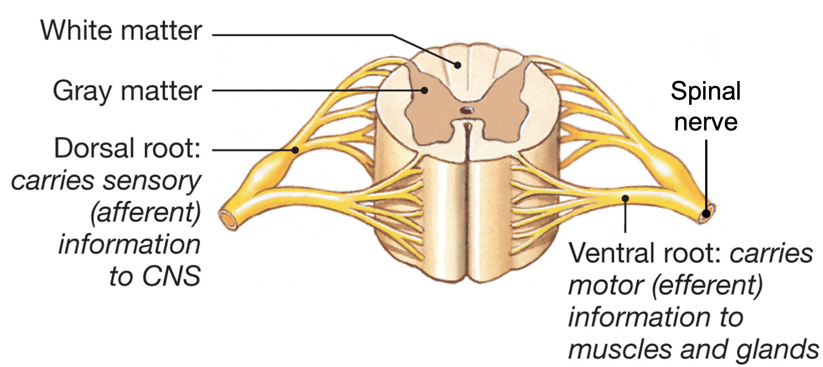
How are each individual segments of the spinal cord defined by?
Ventral and dorsal rootlets from spinal cord area that gives off a spinal nerve.
i.e. This diagram has two spinal segments
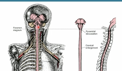
At what point does the medulla oblongata become the spinal cord?
Foramen magnum
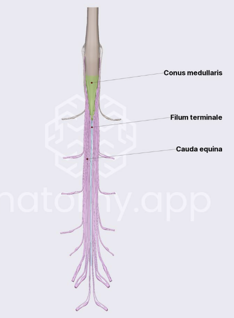
Spinal cord extends all the way down from C1 to its termination point. What is this termination point called?
Conus medullaris
At what level does the spinal cord terminate in an adult?
L1-L2
What vertebral structures surrounds the spinal cord?
vertebral body anterior and a posterior vertebral arch (lamine and spinous process)
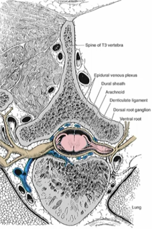
What boundaries make up the intervertebral foramen?
Vertebral bodies and IVD - anterior
Pedicles - superior/inferior
Zygapophyseal joint - posterior
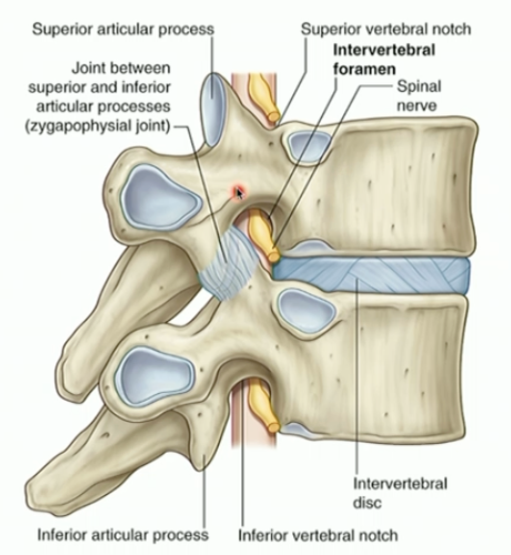
what layers of protection are there to keep spinal cord protected
bone
epidural space
Dura mater
Arachnoid mater
Pia mater
What is the epidural space?
epidural space - space between vertebral body and dura mater. it contains fat and to give an extra layer of prottection the spinal cord.
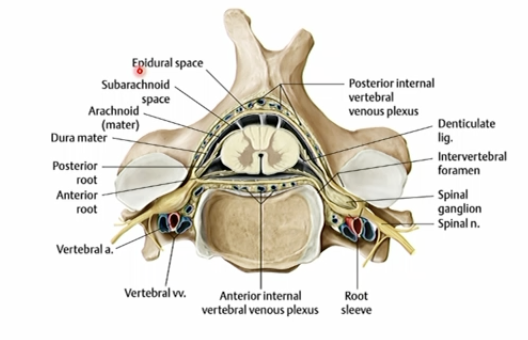
What is Dura mater?
dense connective tissue on the outermost layer of the meninges covering the spinal cord and is continuous with the spinal nerve.
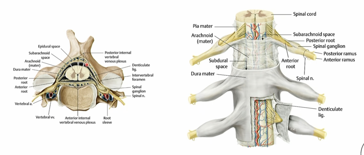
What is arachnoid mater?
Second layer of the meninges
Made up of Arachnoid trabeculae that gives it a spider-web like appearance.
This trabeculae also bridges the arachnoid mater to the pia mater.
Pressed up against the dura mater due to CSF pressure.
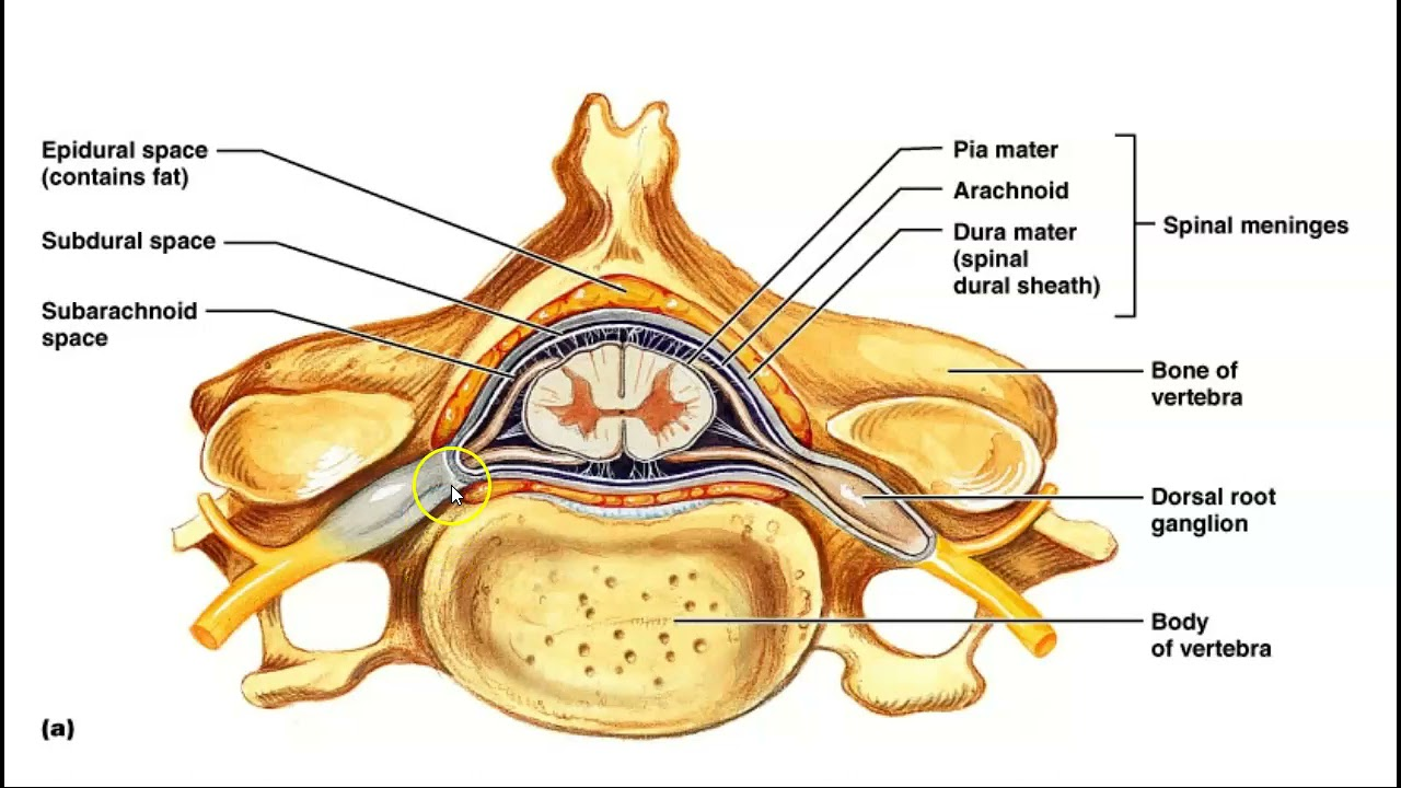
What is the pia mater?
Delicate innermost layer of the meninges that tightly hugs the surface of the brain and spinal cord. It carries blood vessels to nourish neural tissue.
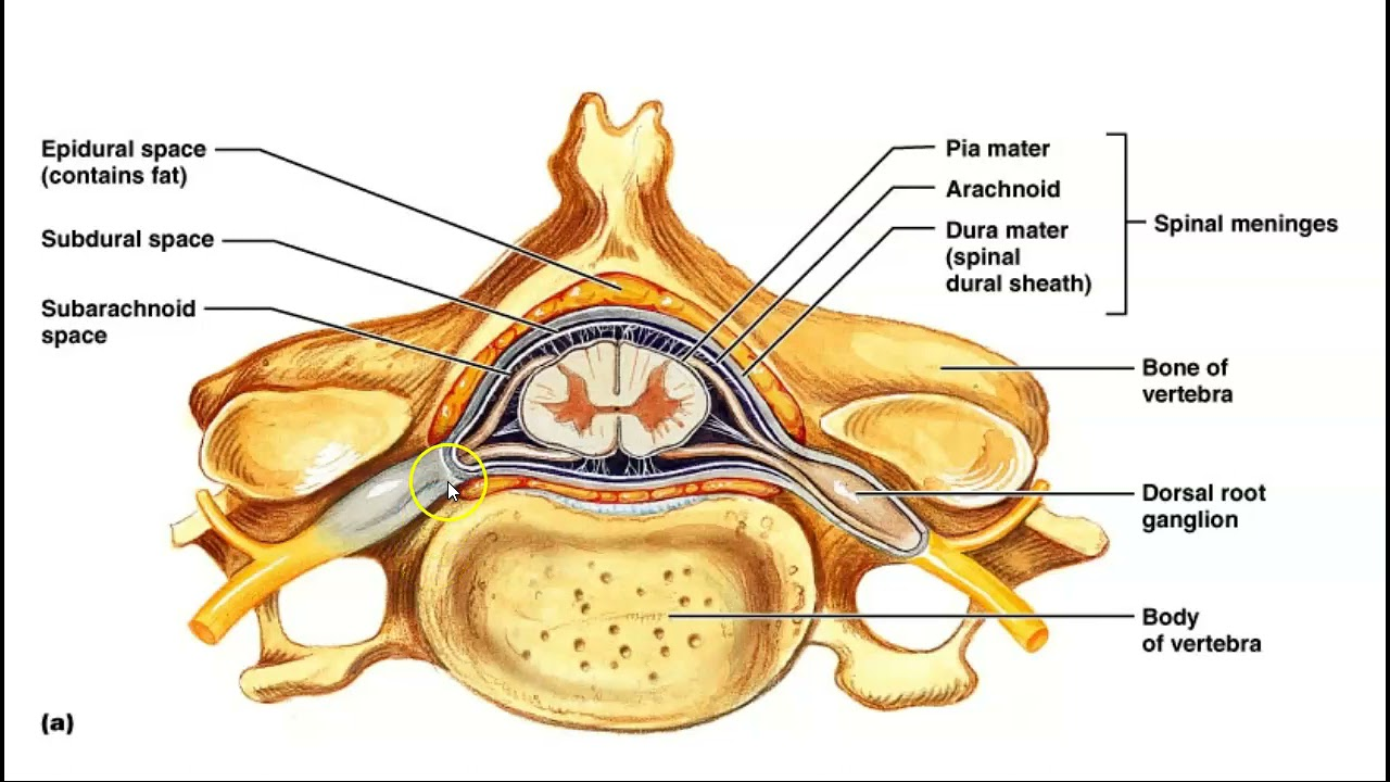
What is inside the subarachnoid space?
Cerebrospinal Fluid
Blood Vessels
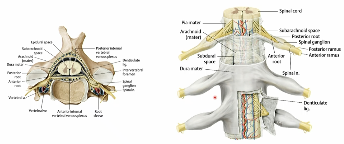
Cauda euina is the structure that continues at the end of the spinal cord. Explain how spinal nerves are still able to exit at their respective site?
The series of anterior and posterior roots descend to their respective IV foramen and then join to create the spinal nerve just before exiting.
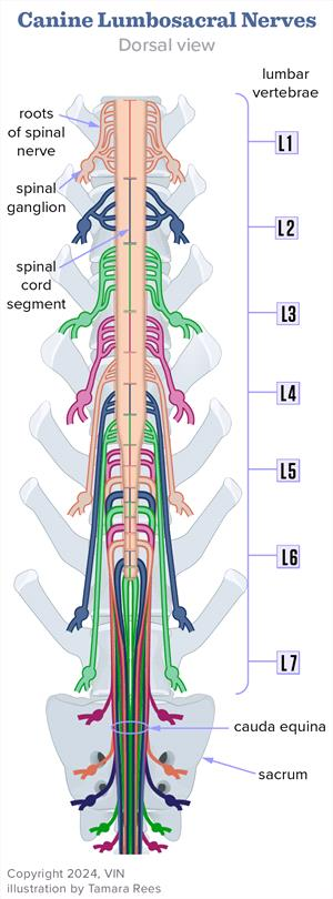
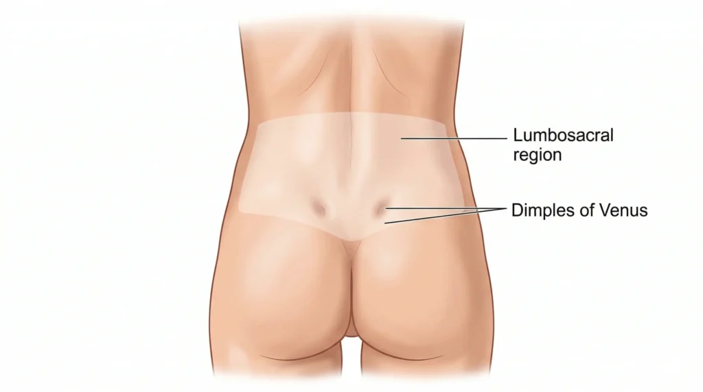
2cm below the dimples of venus are at the level of…? What ends around this level?
PSIS/S2 - termination of dura and arachnoid mater and CSF
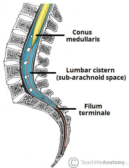
Pia mater continues down until the coccyx surrounding what structure. Describe the purpose of this structure.
It continues coating the filum terminale which is a slender, fibrous thread of connective tissue that anchors the lower end of the spinal cord to the coccyx
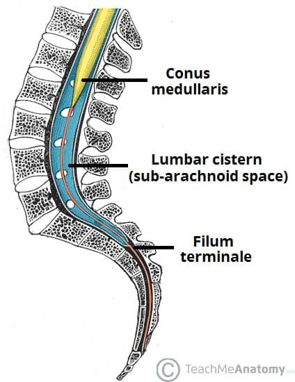
After the dura mater and arachnoid mater ends while the pia mater continues down, it creates this big space filled with CSF. What is this space called?
Lumbar cistern.
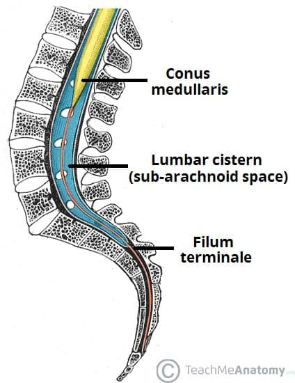
Lumbar puncture is a procedure where a needle is inserted into the lower back to collect CSF. Why is it important for this procedure to happen at L3 and below?
To reduce the risk of the needle hitting the spinal cord.
Roots of the caudal equina can just move out of the needle’s way.
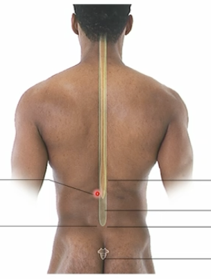
Neonates’ spinal cords ends around…
L3-4
In the spinal cord, white matter is organised… while gray matter is organised….
peripherally, centrally
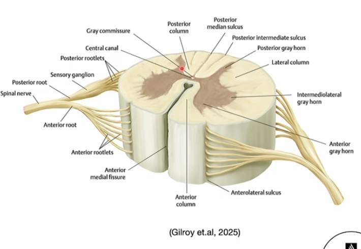

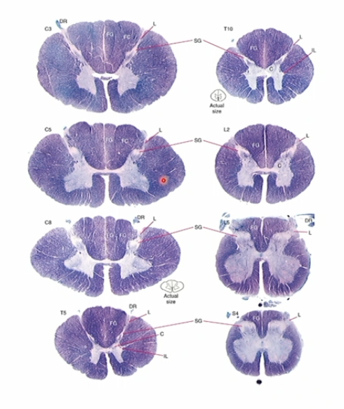
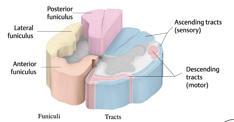
Label the cross sectional anatomy of the spinal cord
instead of saying columns → use funiculus
What is the functional difference of grey and white matter in the spinal cord?
gray matter, consisting of mostly neurons, acts as the processing and integration center in the spinal cord
White matter, consisting of mostly myelinated axons, acts as the transmission site to the brain.
In grey matter, anterior horns houses which neuronal cell bodies?
Motor neurons (Lower Motor Neurons)
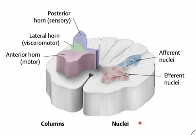
In grey matter, posterior horns is associated which neurons?
Sensory neurons (afferent neuron).
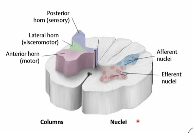
Why are the anterior horns of the thoracic region of spinal cord so thin compared to other regions?
Because the thoracic region only innervates the intercostal muscles so less motor neurons are used.
In grey matter, lateral horn is associated with which neurons?
Autonomic nervous system neurons (preganglionic neurons)
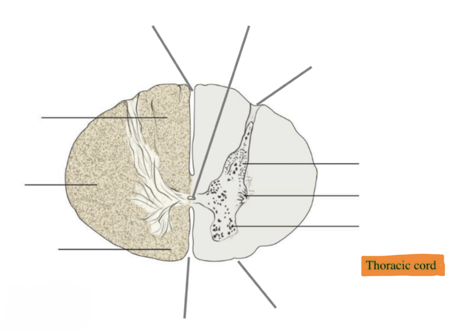
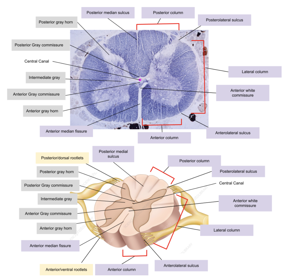
Label this cross sectional image of the spinal cod in the thoracic region.
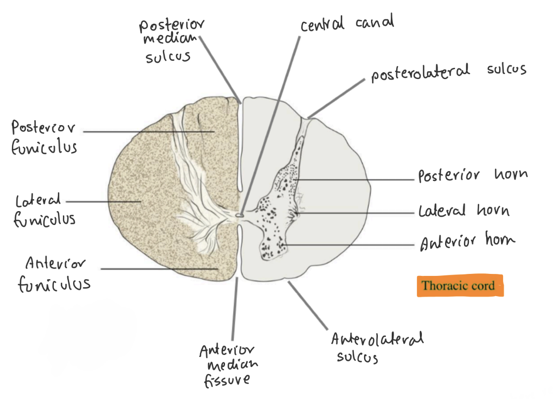
Why might the thoracic, upper lumbar and sacral region of the spinal cord have very evident lateral horns but not in cervical?
Because the lateral horn houses autonomic neurons and those regions supply the autonomic nervous system.
Anterolateral sulcus of the spinal cord is where… attaches.
anterior/ventral rootlets
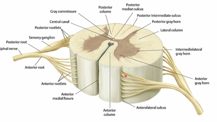
Posterolateral sulcus of the spinal cord is where… attaches.
posterior/dorsal rootlets attach
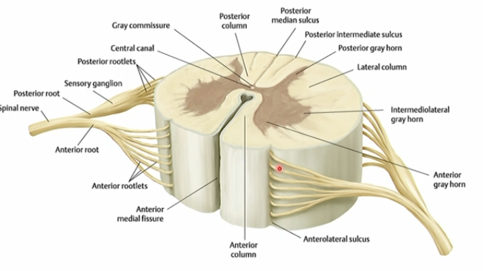
what boundaries make up the anterior, lateral and posterior columns of the spinal cord?
anterior - anterior median fissure to anterolateral sulcus
lateral - anterolateral sulcus to posterolateral sulcus
posterior - posterolateral sulcus to posterior median fissure
If the posterior column of the white matter in the spinal cord is damaged, what would happen?
Posterior column:
Sensory → Touch, pressure, vibration and proprioception information going up to the brain
Touch and pressure information is unable to reach the brain as the pathway is damaged.

If the lateral column of the white matter in the spinal cord is damaged, what would happen?
Lateral column:
Spinothalamic → temperature and pain (Nociception)
Corticospinal tract → voluntary muscle movement
Temperature and pain information is unable to reach the brain as the pathway is damaged.
Voluntary muscle contraction is affected

What does grey and white commissures do?
Gray commissures:
links the left and right horns of the spinal cord.
contains unmyelinated interneurons that coordinate local reflex activities and pass information between the two sides.
White commissures:
crossing between the two sides and helps transmits sensory signals and motor instructions from the brain to the opposite sides of the body.
Where does pain and temperature sensation decussate?
At the level of the spinal cord

Where does light touch, proprioception and vibration sensation decussate?
In the brainstem
Where does motor axons decussate?
In the brainstem
why is the cervical segment of the spinal cords thicker?
thicker spinal cord → bigger anterior horns → more volume of motor neurons → innervates a large no. of muscles in the upper limbs
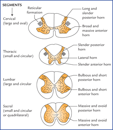
why is the thoracic segment of the spinal cords thinner? But there is a special white matter feature that is bigger in this region, what is it and why is it?
thinner spinal cord → smaller anterior horns → less volume of motor neurons → innervates a small no. of thoracic muscles
Intermediate gray bigger → more autonomic nerve cell bodies → sympathetic nervous system in the thoracic region
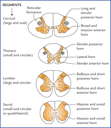
why is the lumbar segment of the spinal cords thicker?
thicker spinal cord → bigger anterior horns → more volume of motor neurons → innervates a large no. of muscles in the lower limbs
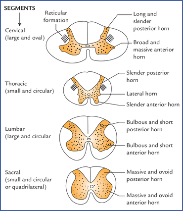
How does the volume of white matter change from superior (cervical) to inferior (sacral).
Increase in white matter as we travel superiorly because more information has to travel through the superior segments compared to the inferior segments of the spinal cord.
What are the 2 types of reflexes and how are they different?
Somatic - affects voluntary skeletal muscles
deep tendon reflex
Superficial
withdrawal reflex
Visceral/autonomic - Affects heart, glands, and smooth muscle
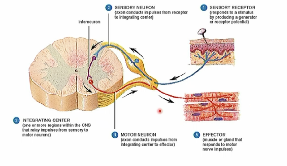
Describe the process of a somatic reflex
sensation/stimuli/receptor
sensory information is transmitted to the integration centre (gray matter) via sensory neuron through the dorsal root
sensory info gets integrated and turned into a motor command
motor command gets transmitted via the anterior root
Effectors like skeletal muscles take action of the command
What are monosynaptic reflexes?
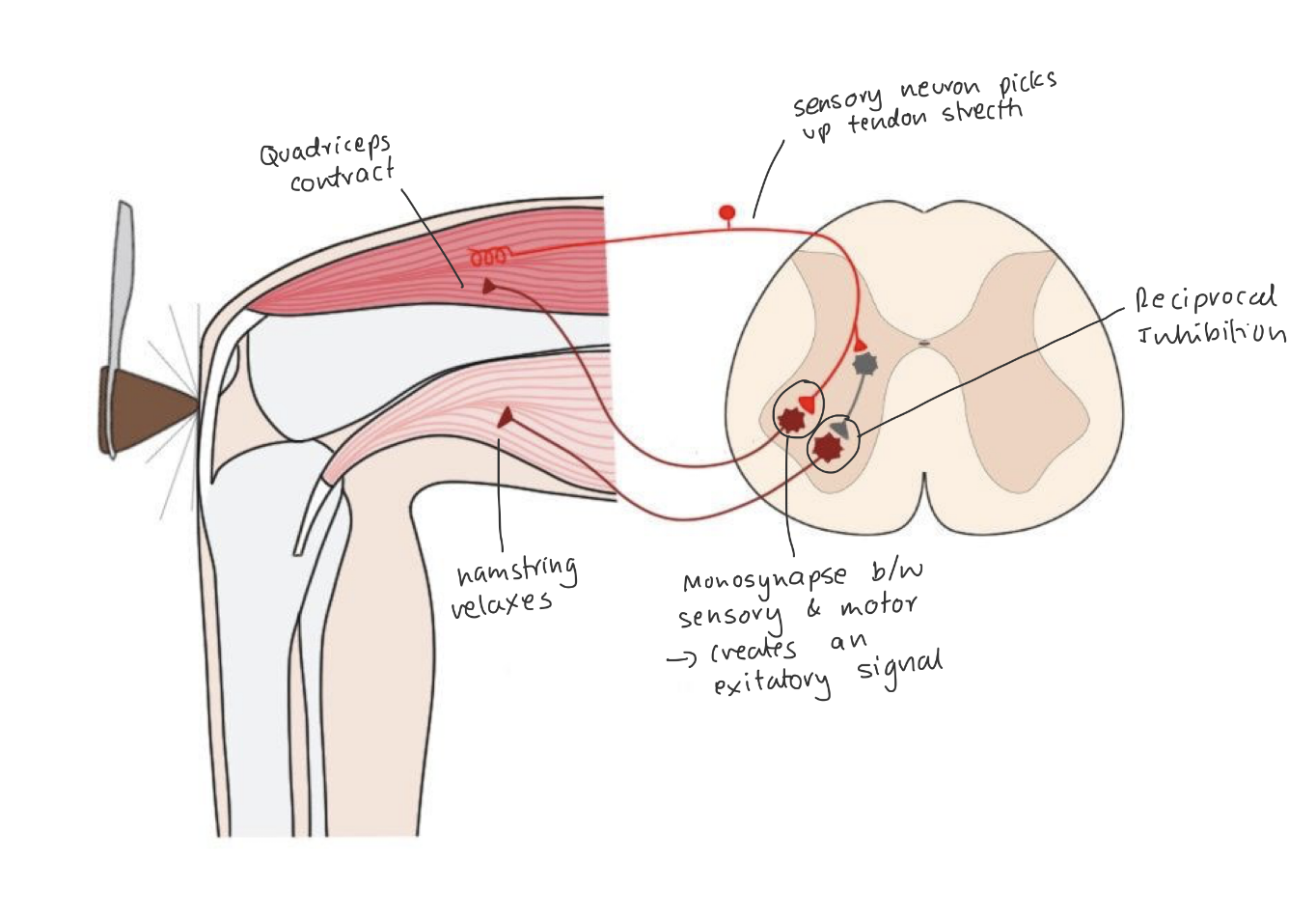
Fast, automatic muscle action that has only one connection point, called a synapse, between a sensory nerve and a motor nerve.
Deep tendon reflex such as the knee jerk is an example.
the brain can adjust this sensitivity
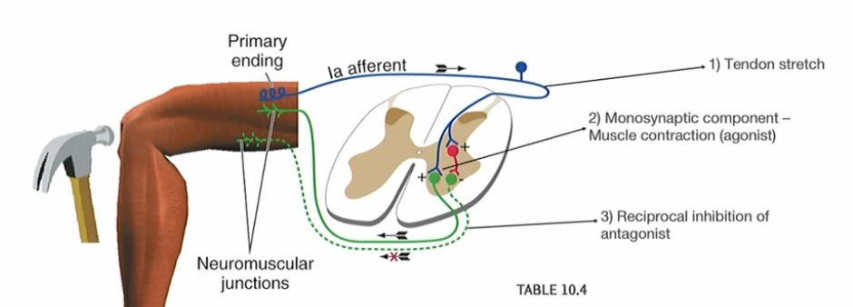
Exaplain the knee jerk reflexes in terms of sensory and motor neurons.
What are polysynaptic reflexes? Can you give an example?
reflex pathways that involve one or more interneurons and facilitate coordinated muscle activity in response to sensory.
i.e Withdrawl reflex
the brain can adjust this sensitivity
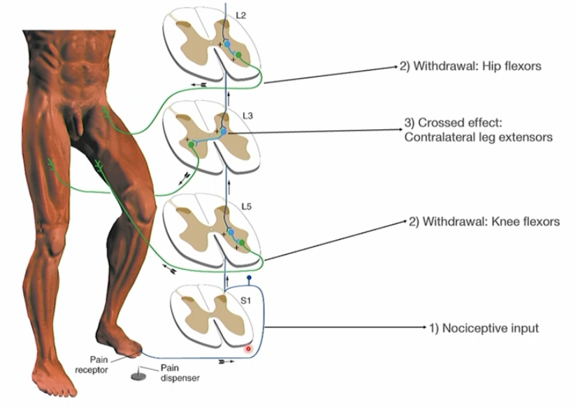
Explain withdrawl reflex
Communication from multiple spinal segments
info gets into the spinal cord and sends it through ascending tracts
Multiple different muscles are active
In Gray commissures, interneurons talk to the opposite side of the spinal cord, activating muscles in your other limb to maintain balance.
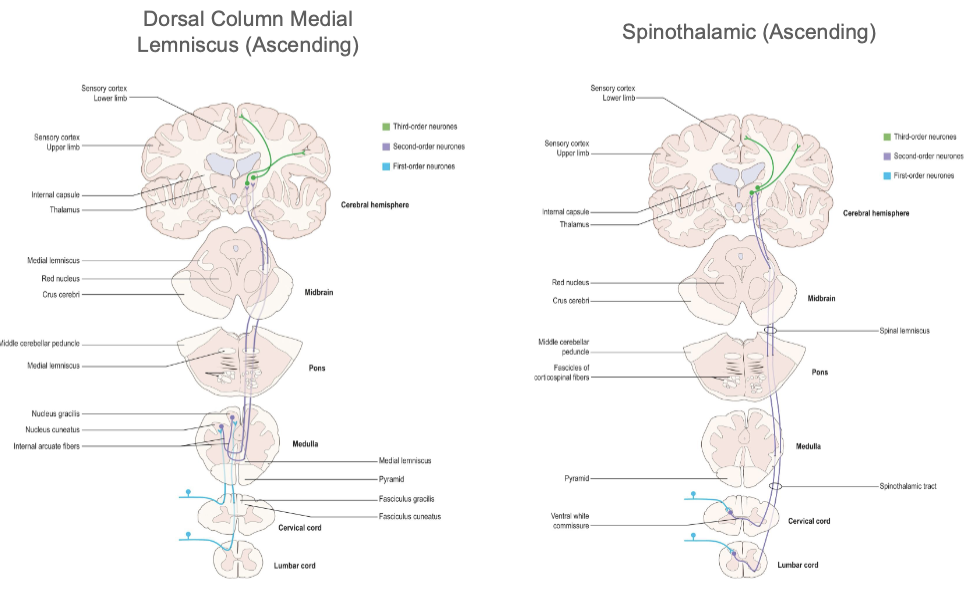
What are the two ascending tracts?
Dorsal column medial lemniscus - light, touch, proprioception and vibration
travels through posterior column
Spinothalamic - pain and temperature
sensory info crosses from the posterior horn → the white commissure → to the front part of the lateral column
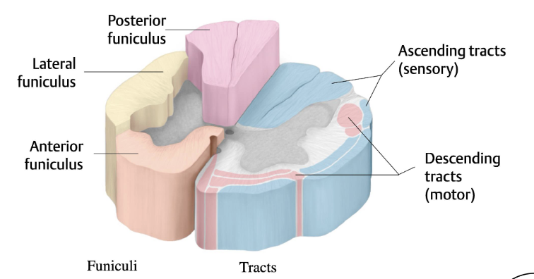
What is the descending tract?
Lateral Coticospinal - motor commands
Lateral column
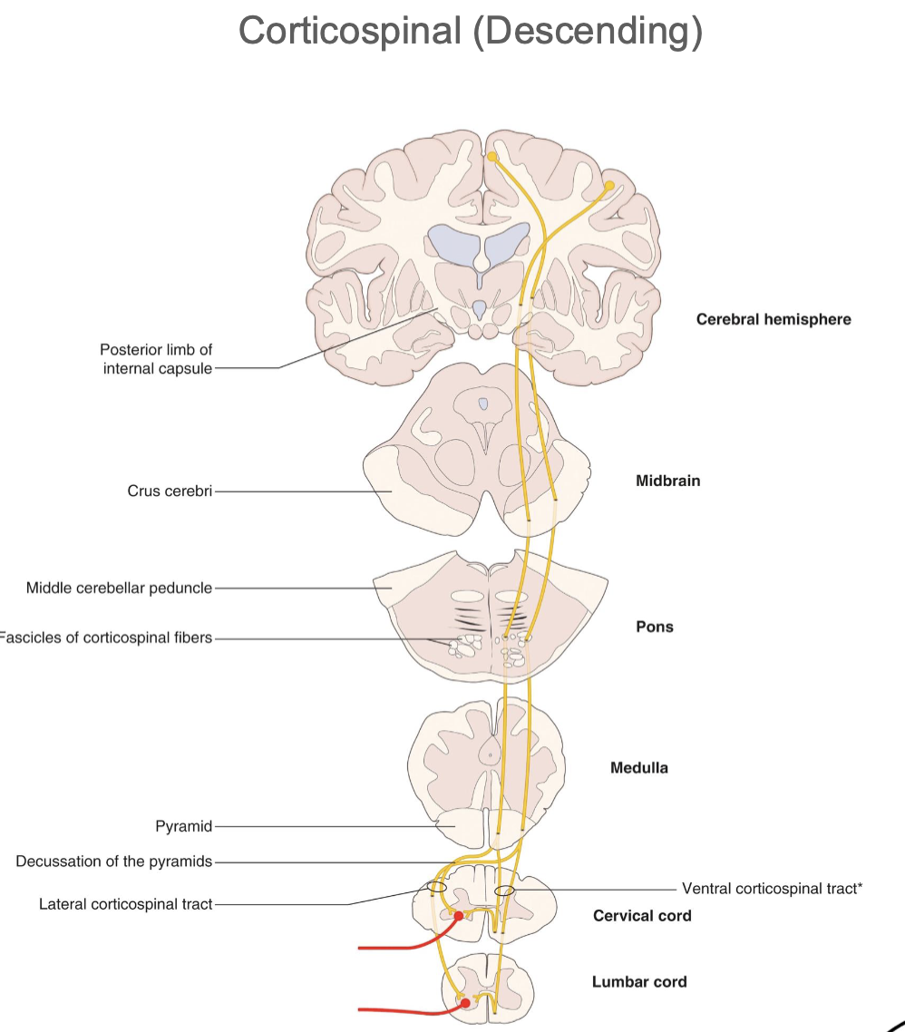
If the lateral column was damaged, what clinical symptoms wld patients face?
loss of pain and temperature sensation
Loss of voluntary control of the limbs
There are two main arteries that supply the spinal cord, anterior and posterior spinal arteries.
Explaing what areas of the spinal cord does the anterior one supply, where it branches off from and where it travels through.
Supplies the anterior 2/3 of the spinal cord (anterior and lateral column and anterior horns)
Branches off the vertebral artery
travels thrugh the anterior median fissure.
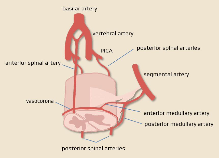
There are two main arteries that supply the spinal cord, anterior and posterior spinal arteries.
Explaing what areas of the spinal cord does the posterior one supply, where it branches off from and where it travels through.
Supplies the posterior columns and posterior horn
Branches off the posterior inferior cerebellar arteries (PICA)
travels through the posterolateral sulcus
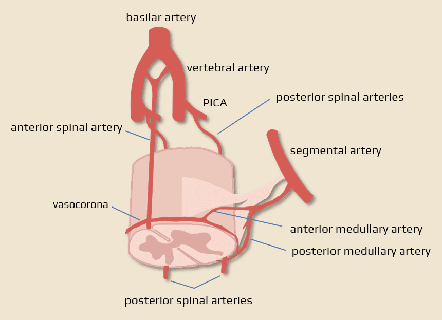
The two spinal arteries are very tiny and need to supply the huge spinal cord, how does the body ensure enough blood supply?
the arteries require a boost from segmental spinal arteries from different regions:
cervical - vertebral arteries
thoracic - posterior intercostal
lumbar - lumbar arteries
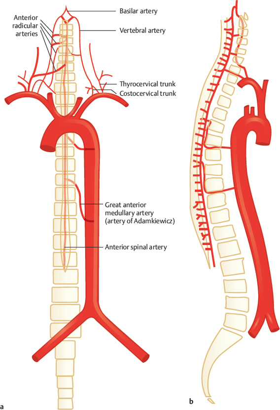
What is a significantly large segmental spinal artery in the lumbar region? Why is this significant?
Artery of Adamkiewicz.
Supplies blood to the lumbar enlargement and conus medullaris of the spinal cord
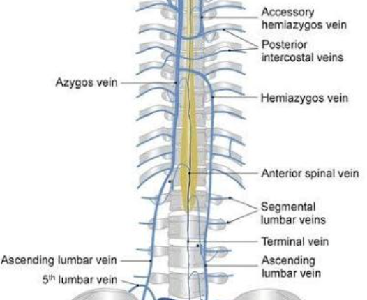
Venous drainage in the spinal cord
6 veins drain the spinal cord:
One anterior & posterior
Two for each posterolateral and anterolateral sulcus
these then drain intto → internal and external venous plexuses → azygous vein
What is a dermatome?
area of skin innervated by a single spinal nerve
Which dermatome is associated with the upper side of the upper limb and thumb?
C4,5,6.

Which dermatome is associated with the middle finger?
C7.
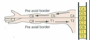
Which dermatome is associated with the lower finger region/little finger side?
C8
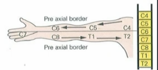
Which dermatomes are found toward the axilla?
T1–T2.

Which dermatomes are represented more posteriorly and in the sole of the foot?
Sacral dermatomes.

The umbilicus is approximately at what dermatome level?
T10.

Sensory loss below the umbilicus suggests injury around which spinal cord segment?
T10.

The nipple line is approximately at what dermatome level?
T4–T5.

What is a myotome?
A predictable muscle movement contribution associated with a spinal cord/spinal nerve level.
What do lesions at C3 and above threaten?
Results in paraplagia due to a loss of innervation to the upper and lower limbs and below the lesion.
Requires respiratory assistance due to the inability of motor signal to reach the phrenic nerve of the diaphragm.
What spinal levels contribute to the phrenic nerve?
C3, C4, and C5.
What is typically seen with lower cervical cord lesions? (i.e. C7)
Quadriplegia, but respiration may be preserved.
What is usually preserved in thoracic cord lesions?
Ventilation and upper limb function as this usually innervated by the cervical region.
What happens in lumbar cord lesions?
Lower limb deficits plus possible pelvic organ dysfunction like bladder and bowel function as well as sexual function.
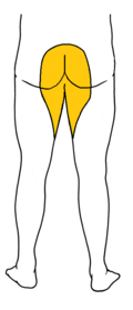
Cauda equina syndrome is when the cauda equina is squeezed or compressed and prevents signals from traveling to your legs, feet, bladder, and bowel.
What sensory loss is clasically associated with cauda equina syndrome?
Saddle sensory loss
What is radiculopathy?
Signs and symptoms caused by a lesion of a spinal nerve, often from foraminal narrowing, disc pathology, or osteophytes.
In order from proximal to distal, what is the basic spinal nerve pathway?
Rootlets → roots → spinal nerve (mixed nerve) → rami.
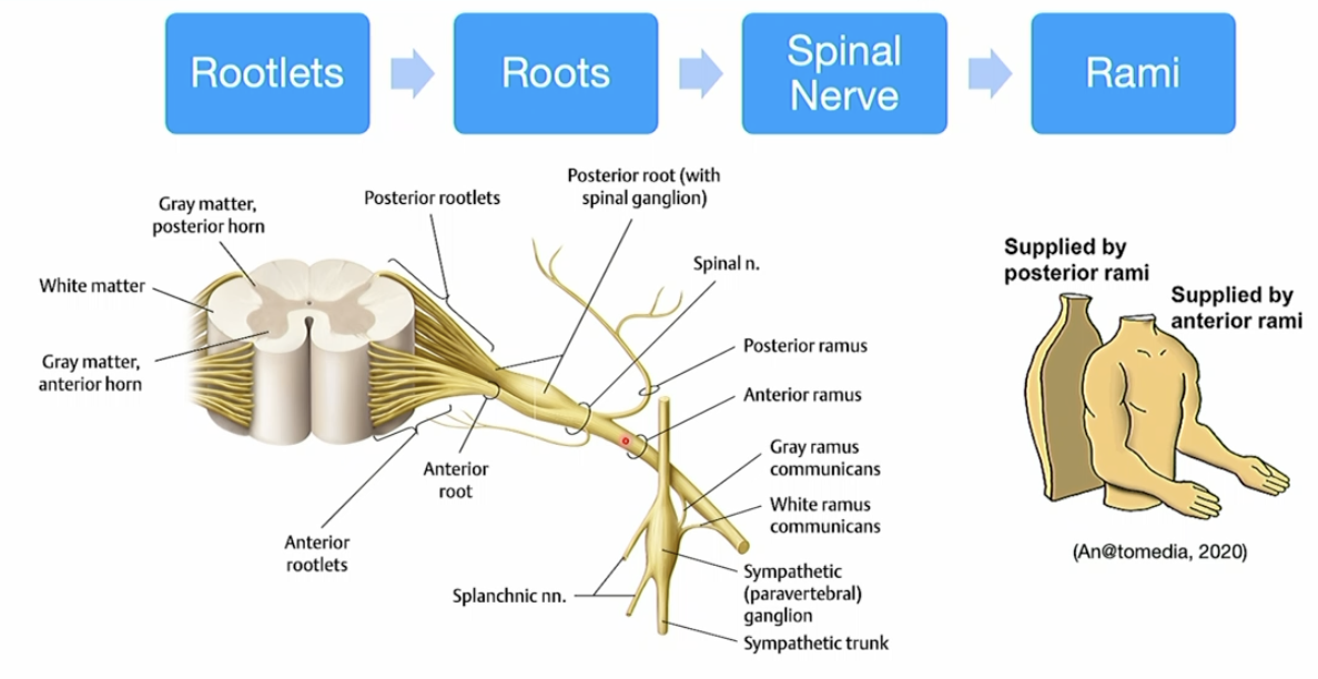
On the drosal root, there is a structure called the dorsal root ganglion. Define the term 'ganglion' and describe the type of cell bodies of which division of the nervous system are found within the dorsal root ganglion?
A ganglion contains the cell bodies of neurons. The dorsal root ganglion contains cell bodies of the pseudounipolar neurons from the somatic and visceral sensory division.
What kind of information travels in the dorsal/posterior and ventral/anterior root?
Dorsal - Sensory information.
Ventral - Motor information.
Why is the spinal nerve called a mixed nerve?
Because it carries both sensory and motor fibers.
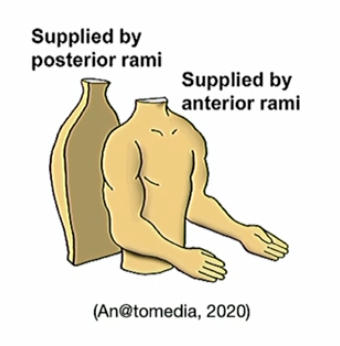
Do both anterior and posterior rami carry motor and sensory fibers? And where do the supply?
Yes.
Posterior - structures at the back
Anterior supplies - structures at the front and limbs
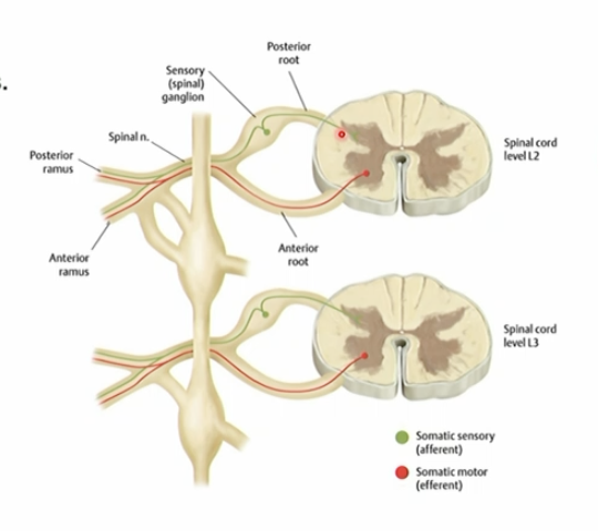
Where does somatic sensory information travel compared with motor information in the spinal nerve pathway?
Sensory information travels through the rami → spinal nerve → dorsal root ganglion/dorsal root → spinal cord
Motor information travels from the ventral horn → ventral root → spinal nerve → rami → target.
How does the sympathetic motor division exit?
It exits from the intermediate gray → anterior root → travels into the spinal nerve → uses the white rami to enter the paravertebral ganglionic chain → synapses with post ganglionic neurons → postganglion exits via the grey rami and towards the target
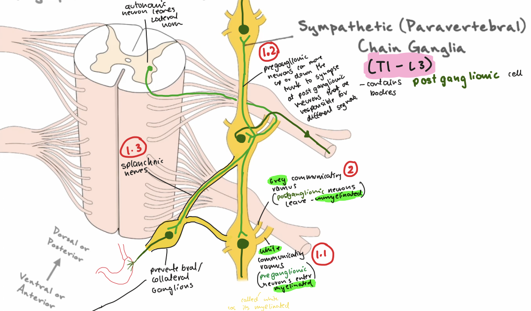
Splanchnic nerves connect to the paravertebral chain so that preganglions can…
connect to prevertebral ganglions.

What is the difference of paravertebral vs prevertebral ganglia?
Paravertebral ganglia sit in chains along the sides of the spine to control the body wall, limbs, and chest.
Prevertebral ganglia sit closer to abdominal and pelvic organs to innervate them.

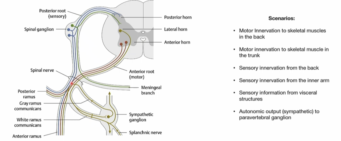
Go through each of these scenarios and trace the pathways.
Motor output starts in the ventral horn, exits via the ventral rootlets and ventral root, enters the mixed spinal nerve, then travels through the posterior ramus to the back muscles.
Motor output starts in the ventral horn, exits via the ventral rootlets and ventral root, enters the mixed spinal nerve, then travels through the anterior ramus to trunk muscles.
Sensory input from the back travels through the posterior ramus, spinal nerve, dorsal root, and dorsal rootlets into the spinal cord, with the sensory neuron cell body in the dorsal root ganglion.
Sensory input from the inner arm travels through the anterior ramus, spinal nerve, dorsal root, and dorsal rootlets into the spinal cord, with the cell body in the dorsal root ganglion.
Visceral sensory information hitches a ride through the autonomics, in this case its a meningeal branch. This info still enters through the dorsal root as it’s sensory.
Autonomic output comes from the intermediate grey → enters the dorsal horn because it is an output → spinal nerve → respective ganglia
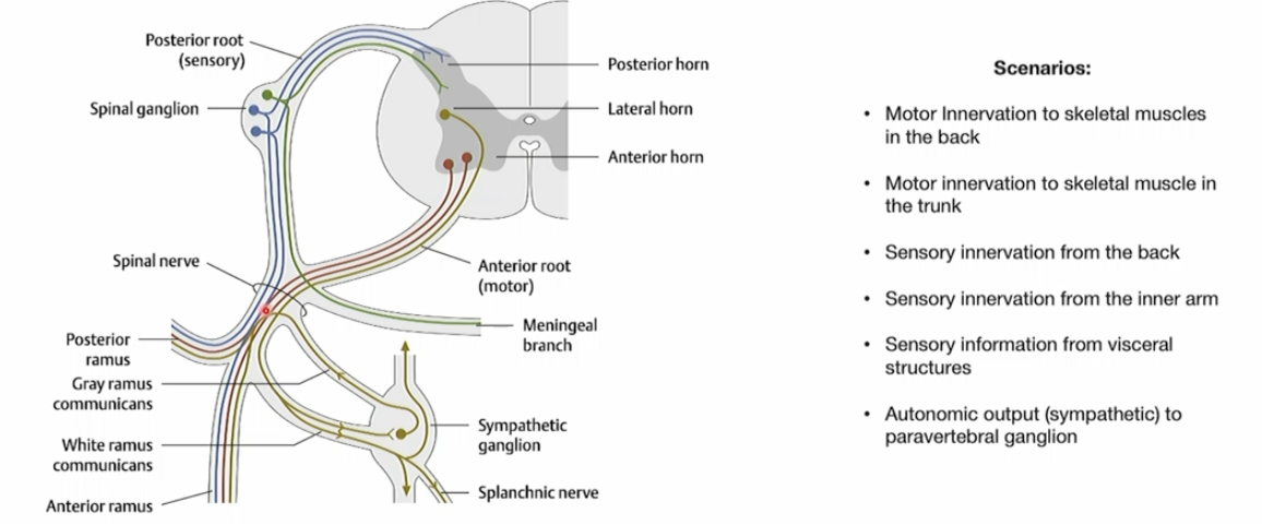
What are the cell bodies in the dorsal root ganglion?
Somatic and viscerael sensory neurons
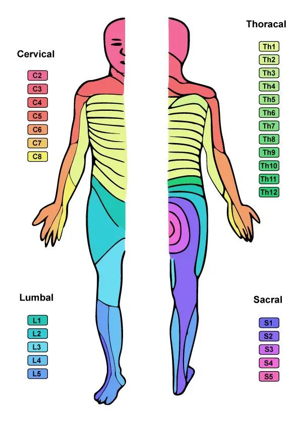
In C5–C6 exit foraminal stenosis, which spinal nerve is affected and what symptom pattern would you expect?
The C6 spinal nerve is affected and compression can cause proximal lateral forearm pain.
How is a dermatome different from a peripheral nerve sensory distribution?
A dermatome is the sensory territory of a single spinal nerve, while a peripheral nerve sensory distribution reflects the area supplied by a nerve formed from multiple spinal cord levels.
How is a myotome different from a peripheral nerve motor distribution?
A myotome is the muscle action associated with a spinal cord segment, while a peripheral nerve motor distribution is the group of muscles supplied along the course of a specific peripheral nerve
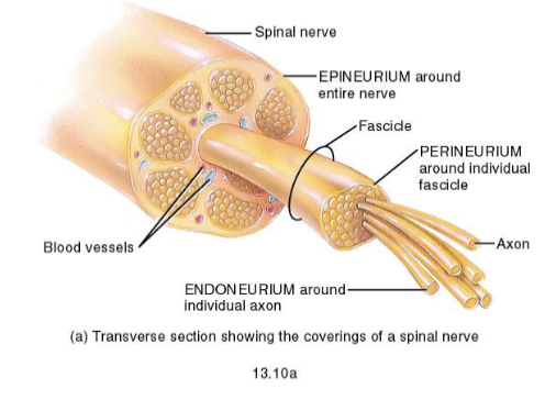
How is a peripheral nerve structurally organized?
Individual axons are wrapped by endoneurium, bundles of axons form fascicles wrapped by perineurium, and multiple fascicles are wrapped together by epineurium.
PS. Epineurium is a continuation of dura mater
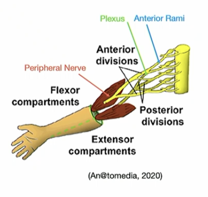
What rami forms plexuses?
Anterior rami
What forms the cervical plexus and what does it supply?
The cervical plexus is formed by ventral rami C1–C4
supplies skin and muscles of the neck.
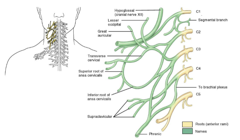
What branch of the cervical plexus is especially important, and why?
The phrenic nerve is especially important because it arises from C3–C5
What forms the brachial plexus?
The brachial plexus is formed by the anterior rami of C5–T1.

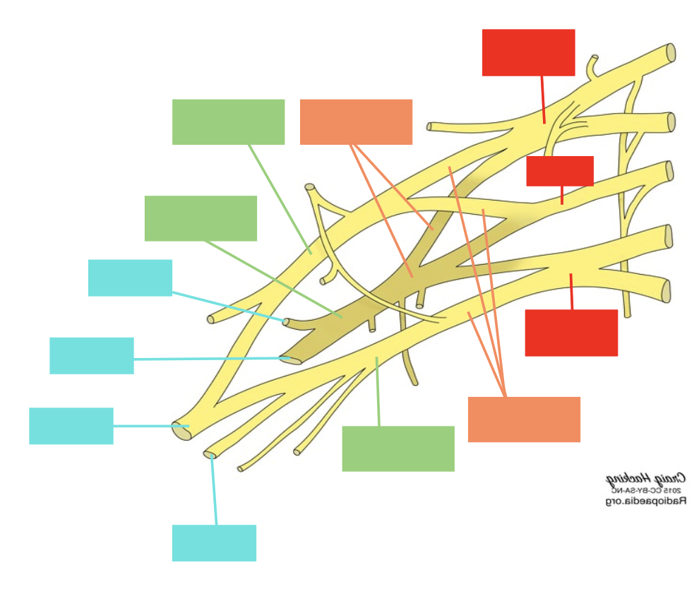
Label the roots, trunks, divisions, cords and branches of the brachial plexus.
(Real truckers drink cold beer)
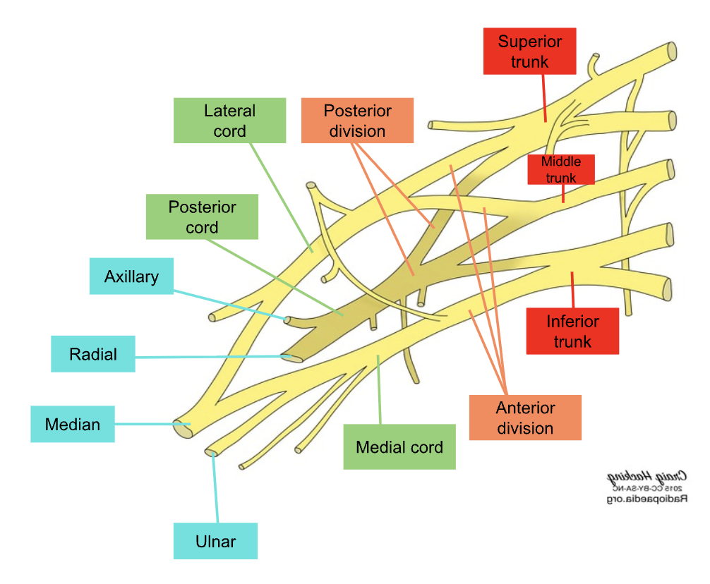
Explain the relationships of brachial plexus with surrounding structure that help to name it.
Roots: b/w anterior and middle scalene
Trunks: In posterior triangle of neck
Divisions: Behind clavicle
Cords: around axillary artery
Branches: emerging from axilla

What are the cords of the brachial plexus and how are they named?
The cords are the lateral, posterior, and medial cords; they are in the axilla and are named by their relationship to the axillary artery.
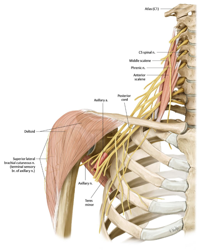
What are the key features of the axillary nerve?
passes around the surgical neck of the humerus under the glenohumeral joint
supplies deltoid, helps with arm abduction
Supplies sensation to lateral deltoid
Vulnerable in surgical neck fractures and anterior shoulder dislocations.
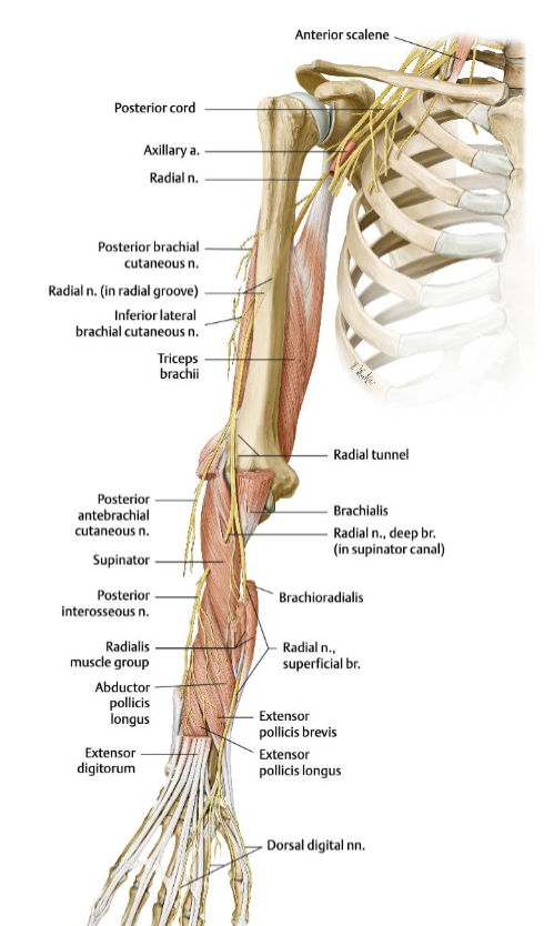
What are the key features of the radial nerve?
travels posteriorly through the spiral groove of the humerus, then the cubital fossa laterally into the extensor compartment of the forearm
Extension of shoulder, elbow, wrist and fingers
Supplies sensory to dorsum of hand and 3 lateral digits
Vulnerable in midshaft humeral fractures
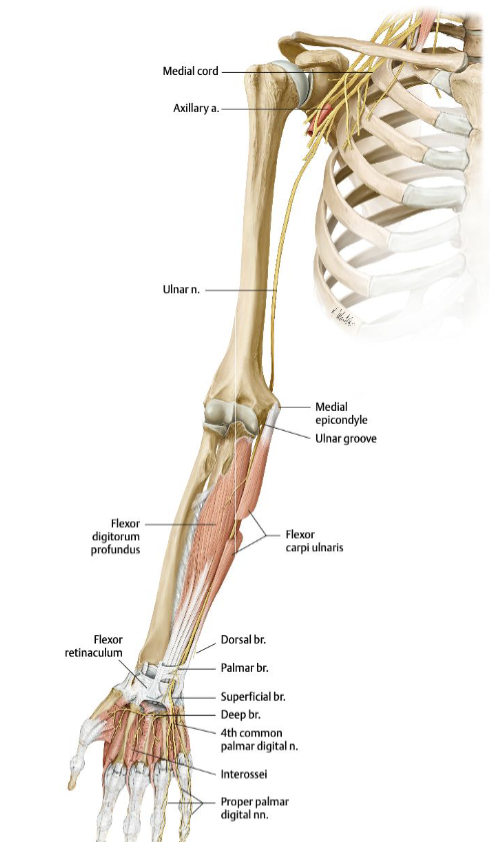
What are the key features of the ulnar nerve?
Descends the arm medially
Wrist flexion, flexion of medial 2 digits at the knuckles
Provides sensation to the medial part of the arm
Vulnerable to fractures to the medial epicondyle
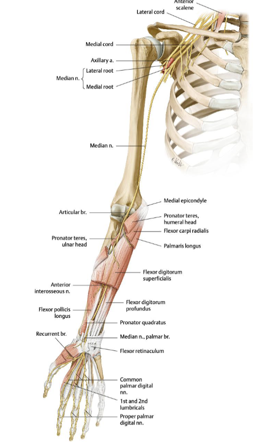
What are the key features of the median nerve?
Descends the arm medially, crosses the elbow, pierces the pronator teres, travels down the forearm, and enters the hand through the carpal tunnel
wrist flexion, thumb movement
sensory information of palm
A supracondylar humeral fracture can tear or stretch the nerve, causing sensory loss and preventing thumb movement and index/middle finger flexion
What are the key features of the lumbosacral plexus?
lies on the posterior abdominal wall
includes the major branches: femoral, obturator, and sciatic, and overall spans L4–S4.


What are the key features of the femoral nerve?
The femoral nerve supplies the anterior thigh
Innervates quads for knee extension
Sensory information of medial lower leg
Injury causes loss of quadriceps function/knee extension plus sensory loss to the medial lower leg.