Unit 2: Bones of the Lower Limb
1/120
There's no tags or description
Looks like no tags are added yet.
Name | Mastery | Learn | Test | Matching | Spaced |
|---|
No study sessions yet.
121 Terms
At the knee the femur articulates with the…
tibia

bones of pelvic girdle and lower limb - 1
pelvic girdle
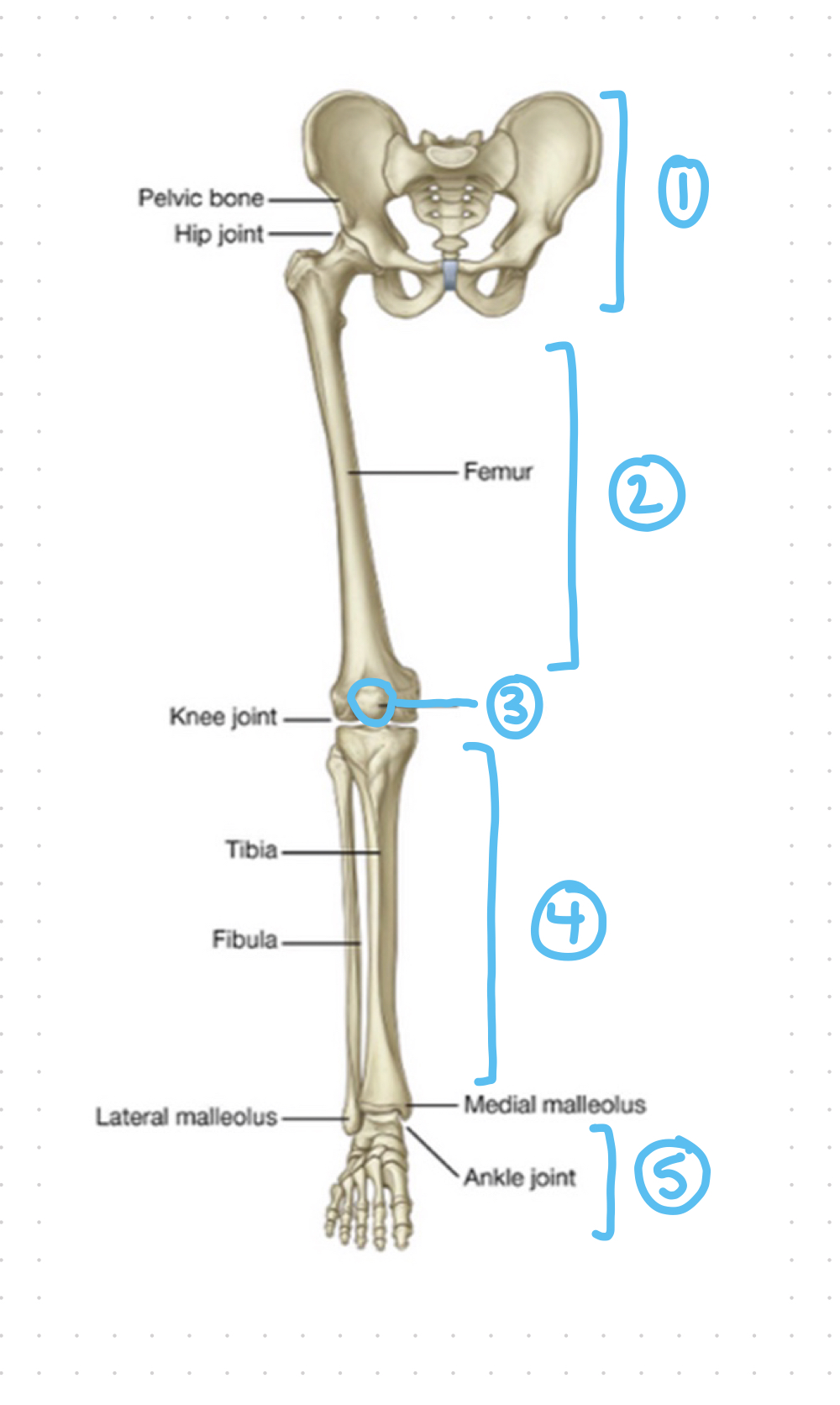
bones of pelvic girdle and lower limb - 2
thigh

bones of pelvic girdle and lower limb - 3
patella

bones of pelvic girdle and lower limb - 4
leg

bones of pelvic girdle and lower limb - 5
foot

pelvic girdle - 1 & 2
ossa coxae
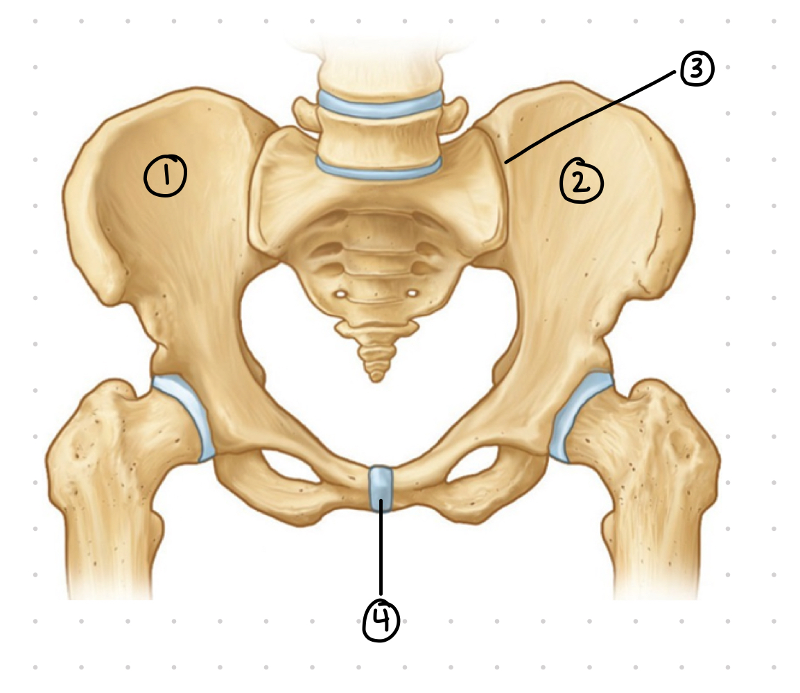
pelvic girdle - 3
sacroiliac joint

pelvic girdle - 4
pubic symphysis; fibrocartilage joint where 2 ossa coxae join
what two structures make up the pelvic girdle
ossa coxae (2)
sacrum
sacrum is part of the axial/appendicular skeleton
axial
ossa coxae is part of the appendicular skeleton
appendicular
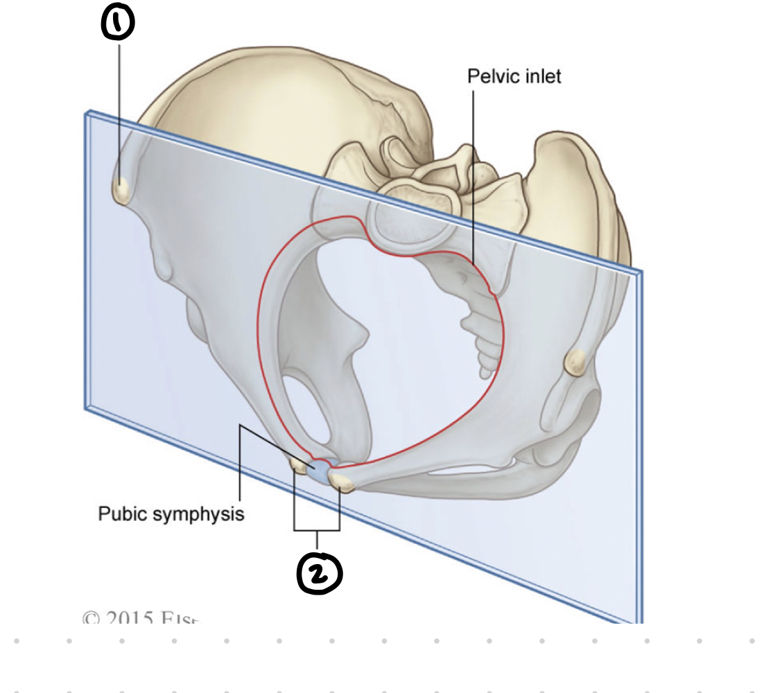
pelvic girdle coronal plane - 1
anterior superior iliac spine (asis)

pelvic girdle coronal plane - 2
pubic tubercles

true vs. false pelvis - 1 black
false pelvis; where abdominal structures are found

true vs. false pelvis - 2 black
plane of pelvic brim; runs between sacral promontory and pubic symphysis which divides the true and false pelvis
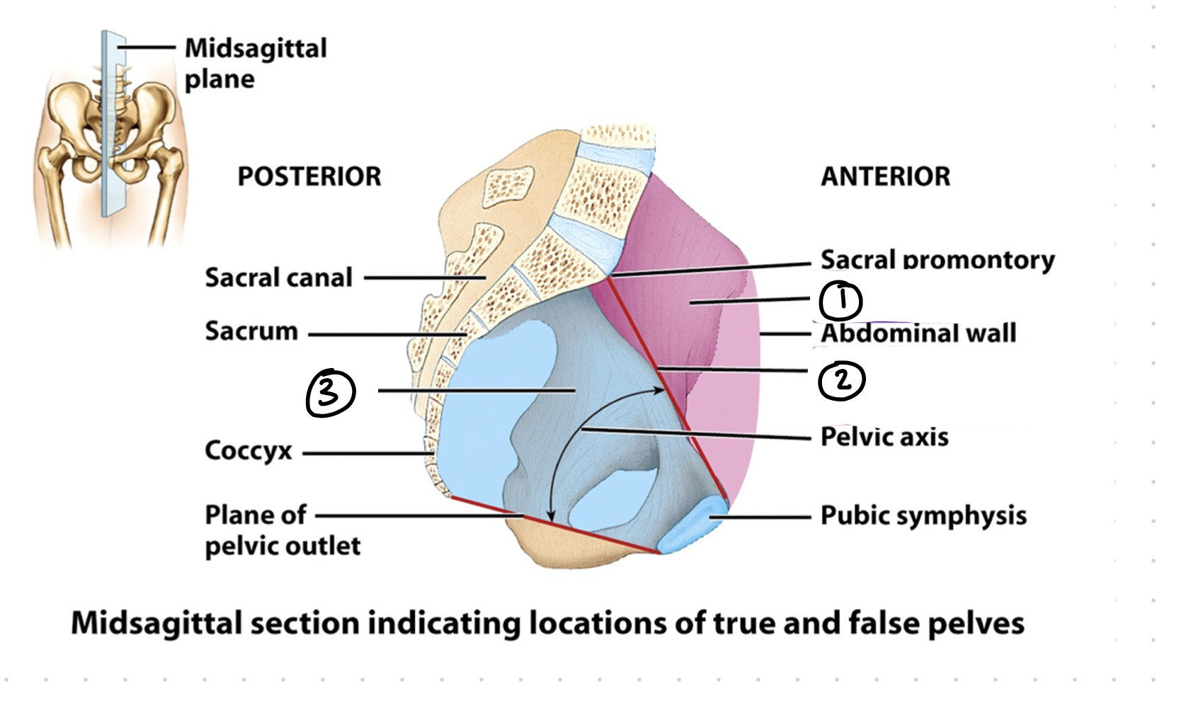
true vs. false pelvis - 3 black
true pelvis; where urogenital structures are found

true vs. false pelvis - 1 purple
sacral promontory
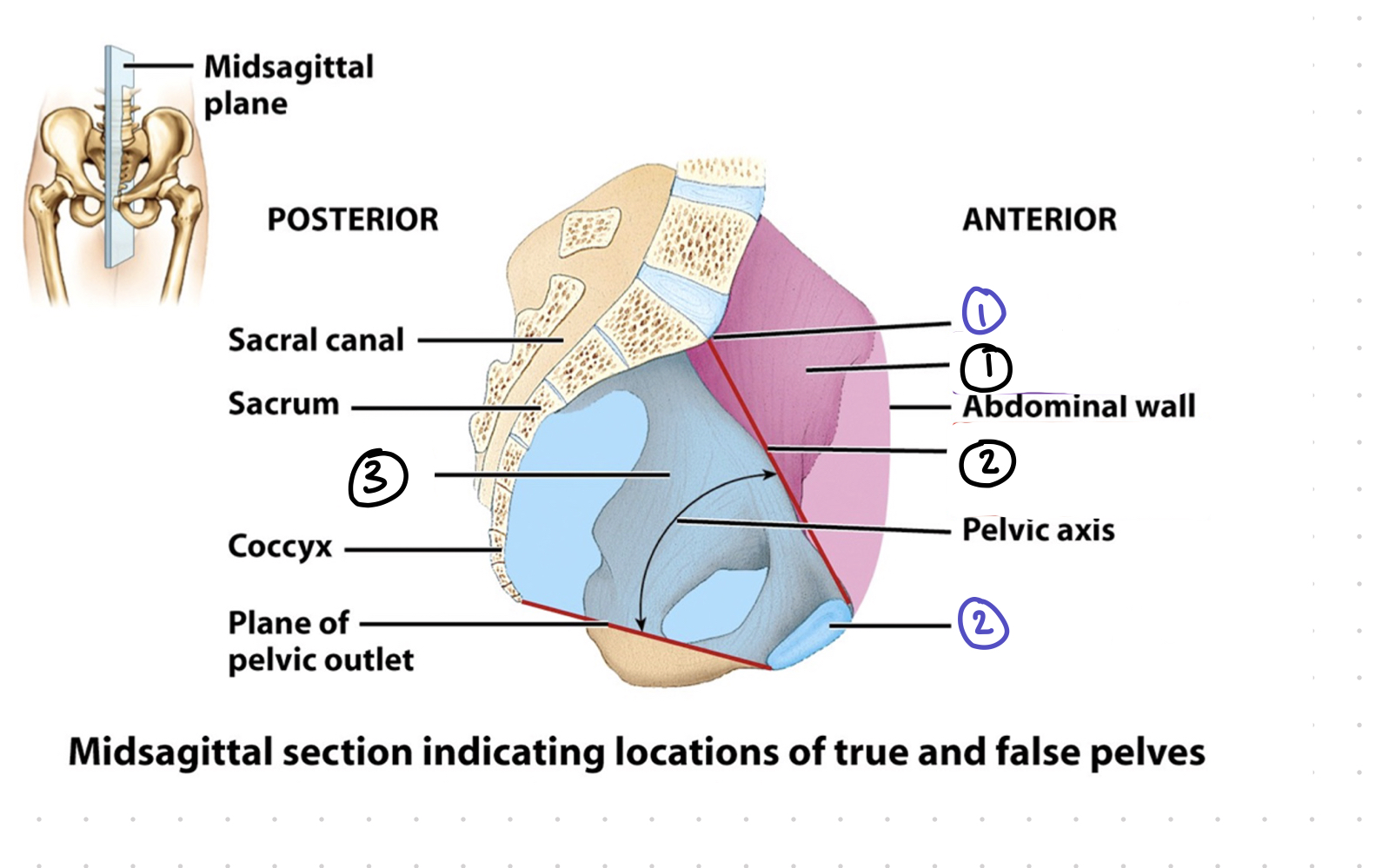
true vs. false pelvis - 2 purple
pubic symphysis
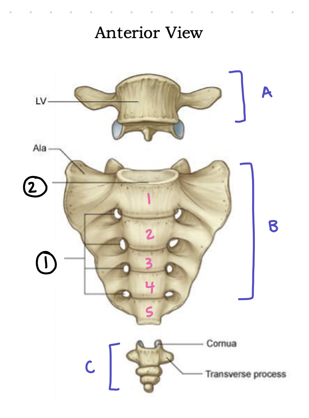
anterior sacrum - A
lumbar 5

anterior sacrum - B
sacrum

anterior sacrum - C
coccyx

anterior sacrum - 1
anterior sacral foramina; where spinal nerves pass through

anterior sacrum - 2
promontory; anterior-most point of sacrum
what is a foramen?
1 hole
what are foramina?
many holes

posterior sacrum - 1
sacral canal; where the spinal cord passes through

posterior sacrum - 2
median sacral crest; ridge of bone on the posterior side of sacrum

posterior sacrum - 3
auricular surface; “ear” where sacrum connects to ossa coxae
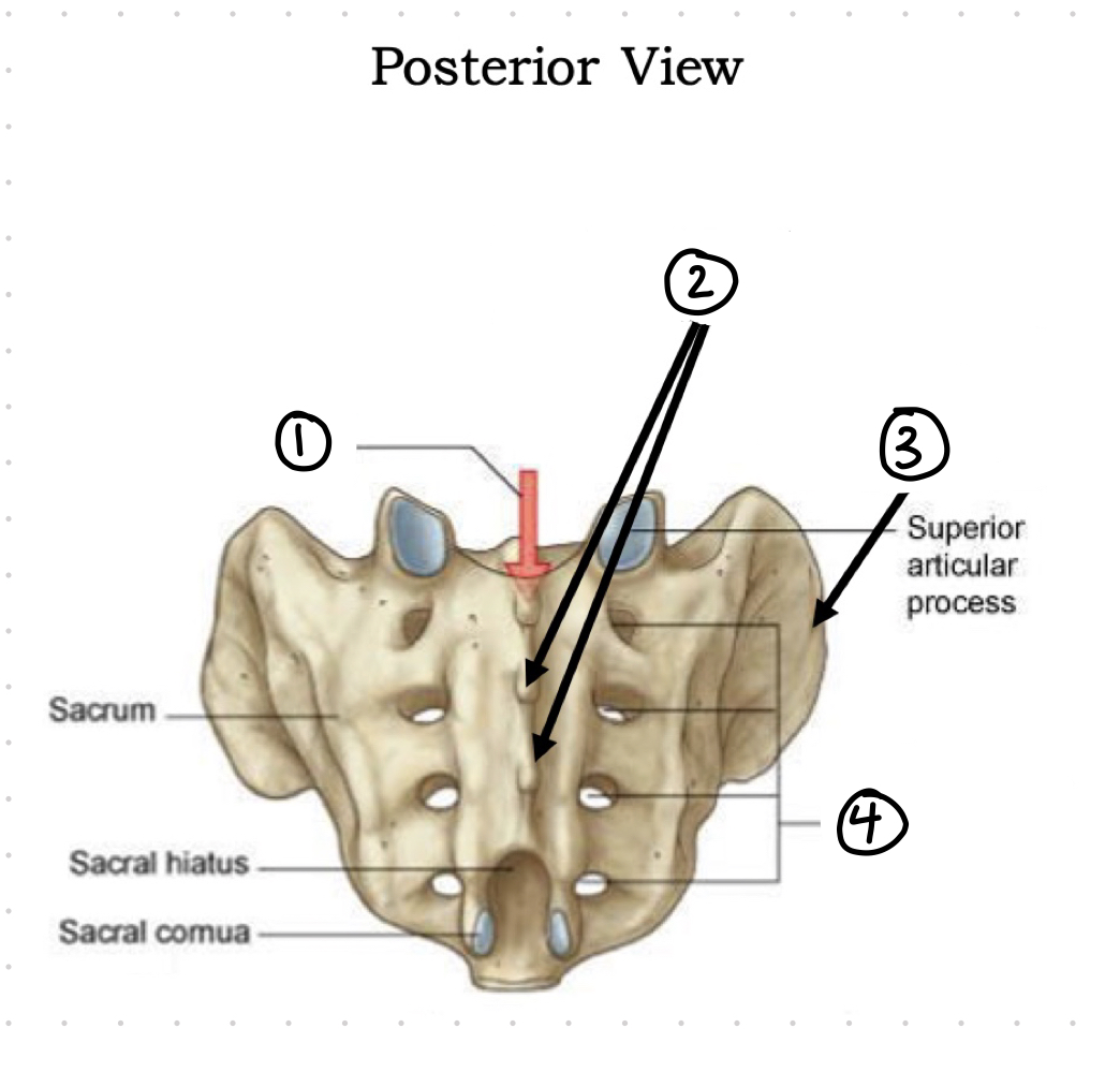
posterior sacrum - 4
posterior sacral foramina; where spinal nerves pass through
T or F: the sacrum is part of the vertebral column
T: the sacrum is a continuation of the vertebral column

os coxae lateral view - right or left os coxae?
right

os coxae - beige section (pink numbers)
ileum

os coxae - purple section (green numbers)
ischium

os coxae - red section (blue numbers)
pubis

os coxae lateral view - 1 pink
ileum

os coxae lateral view - 2 pink
ileac crest

os coxae lateral view - 3 pink
anterior superior ileac spine
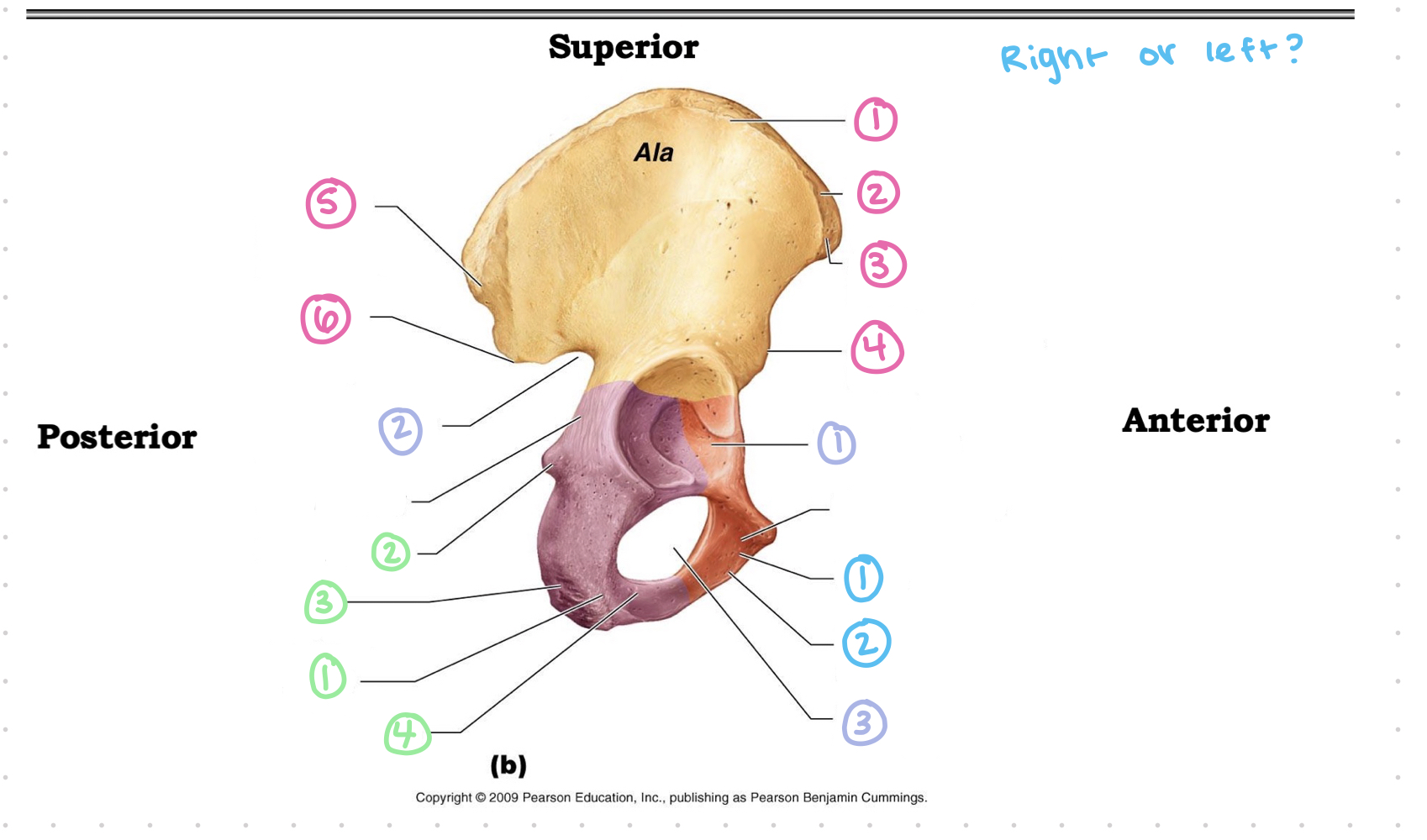
os coxae lateral view - 4 pink
anterior inferior ileac spine

os coxae lateral view - 5 pink
posterior superior ileac spine

os coxae lateral view - 6 pink
posterior inferior ileac spine
3 parts of the os coxae
ileum
ischium
pubis

os coxae lateral view - 1 purple
acetabulum; “socket” where head of femur sits

os coxae lateral view - 2 purple
greater sciatic notch

os coxae lateral view - 3 purple
obturator foramen

os coxae lateral view - 1 green
ischium

os coxae lateral view - 2 green
ischial spine

os coxae lateral view - 3 green
ischial tuberosity

os coxae lateral view - 4 green
ischial ramus

os coxae lateral view - 1 blue
pubis

os coxae lateral view - 2 blue
inferior ramus of pubis

os coxae medial view - right or left?
right

os coxae medial view - pink 1
ileum

os coxae medial view - pink 2
ileac crest

os coxae medial view - pink 3
anterior superior iliac spine

os coxae medial view - pink 4
anterior inferior ileac spine

os coxae medial view - pink 5
ileac fossa; scooped out portion

os coxae medial view - pink 6
posterior superior iliac spine

os coxae medial view - pink 7
posterior inferior iliac spine

os coxae medial view - pink 8
auricular surface; “ear” where sacrum joins ileum

os coxae medial view - purple 1
greater sciatic notch

os coxae medial view - purple 2
lesser sciatic notch

os coxae medial view - purple 3
obturator foramen; covered with connective tissue and where arteries pass through

os coxae medial view - blue 1
pubis; most anterior part of os coxae

os coxae medial view - blue 2
superior ramus of pubis

os coxae medial view - blue 3
inferior ramus of pubis

os coxae medial view - blue 4
pubic tubercle
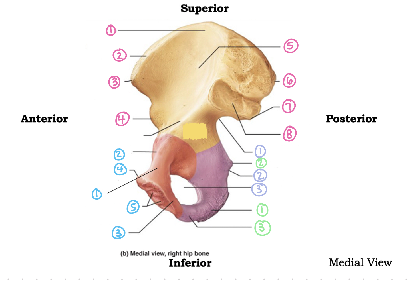
os coxae medial view - blue 5
articular surface of pubis (at pubic symphysis)

os coxae medial view - green 1
ischium
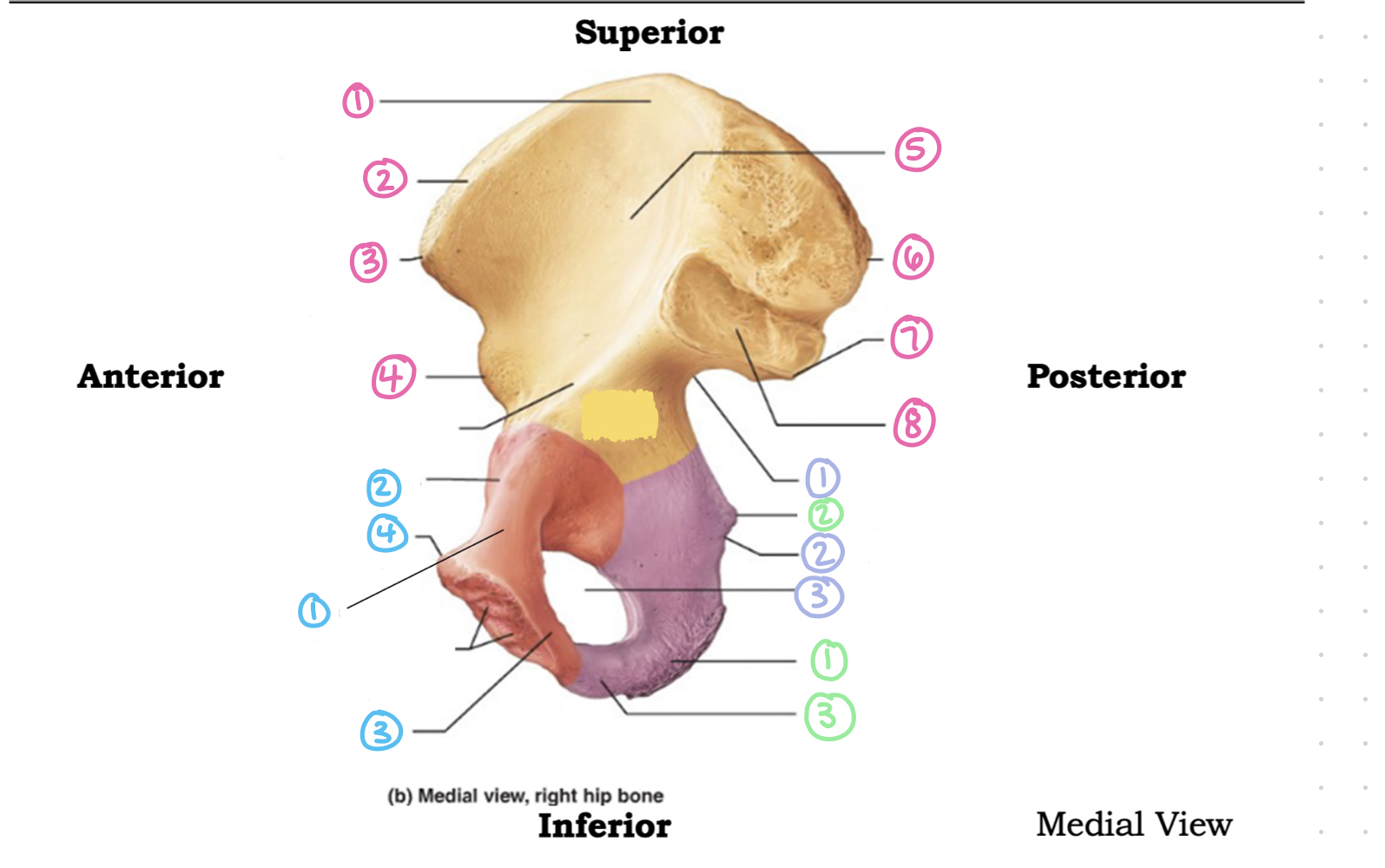
os coxae medial view - green 2
ischial spine; bony point

os coxae medial view - green 3
ischial ramus
what is a rami/ramus?
branch(es)
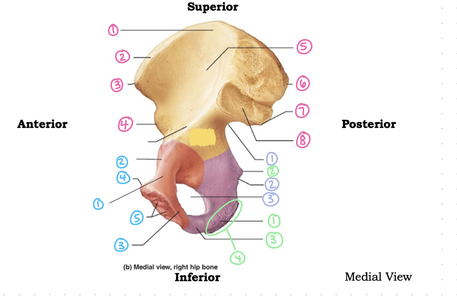
os coxae medial view - green 4
ischial tuberosity (rough patch); sitting bone and most inferior point of os coxae

femur - right or left?
right

femur - 1
neck; commonly called “hip” fracture

femur - 2
greater trochanter

femur - 3
lesser trochanter
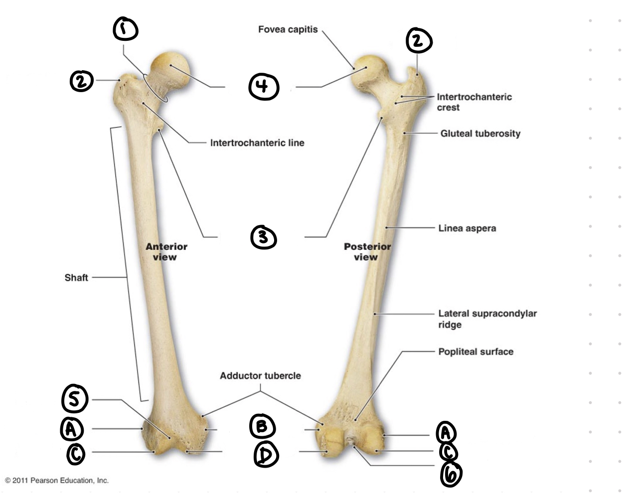
femur - 4
head; ball of the bone and should always point medially

femur - 5
patellar surface

femur - A
lateral epicondyle; rough so that muscles can attach

femur - B
medial epicondyle

femur - C
lateral condyle; easier to see on posterior side of femur because there are two of these smooth patches

femur - D
medial condyle

femur - 6
intercondylar fossa; scooped out region
what connects at smooth bone sites?
bone to bone

tibia and fibula - right or left?
right

tibia and fibula - A
lateral condyle

tibia and fibula - B
medial condyle

tibia and fibula - 1
head of fibula

tibia and fibula - 2
fibula; skinny bone
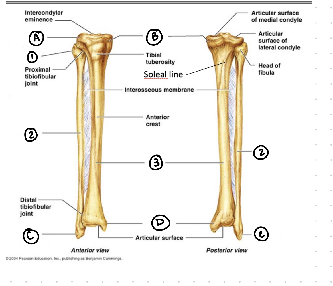
tibia and fibula - 3
tibia; thicker weight bearing bone

tibia and fibula - C
lateral malleolus

tibia and fibula - D
medial malleolus
what are the tibia and fibula like?
tongs that grab onto the ankle bone
how many tarsal bones are in the foot?
7
how many metatarsals are in the foot?
5
how many phalanges are in the foot?
14

bones of the foot - 1 black
phalanges

bones of the foot - 2 black
metatarsals
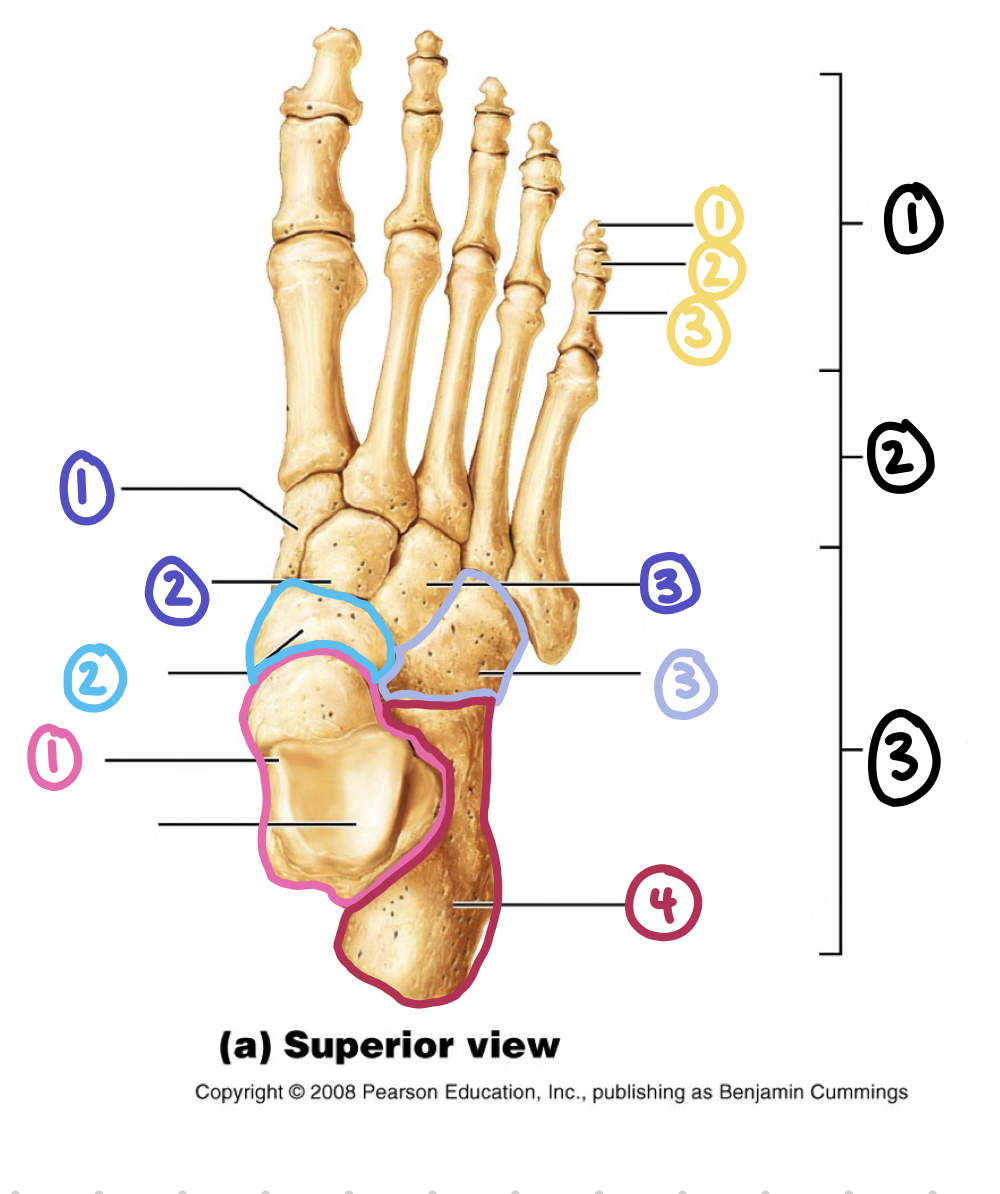
bones of the foot - 3 black
tarsals (ankle bones)