Complex Medical Oral
 Call Kai
 Learn
Learn Practice Test
 Spaced Repetition
 Match
Match Flashcards
 Knowt Play
1/115

There's no tags or description
Looks like no tags are added yet.
Name | Mastery | Learn | Test | Matching | Spaced | Call with Kai | Chat |
|---|
No analytics yet
Send a link to your students to track their progress
116 Terms
Explain the pathophysiology for coronary artery disease:
what is it
cause
effects
What: atherosclerosis of the coronary arteries
plaque build up in coronary arteries
Cause:
excess cholesterol
too many LDLs
Effect:
blood flow restriction
ischemia (plaque, thrombus formation)
ischemia (lack of O2) —> infarction (tissue DEATH) —> MI (heart attack)
Explain the difference between stable and unstable angina and its clinical relevance
stable
comes on during exertion but goes away at rest
predictable, doesn’t last
patient okay to treat/work with
unstable
comes on with exertion but continues after activity stops
MEDICAL EMERGENCY —> get this checked out!
What are the stages of a myocardial infarction?
ischemia
lack of oxygen due to plaque build up, thrombus, O2 not getting to heart tissue = bad!
injury
ischemia has gone on too long and is starting to damage tissue
infarction
NON-REVERSIBLE cell death in cardiac tissue
What are characteristics of a STEMI and what does this mean for us as PTs?
STEMI = ST segment elevated myocardial infarction
severe type of heart attack where ST on EKG looks like a tomb stone
PATIENT IS NOT SAFE TO WORK WITH
Which coronary artery causes major issues if blocked and why?
LAD: left anterior descending artery
supplies blood to L ventricle
if there is occlusion, then this will cause MI in this area of the heart, L ejection fraction will decrease which means blood is not being pumped out effectively to the rest of the body
leads to cariogenic shock and acute heart failure
What is cardiogenic shock?
LIFE THREATENING CONDITION
when there is too much cardiac dysfunction for the heart to pump adequately to perfuse blood
MI —> not enough blood perfusion —> widespread organ failure
How does the body try to compensate in response to cardiogenic shock? Which organ is most sensitive to loss of perfusion?
body will increase sympathetic activation
tachycardia
vasoconstriction
this all causes even more stress on the heart which makes the symptoms worse
kidneys are most sensitive to loss of perfusion and hypotension
look at BUN and creatinine levels which indicate kidney dysfunction or failure
What is a normal left ventricular ejection fraction percentage? What is the LVEF for patients with L-sided heart failure?
normal: 55-70%
L-sided heart failure: < 40%
What are the normal lab values for troponin? What is the significance of it being elevated?
normal: < 0.04
elevation: > 0.04 during MI
PATIENT NOT SAFE FOR THERAPY
immediately refer to doctor or nurse
What are the normal lab values for CK-MB? What is the significance of it being elevated?
Normal: 0-5
elevation: 4-6hrs post MI (so an MI has occurred)
Need to confirm stabilization prior to functional mobilization
What are the normal lab values for BNP? What is the significance of it being elevated?
normal: < 100
elevated: > 400 = acute heart failure from fluid overload
exercise intensity should be adjusted + monitor lung sounds for crackles during inspiration
What are the normal lab values for creatine kinase? What is the significance of it being elevated?
normal: 22-220
elevated: muscle damage; during MI this means heart muscle damage
look for signs and symptoms of rhabdomyolysis
LIFE THREATENING CONDITION
muscle pain, weakness, cola-colored urine, nausea, vomiting, fever, rapid HR
What are the normal lab values for lipids? What is the significance of it being elevated?
normal:
LDL < 100
HDL > 40-50
elevated:
> 200 total cholesterol
> 150 triglycerides
elevated cholesterol can lead to atherosclerosis which can lead to MI (if thrombus/ischemia occurs)
patient education on risk factors and prevention/lifestyle modification
What does MONATASS stand for and what is it an acronym for?
medications a person who has experienced a heart attack might be taking
M: morphine; pain reliever
O: oxygen
N: nitroglycerin-vasodilators; relaxes smooth muscle in vessels, reduces heart workload, helps w/ angina
A: aspirin; prevents formation of new clots
T: thrombolytic; clot buster
A: anticoagulant; also prevents blood clots
S: stool softener; prevents straining which could provoke arrhythmia
S: sedatives; patient might be lethargic
What are sternal precautions (modern) following heart surgery?
move within your tube
patients can move as if they are hugging a pillow, just need to move sternum and arms together
some mobilization is necessary to prevent shoulder ROM issues like frozen shoulder
no lifting > 5-10lbs
no pushing or pulling
can use hand for BALANCE during transfers
likely will NOT have patient use arm to push self up/pull to side of bed during bed mobility (teach log roll method with modification)
no OH arm elevation > 90*
no reaching behind back
As a PT what should you be aware of when it comes to information presented in an EKG?
be able to recognize new symptoms or rhythm changes
if arrhythmia present ask: IS IT NEW? and CHECK BP
knowing when to stop activity if changes in patient status occur
As a PT what should you be aware of when it comes to information presented in an echo?
understand ejection fraction values/indications
expect with lower LV function (like with heart failure) patient will have reduced exercise reserve/capacity
monitor patients for symptoms like angina, dyspnea, and arrhythmia
As a PT what should you be aware of when it comes to information presented in a angiography?
understand post-procedure restrictions based on access site used during the procedure
radial artery vs. femoral
monitor hemodynamic stability
Explain the pathophysiology for heart failure:
what is it
cause
effects
what: heart is not pumping or filling up with blood properly to keep up with body’s needs
causes:
CAD
HTN
cardiomyopathy
valvular disease
effects
hypertrophy (not the good kind; heart is working harder and not smarter)
fluid retention in kidneys —> kidney failure
pleural effusion (L-sided HF)
Explain reduced ejection fraction HF vs. preserved ejection fraction HF
reduced
heart can’t pump
heart can fill
heart fills —> weak pump —> less blood pumped out
results in lower EF; < 40%
S3 heart sound
preserved
heart can pump
heart can’t fill
heart starts with less blood —> less blood pumped out
S4 heart sound
Explain L-sided HF vs. R-sided HF
L-sided (L —> LUNG)
causes fluid to back up into lungs (pleural effusion)
R-sided (R —> Rest of body)
causes fluid to back up into body
edema in legs/ankles, ascites in abdomen, weight gain from fluid retention
What are common symptoms of L sided HF?
SOB
orthopnea
cough or wheezing
* mainly causes breathing problems because fluid backs up into the Lungs
What are common symptoms of R sided HF?
peripheral edema
weight gain
ascites
*mainly causes body swelling because fluid backs up into the Rest of the body
What are the AHA stages of HF?
A: at risk
no symp
HTN, CAD, DM, obesity, cardiomyopathy
B: pre-HF
no symp
structural heart disease or increased filling pressures
C: HF
symptomatic
current or previous symp
D: advanced HF
symp disrupt life function or cause hospitalization
What is the NYHA classifications of HF? Which stages of the AHA do they correspond to?
correspond to stages C + D from AHA stages of HF
Class I:
no limitation in PA
no symp (palpitations, SOB, fatigue)
Class II:
slight limitation in PA
no symp at rest
ordinary PA causes symp
Class III:
marked limitation of PA
no symp at rest
less than ordinary PA causes symp
Class IV:
symp of HF at rest
any PA causes further symp
Which lab value if trending upward is highly indicative of HF? (what is normal; what value would indicate HF)
BNP (B-type natriuretic peptide)
normal: < 100
HF > 200
When it comes to electrolyte lab values, what would you expect in a patient with HF for sodium and potassium + signs/symp that might present? What is the significance of patients taking diuretics and its effect on potassium?
sodium
lab values may be LOW (< 135)
S/S of lower sodium levels: headache, nausea, vomiting, muscle cramps, fatigue, dizziness
potassium
lab values may be LOW (< 3)
S/S: fatigue, lethargy, heart palpitations, numbness/tingling
if a patient is taking diuretics for Tx for fluid accumulation, this might cause lower potassium levels
need to remind patients to take supplemental potassium to avoid risk of arrhythmias
What are normal lab values for BUN and creatinine? What does elevated levels indicate? Which heart conditions might cause this?
normal BUN: 5-20
normal creatinine: 0.6-1.2
elevated: kidney dysfunction or failure
heart conditions: HF
What is the treatment for HF and what might you need to be aware of as a PT?
typical medical management involves:
diuretics: watch out for potential low potassium levels + S/Sx
vasodilators
beta blockers: use RPE for exercise tolerance rather than HR
pacemaker
lifestyle changes: smoking cessation, low sodium diet, regular PA, monitoring weight for fluid retention
___1___ HF is more common and is either __2__ HF or __3__ HF. It causes fluid back up in the___4___
L sided
reduced EF
preserved EF
lungs
___1___ HF is often caused by __2__, ___3__, and __4__. It causes fluid back up in the ____5____
R sided
L-sided HF
pulmonary HTN
COPD
body
__1__ results from reduced EF HF while ___2__ results from preserved EF HF
systolic failure
diastolic failure
In reduced EF HF, the heart can _______ but it can’t_______ so EF is _______
FILL
PUMP
EF > 40%
In preserved EF HF, the heart can _______ but it can’t_______ so EF is _______
PUMP
FILL
EF normal: 50-70%
True or false: For patients in the Class I NYHA classification for heart failure ordinary PA causes symp
FALSE; ordianry PA does NOT cause symp for patients considered a class I
True or false: For patients in the Class II NYHA classification for heart failure ordinary PA causes symp
TRUE; ordinary PA does cause symp for patients considered a class II
For patients in the class III NYHA classification for HF, _______ causes symp
less than ordinary PA
When doe patients in the class IV NYHA classification for HF experience symptoms?
all the time, even at rest
symp get worse with PA
________ is when there is narrowing of a valve causing it to fail to open completely
stenosis
_____ results from a valve not closing completely. Blood back flows into previous space
regurgitaiton
______ is when a valve bulges backward into its chamber. The gate is supposed to be closed but it is open. This can lead to _______
prolapse
regurgitation
______ is turbulent blood flow in or near the heart. It can be caused by: ______, _______, or ________.
murmur
stenosis
regurgitation
high output stakes
Explain the pathophysiology for valvular stenosis:
what is it
cause
effects
what
increase in pressure due to lack of blood flow through the valves
cause
calcific: damage to fibrosa layer eventually leads to deposition of calcium crystals + type I collagen
rheumatic: common with mitral stenosis; chordae tendinae fuse together and shorten
effect
ventricular hypertrophy
increased outflow resistance and afterload
(pressure/resistance heart must overcome to eject blood out of ventricles during contraction)
ischemia
HF
You can typically hear a ________ upon auscultation of mitral stenosis at the L 5th intercostal space
low-pitched rumbling murmur
What are red flags related to valve stenosis?
massive spike or drop in systolic BP during exertion
lightheadedness
onset of crackles during inspiration or irregular pulse
What should be avoided with patients with valvular stenosis? Why?
exercises that cause sudden or rapid spike in systolic BP or HR
BP: could increase myocardial oxygen demand which could lead to ischemia and exertion syncope
HR: could cause fluid to back up into the lungs
Explain the pathophysiology for valvular regurgitation:
what is it
cause
effects
what
backflow of blood in the reverse direction usually due to valves failing to close completely (such as with a prolapse)
cause
Mitral
prolapse
rheumatic endocarditis: destruction of valve due to bacteria
L ventricular remodeling
Aortic
leaflet disease
aortic root/ascending pathology
effect
heart failure
atrial fibrillation
pulmonary HTN
What should be avoided with patients with valvular regurgitation?
high after-load exercises: heavy weightlifting, HIIT workouts
UE isometric holds
valsalva maneuver
What are red flags related to valvular regurgitation?
spike in L ventricular end-diastolic BP
massive drop in stroke volume can lead to cardiogenic shock
Explain the pathophysiology for valvular prolapse:
what is it
cause
effects
what
defect in cusp of heart value leads to parachuting of valve backwards into previous chamber (flap opens up)
cause
myxomatous degeneration (typically affects mitral valve)
accumulation of GAGs causing weakening of valve
effects
could eventually lead to regurgitation
What are red flags/signs valvular prolapse has progressed to regurgitation?
fatigue
orthopnea
new onset of arrhythmia
A patient comes in for treatment of his rotator cuff tear but he also has a mitral prolapse. What should you NOT do with the patient?
ISOMETRICS!!!
they can cause a spike in BP
Explain the pathophysiology for infective endocarditis:
what is it
cause
signs + symp
effects
what
infection of inner lining of heart chamber
cause
bacteria or fungi entering and attaching to damaged heart from:
brushing teeth, skin infections, dental work, dirty needles
S/Sx:
fever, chills, new or changed heart murmur, extreme tiredness, small spots on skin or fingernails
effect
heart failure
heart block
stroke
What are red flags related to infective endocarditis?
new or worsening murmur, chest pain
sudden weakness or facial droop (stroke), severe headache, seizure
sudden SOB, chest pain hyoxemia
lesions on palms/sores
Explain the pathophysiology for cardiac tamponade
what is it
cause
effects
what
LIFE THREATENING EMERGENCY
buildup of excess fluid around the heart
cause
pericarditis
pericardial effusion
effects
can press onto heart and cause it to stop filling up with blood
leads to drop in BP (especially during inspiration) and lack of blood flow
What are signs/symp of pericarditis? What can this condition eventually lead to?
chest pain
typically increased with coughing, swallowing, or deep breathing in supine position
relief of sharp stabbing pain when sitting upright/forward or bending over
________ results in elevated pressure and reduced filling of chambers in the heart, causing a decrease in venous return
pericardial effusion
Explain the pathophysiology for aortic aneurysm
what is it
cause
effects
what
permanent dilation of aortic segment
cause
effects
progressive expansion can lead to rupture
Explain the pathophysiology for aortic dissection
what is it
cause
effects
What is worse type A or type B?
what
intima of aortic vessel becomes torn through
cause
effects
rupture
cardiac tamponade
acute aortic regurgitation
TYPE A IS WORSE
effects ascending aorta
has a higher risk for rupture and above effects
What are S/Sx of aortic dissection (type A vs. type B)
type A
anterior chest pain
syncope
hypotensive shock
new aortic regurgitation murmur
cardiac tamponade
type B
back or abdominal pain
HTN
limb, renal, spinal cord ischemia
What are red flags associated with aortic dissection?
pulse and BP difference btw lilmbs
altered mental state
stroke-like symp
unexplained lower extremity ischemia
What two questions should you ALWAYS ask a patient if you see that they have an arrhythmia?
IS IT NEW?
how is their cardiac output? (check their BP)
Explain the pathophysiology for heart blocks
what is it
cause
effects
what
delay or interruption of an electrical signal traveling from the atria to the ventricles
cause
effect
can result in the need for a pacemaker
3rd degree block is most severe can result in damage that is complete and unresponsive to medication
If the R is far from the P then you have… (which heart block)
a first degree (heart block)
Longer, longer, longer, drop! Then you have a … (which heart block)
wenkeback (mobitz I)
If some Ps don’t get through, then you have … (which heart block)
mobitz II
If Ps and Qs don’t agree, then you have a … (which heart block)
third degree
Is it more or less common to have heart block after valve surgery?
more common
need to be cautious of people with temporary pacemakers
What are characteristics of a first degree heart block?
signal slows through AV node but still reaches the ventricles
PR interval > 0.2 seconds
not usually treated
What are characteristics of Mobitz Type I heart block?
successive atrial impulse conductions gradually slow until one atrial impulse fails to conduct to the ventricles
PR interval progressively gets longer
QRS drops (P wave not followed by QRS periodically)
Homer is a slowpoke but he is at least consistent
What are characteristics of Mobitz type II heart block?
wider QRS complex
regular dropped QRS every 2nd, 3rd, 4th P wave
consistent RR pattern
What are characteristics of third degree heart block
no conduction or communication between atria and ventricles; SA node signal doesn’t reach ventricles
MARGE AND HOMER NOT GOING TO MAKE IT
patient needs a pacemaker
What are red flags for the cardio portion of the complex medical case?
conditions
v-tachycardia
v-fib
3rd degree heart block
cardiac tamponade
aortic dissection
torsades de pointes
DVT
heart attack (MI)
signs/symp
unstable angine
new arrythmia
UE pain (referred)
ST segement elevation on EKG
increase in troponin > 0.04
spike in systolic BP
sudden SOB
If a patient is experiencing an arrhythmia, what two things should you ask/do?
is this new?
how is their cardiac output; check BP
What are the phases of a cardiac action potential and how do they relate to an EKG reading?
phase 0: depolarization; Na+ channels open
rising part of QRS complex
phase 1: prolonged action potential; Ca channels open
top of QRS complex (ventricular contraction)
phase 2: outward flow of K+ and prolonged Ca —> plateau
between Q and beginning of T wave
phase 3: closing of Ca channels and opening of K channels (ventricular relaxation)
phase 4: resting phase
right after P wave/atrial contraction
What do the following letters relate to on an EKG?
P wave
PR interval
QRS complex
QRS duration
ST interval
T wave
P wave: atrial contraction (depolarization)
PR interval: time between atrial contraction and ventricular contraction
QRS complex: ventricular contraction (depolarization)
QRS duration: time for ventricular contraction
ST interval: time between ventricular contraction and ventricular relaxation
T wave: ventricular relaxation (repolarization)
Put simply, an EKG reading shows: (3 things)
activation of the atria: P wave
activation of the ventricles: QRS complex
recovery of the ventricles: T wave
Where is the best place to assess for heart blocks on an EKG?
PR interval
if > 0.20 seconds
When it comes to the heart, whatever depolarizes must ________ before it can depolarize again (tip for reading EKGs)
repolarize
What are characteristics of supra ventricular tachycardia (SVT) arrhythmia? (what, symp, EKG)
what
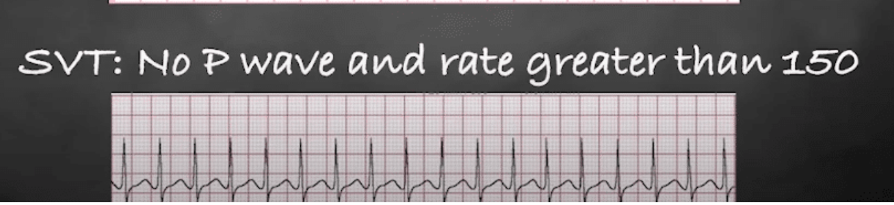
sudden, rapid HR > 150 bpm
onset is random and can last minutes to hours
symp: palpitations, dizziness, SOB, angina, fatigue
EKG:
high rate: 150-250
regular rhythm
no visible P wave, PR not measurable
contraction of ventricles is so strong, you don’t see the atrial contraction (which would be in P wave)
QRS < 0.12 sec
What is the treatment for SVT? Is it usually life threatening?
cardioversion
catheter ablation
not usually life threatening but symp can be significant
What are characteristics of atrial flutter? (what, symp, EKG)
what
rapid atrial activity of 300 bpm
HR sometimes shows up normal if ventricular rate is normal
symp: palpitations, fatigue, lightheadedness, SOB, exercise intolerance, hypotension, syncope
EKG:
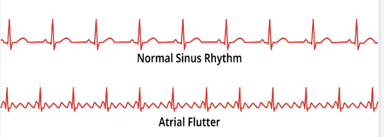
sawtooth P wave
rate 250-450
What is the treatment for atrial flutter? What are serious complications of atrial flutter?
rhythm control: ablation, cardioversion
anticoagulant to prevent stroke
if a flutter causes irregular ventricular response —> blood becoming static and clot formation
Does atrial flutter always show up on EKG? Why or why not? What is the effect of a fib + a flutter on CO?
No, atrial flutter might not show up if ventricular rate appears normal (ex: HR shows 70 but atria are doing 300)
a fib + a flutter = lose atrial kick which decreases CO
What are characteristics of atrial fibrillation? (what, symp, EKG)
what
abn electrical activity results in fibrillation of the atria (rapid, uncoordinated irregular contractions of muscle fibers)
symp
can be asymp; angina, palpitations, SOB, syncope
EKG:
irregular rhythm, chaotic P waves, can lead to decreased CO

What is the treatment for a fib?
anticoagulants: to reduce stroke risk
beta blockers: HR control
Ca channel blockers: HR control
cardioversion: help return rhythm of heart to normal
What are characteristics of premature atrial contraction (PAC)? (what, symp, EKG reading)
what: atrial contraction that doesn’t originate from SA node but is triggered by atrial myocardium
symp: asymp; skipping sensation, palpitations, SOB
EKG: abn P wave, normal QRS,
What is the treatment for premature atrial contraction?
beta blockers
What are characteristics of premature ventricular contraction (PVC)? (what, symp, EKG)
what
additional beat caused by random ventricular contraction
symp: fluttering, pounding, skipped beats
EKG:
no P waves
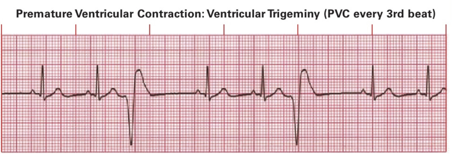
wide, early, random QRS
bigeminy = 1 PVC every beat; trigeminy = 1 PVC every 3rd beat
True or false: every patient might have some premature ventricle contractions (PVC)
true
need to notify nurse if patient is having a lot; likely will have PVC counter (# in the last minute)
When does a PVC become concerning?
if a person is regularly having a lot of them
if a person has a heart condition
What are characteristics of ventricular tachycardia? (what, symp, EKG)
what
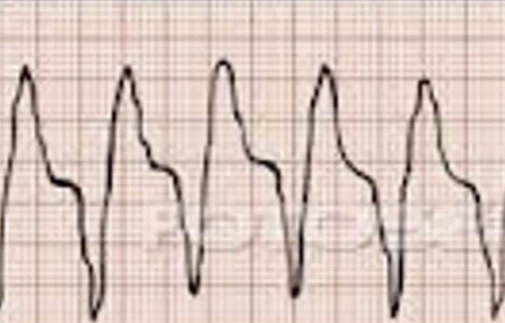
wide complex tachycardia
MEDICAL EMERGENCY due to potential cardiac arrest
symp: palpitations, SOB, dizziness/lightheadedness, angina, cardiac arrest
EKG:
3 or more PVC and HR > 100 bpm
What is the treatment for v-tach?
if cardiac arrest occurs: CPR with defibrillation
no cardiac arrest: cardioversion
What are characteristics of torsades de pointes? (what, symp, EKG)
what
specific form of V-tach with prolonged QT interval
ALSO MEDICAL EMERGENCY
can lead to v fib —> cardiac arrest
EKG
prolonged QT interval

What are characteristics of ventricular fibrillation? (what, symp, EKG)
what
ventricles chaotically quivering rather than pumping
MEDICAL EMERGENCY
can lead to immediate collapse + cardiac arrest
blood no longer being pumped!
symp: sudden collapse, loss of consciousness, no pulse
EKG
no organized rhythm

What is the treatment for ventricular fibrillation?
Use AED + CPR
What is rhabdomyolysis? What is a common symp? What is its typical onset? What lab values do you expect a patient to have regarding creatine kinase?
what:
skeletal muscle breakdown
releases creatine kinase into blood
leads to acute kidney injury, electrolyte disturbances, intravascular coagulation
symp:
severe muscle soreness, cola-colored urine (due to myoglobinuria), local swelling
onset:
symp can start 1-3 days after muscle injury or extreme workout
lab values:
elevated creatine kinase: 5 x normal limit (20-200), so 1000-5000
If a patient has a decreased respiration rate, then this means there is more _____ in the blood, making the pH _____ and blood more ______
CO2
lower
acidic
How do the kidneys respond to the blood having a lower pH level/more acidic due to more CO2?
kidneys will retain more HCO3- (bicarbonate) to neutralize the excess H+ which are making the blood acidic
If a patient has an increased respiration rate, then this means there is less _____ in the blood, making the pH _____ and blood more ______
CO2
higher
basic