blood flow CVS100:ex:3
 Call Kai
 Learn
Learn Practice Test
 Spaced Repetition
 Match
Match Flashcards
 Knowt Play
1/31

There's no tags or description
Looks like no tags are added yet.
Name | Mastery | Learn | Test | Matching | Spaced | Call with Kai | Chat |
|---|
No analytics yet
Send a link to your students to track their progress
32 Terms
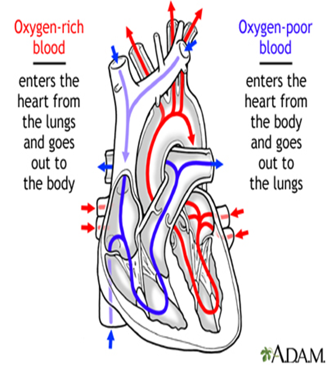
What are the characteristics of the RIGHT HEART?
- Pulmonary Circulation
- Deoxygenated Blood
- Lower Pressure
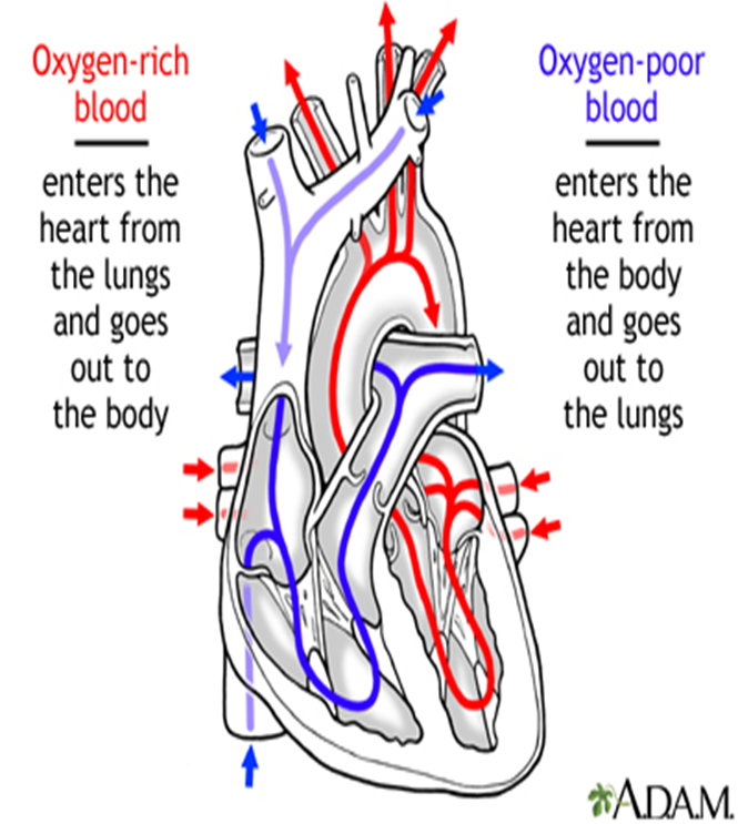
What are the characteristics of the LEFT HEART?
- Systemic Circulation
- Higher Pressure
- Thicker Myocardium than right side
- Oxygenated Blood
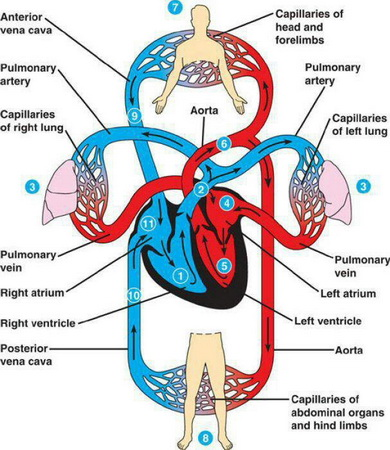
Which major vessels receive oxygen-poor blood from the body and what is their approximate oxygen saturation?
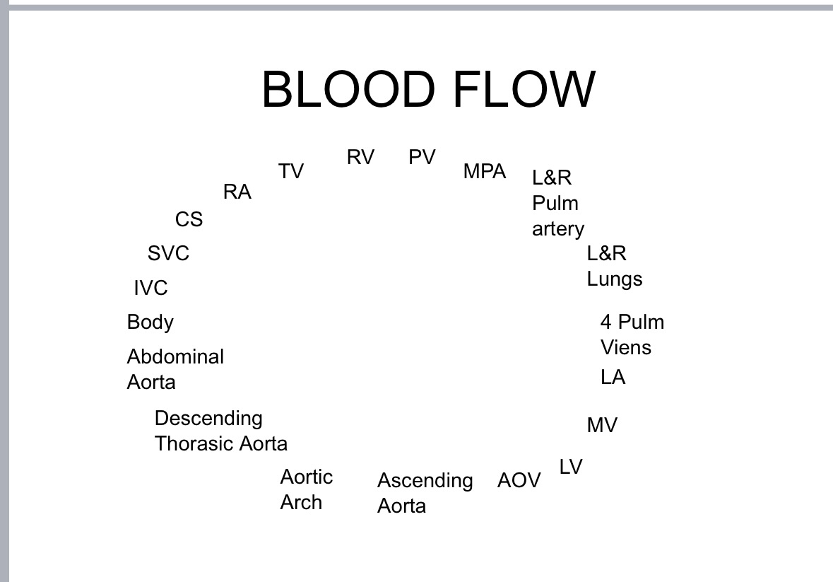
The Superior Vena Cava (SVC): Receives oxygen-poor blood from the upper body (72% oxygen saturation).
The Inferior Vena Cava (IVC): Receives oxygen-poor blood from the lower body (78% oxygen saturation).
The Coronary Sinus (CS): Receives oxygen-poor blood from the heart (60% oxygen saturation).
Oxygen-poor blood is sent to the Right Atrium (RA).
Through the Tricuspid valve (TV) and into the Right Ventricle (RV).
Through the Pulmonic valve (PV).
To the Main Pulmonary Artery (MPA).
Then through the Right and Left Pulmonary Artery to the lungs.
What is the Coronary Sinus (CS)?
The gathering point for oxygen-poor blood collected from the cardiac veins.
What is unique about pulmonary arteries?
Pulmonary arteries are the only arteries with oxygen-poor blood.
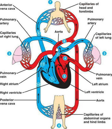
Describe the path of oxygen-rich blood from the lungs, through the left side of the heart, and to the body.
- Oxygen-rich blood returns to the heart through the Four Pulmonary Veins.
- Flows into the Left Atrium (LA).
- Through the Mitral Valve (MV) and into the Left Ventricle (LV).
- Travels out the Aortic Valve (AOV) through the Ascending Aorta.
- To the Aortic Arch (which has 3 branches: Brachiocephalic/Innominate, Left Common Carotid Artery, Left Subclavian Artery).
- Blood travels down the Descending Aorta, which becomes the Abdominal Aorta, and then travels to all different parts of the body.
What is unique about pulmonary veins?
Pulmonary veins are the only veins carrying oxygen-rich blood.
What is the cardiac cycle?
The precisely timed electrical and mechanical events responsible for rhythmic atrial and ventricular contractions, causing blood to be pumped through the heart chambers and out through the great vessels.
What is systole?
The forceful contraction of the cardiac chambers.
What is diastole?
The relaxed phase of the cardiac chambers.
Describe the events during the initial phase of Ventricular Diastole.
- Venous blood enters the Right Atrium from the Superior Vena Cava, Inferior Vena Cava, and coronary sinus.
- Arterial blood returns from the lungs through the four pulmonary veins and enters the Left Atrium.
- The Atrioventricular (AV) valves (Tricuspid and Mitral) are open, allowing blood to flow from the atria into the ventricles.
What happens during Atrial Systole?
Atrial contraction squeezes the remaining blood from the atria into the ventricles. This occurs during ventricular diastole, and a combination of atrial contraction and increased atrial pressure drains atrial blood into the ventricles.
What is Isovolumic contraction?
During ventricular systole, when ventricular pressure exceeds atrial pressure, the Atrioventricular valves close. For about 0.05 seconds, all four heart valves are closed, and pressure increases in the ventricles without changing volume.
What happens after isovolumic contraction during Ventricular Systole?
As pressure increases in the ventricles, the Semilunar valves (Pulmonary and Aortic) open, forcing blood into the lungs and body, respectively.
What is Isovolumic relaxation?
After ventricular contraction is completed, the ventricles relax. When aortic pressure exceeds ventricular pressure, the semilunar valves close, and once again, all four valves are closed. This is part of ventricular diastole.
How do Semilunar valves prevent backflow into the ventricles?
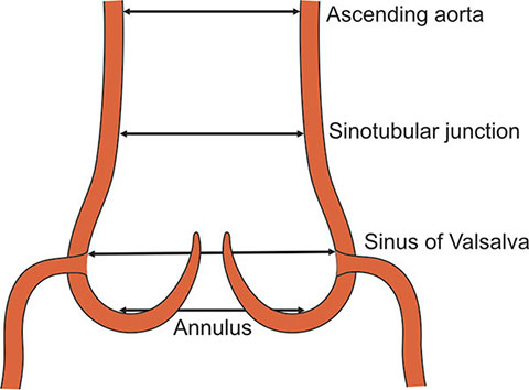
When the ventricles relax and aortic pressure is high, the Semilunar valves close, preventing blood from flowing backward into the ventricles. The blood filling the sinuses of Valsalva also helps force the Aortic valve to close.
What happens at the very end of the cardiac cycle, leading to the next cycle?
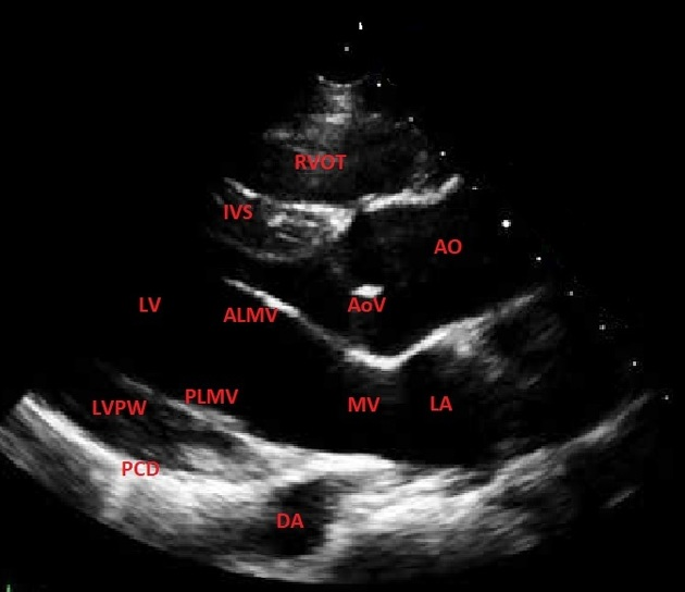
During ventricular contraction, the atria relax and fill with venous blood. When the ventricles are completely relaxed, the Atrioventricular valves open, allowing blood to flow into the ventricles to begin the next cardiac cycle.
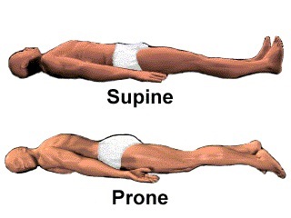
Describe the Supine patient position.
Lying on his or her back.
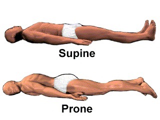
Describe the Prone patient position.
Lying face down.
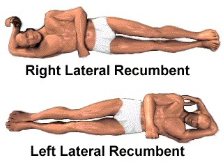
Describe the Right Lateral Decubitus (or Recumbent) patient position.
Lying on their right side.
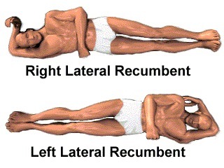
Describe the Left Lateral Decubitus (or Recumbent) patient position.
Lying on their left side.
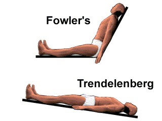
Describe the Fowlers’ patient position.
Sitting straight up or leaning slightly back.
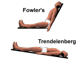
Describe the Trendelenberg patient position.
Lying supine with their head slightly lower than the feet.
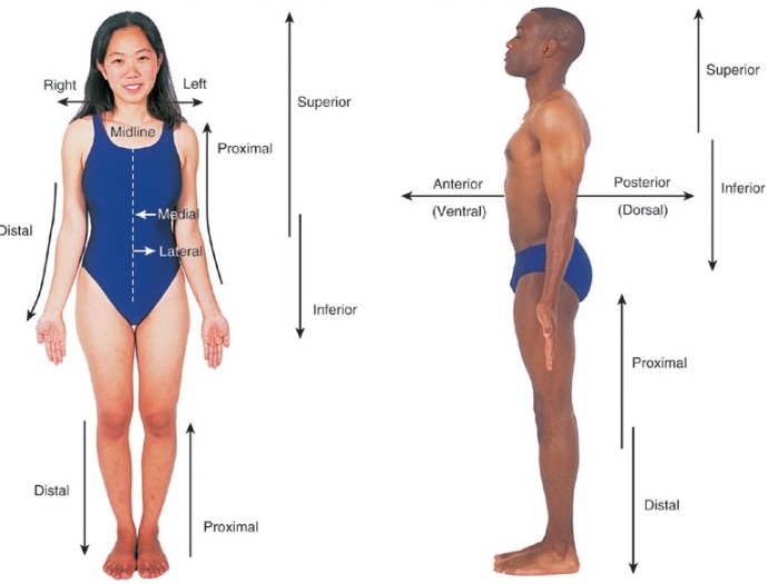
Define Dorsal/Posterior.
Pertaining to the back portion of the body (from Latin 'dorsum' for back).
Define Ventral/Anterior.
Pertains to the front or belly surface of the body (from Latin 'venter' for belly).
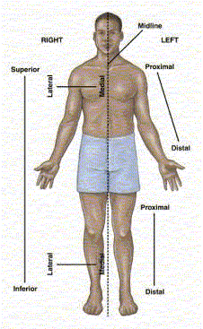
Define Superior.
Above or towards the head.
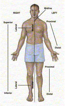
Define Inferior.
Below or away from the head.
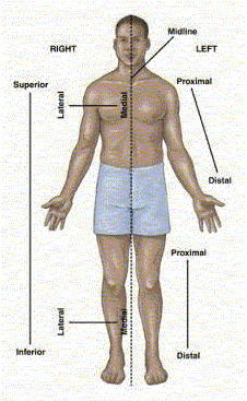
Define Medial.
Relating to the middle or center; towards the median or mid sagittal plane (e.g., the nose is medial to the ears).
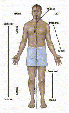
Define Lateral.
Towards the side structure of the body, away from the median plane (from Latin 'latus' meaning side).
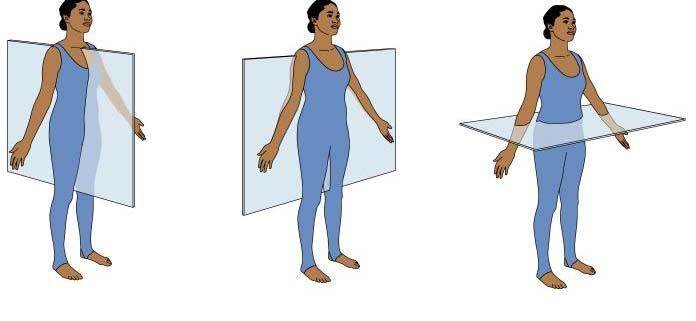
What is the Median sagittal plane?
This plane divides the body into left and right sides.
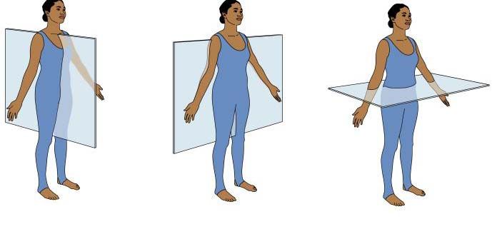
What is the Frontal (or coronal) plane?
Divides the body into front (anterior) and back (posterior).
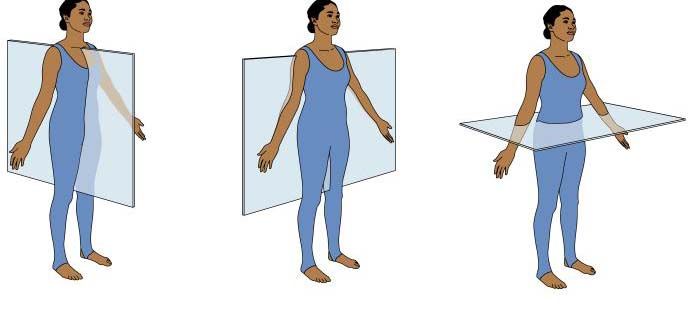
What is the Transverse plane?
This plane is parallel to the ground and divides the body into up (cranial or head) and down (tail or caudal).