Looks like no one added any tags here yet for you.
central nervous system
brain
*protected by the skull
*controls all activities in the body
spinal cord
*protected by the backbone
*controls reflexes
peripheral nervous system
cranial nerves
*12 pairs of cranial nerves
*connects the brain to the sensory and internal organ
spinal nerves
*31 pairs of spinal nerves
*connects the spinal cord to the skeletal muscles
functions of the human nervous system
—the human nervous system controls and coordinates organs and parts of the body
detects stimuli→sends info in the form of impulses→interprets impulses→produces appropriate responses
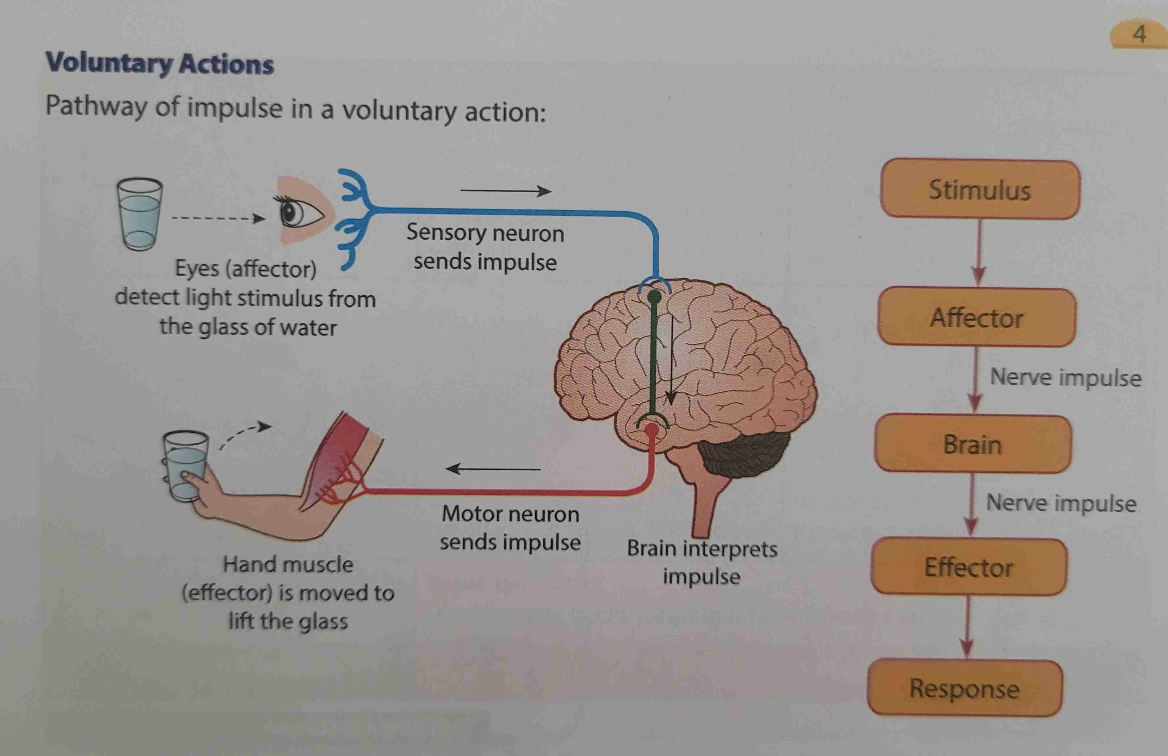
voluntary actions
—concious actions and conducted under one's will
—controlled by the brain
—e.g., reading, writing, etc.
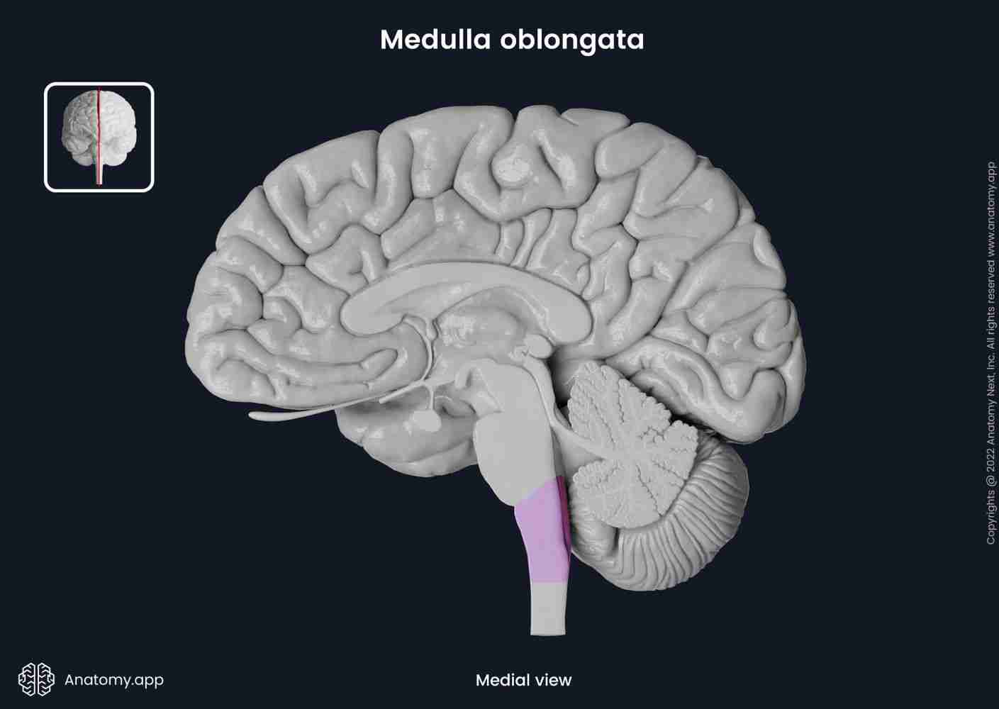
involuntary actions(1)
—actions that occur immediately without concious control or prior thoughts
—involves the medulla oblongata
—e.g., heartbeat, breathing, peristalsis and secretion of saliva
involuntary action(2)
—involves the spinal cord(reflex actions)
—e.g., withdrawing hand when it accidentally touches a hot or sharp object, sneezing when dust enters the nose
pathways of impulse in voluntary action:
pathways of impulse in involuntary action:
importance of the network of human nervous system in daily life
functions of the nervous system
*controls and coordinates movement of the body
*monitors the physiological processes in the body
effects of a damaged nervous system
*paralysis
*depended on machines to carry out physiological processes
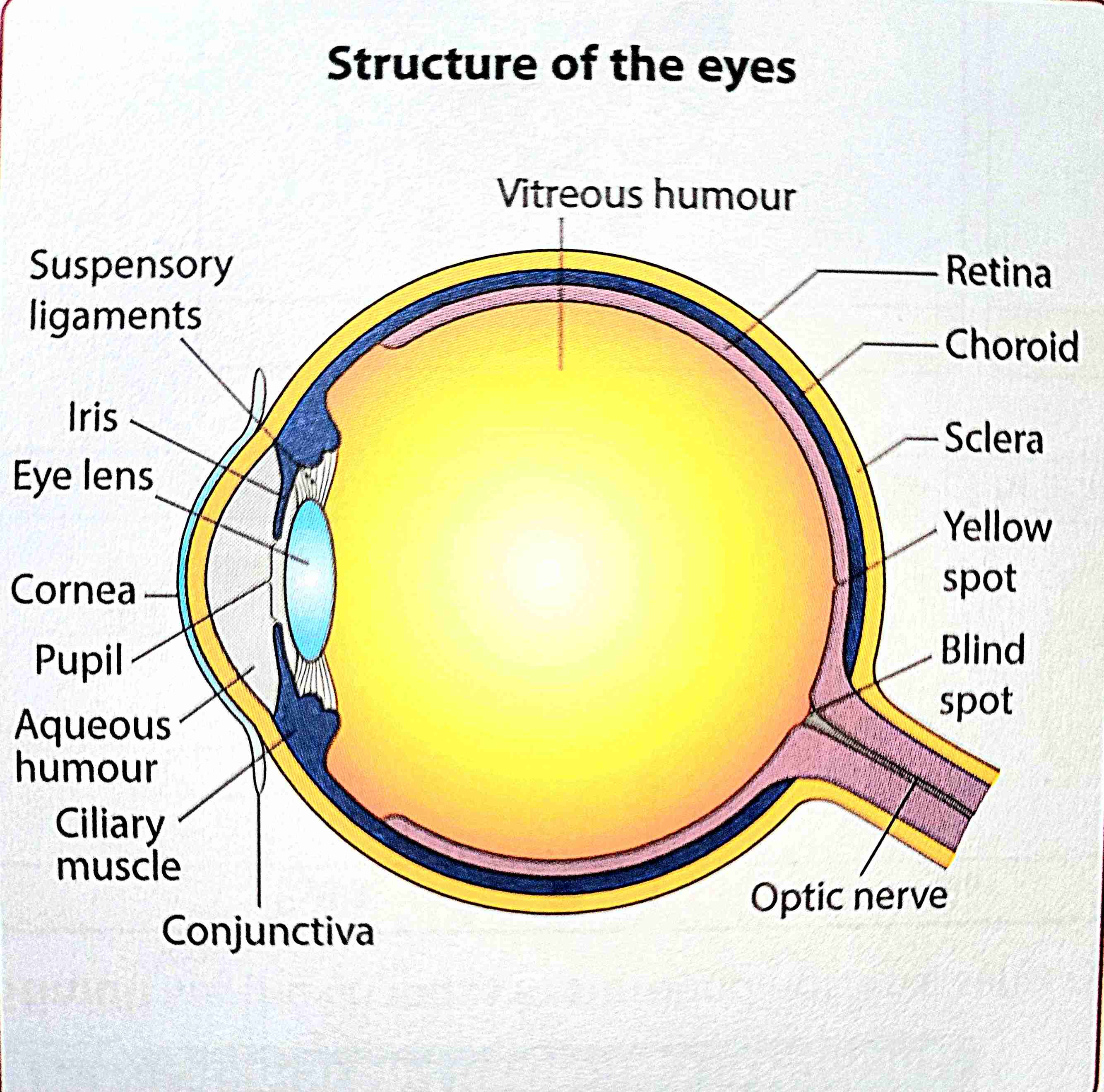
sclera
a strong layer that maintains the shape of the eye and protects it
conjunctiva
transparent membrane that protects the front part of the sclera
iris
the coloured part of the eye which controls the size of the pupil
pupil
opening in the center of the iris which controls the quantity of light entering the eye
ciliary muscle
muscle that changes the thickness of the lens thru contractions and relaxations
cornea
transparent layer that refracts and focuses light onto the retina
aqueous humour
transparent fluid which maintains the shape of the eyeball and focuses light onto the eyes
eye lens
transparent and elastic convex lens which focuses light onto the retina
vitreous humour
transparent jelly-like substance which maintains the shape of the eyeball and focuses light onto the retina
suspensory ligaments
strong fibres which hold the eye lens in its position
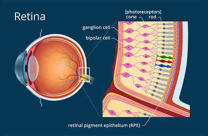
retina
layer containing photoreceptors(cone cells and rod cells) that detect light and produce nerve impulses
yellow spot
Part of the retina which is most sensitive to light as it has many photoreceptors
optic nerve
Nerve fibres which carry nerve impulses from the retina to the brain to be interpreted
choroid
Black layer that prevents reflection of light in the eye and supplies oxygen and nutrients to the eye
blind spot
Part of the retina which is not sensitive to light as there are no photoreceptors. It is the exit point for all optic nerve fibres
functions of the two types of photoreceptors in the retina;
*rod cells are sensitive to different light intensities including faint light
*cone cells are sensitive to the colours of light(red, green and blue) under bright conditions
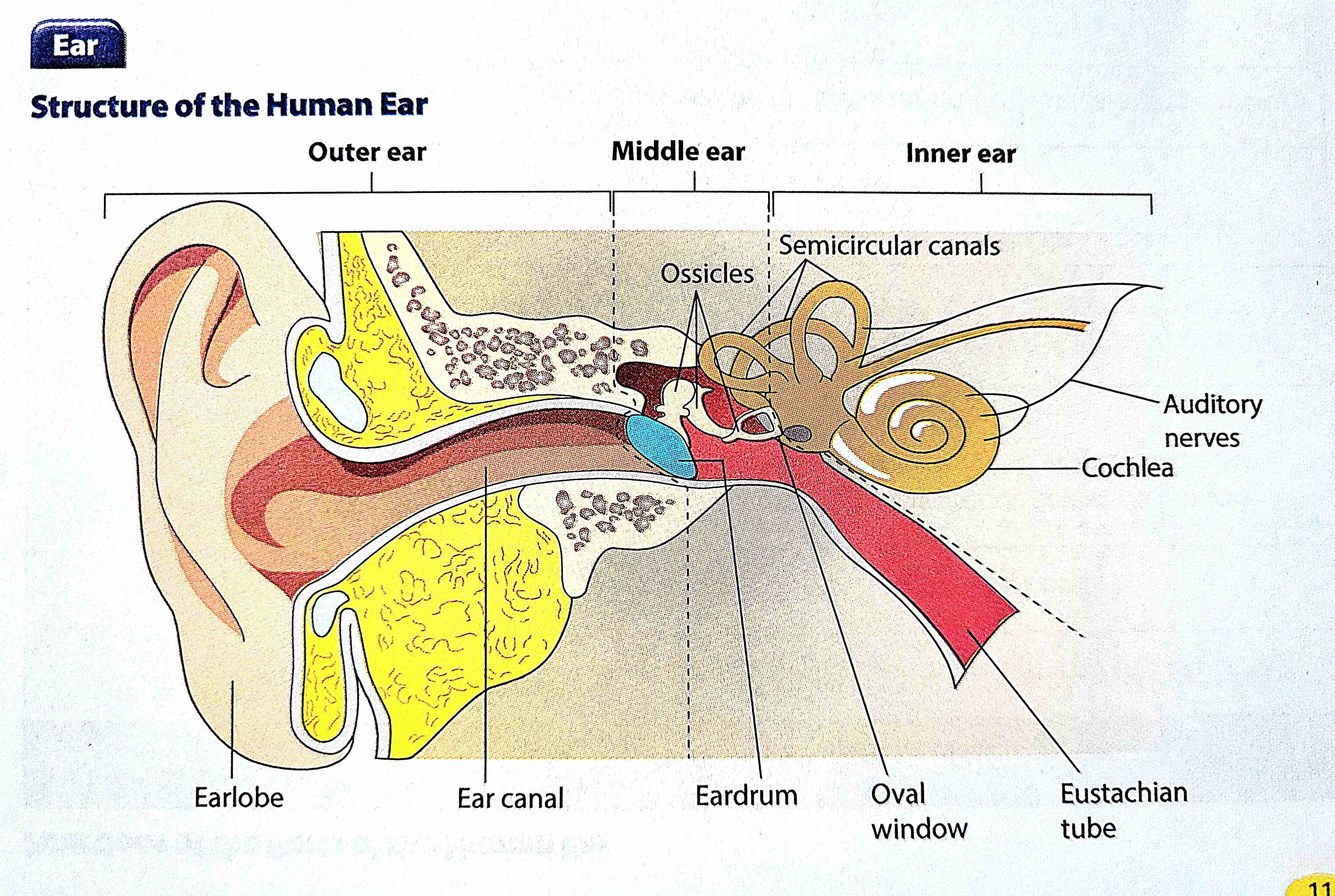
state the parts and functions in the human outer ear
earlobe
—collects and directs sound waves into the ear canal
ear canal
—directs sound waves to the eardrum
state the parts and functions in the human middle ear
eardrum(thin membrane)
*vibrates according to the frequency of the sound waves received and transfers the vibrations to the ossicles
ossicles(three small bones)
*amplify sound vibrations and transfers them to the oval window
oval window
*collects and transfers sound vibrations from the ossicles to the cochlea
eustachian tube
*balances the air pressure on both sides of the eardrum
state the parts and functions in the human inner ear
cochlea(contains fluid)
*detects and converts sound vibration into nerve impulses
semicircular canals(contain fluid)
*detects the position of the head and help to balance the body
auditory nerve
*sends nerve impulses from the cochlea to the brain to be interpreted
parts of the ears that are not involved in the hearing mechanism
eustachian tube(mid ear)
semicircular canals(inner ear)
nose
the nasal cavity has about 10 million sensory cells
theyre very tiny and are covered with a layer of mucus
smells, the chemical substances in the air, will dissolve into the mucus and stimulate the sensory cells, hence producing nerve impulses
these nerve impulses are sent to the brain to be interpreted on what kind of smell it is
tongue structure
a. the tongue has a bunch of these things called papillae
b. the papillae has hundreds of taste buds
c. the taste buds have around 10-50 taste receptors to detect tase
tongue(how it works lol)
a part of or all of the chemical substances in food will be dissolved by the saliva
the dissolved chemical substances will diffuse into the taste buds through the pores, stimulating the production of nerve impulses
then they are transferred to the brain to be interpreted on which kind of taste it is(B/So/Sa/Sw/U)
all parts of the tongue are able to detect all kinds of taste, but different parts of the tongue are more sensitive towards specific tastes
why do we lose our appetite when we're sick?
its bc our sense of smell enhances the humans perception of taste in food. meaning that if our nose are clogged/stuff(due to the flu), we will lose our appetite .
layers of the skin
epidermis→dermis→fat lines
receptors
hot, cold, touch, pressure and pain receptors
factors that influence sensitivity of the skin towards stimuli
the higher the number of receptors, the more sensitive the skin will be
the thinner the epidermis layer is, the more sensitive the skin will be
sensitive and less sensitive parts of the body
fingers, lips, nose, etc
soles of the feet, elbows, back
the ear flowchart
earlobe(sw)
ear canals(sw)
eardrums(v)
ossicles(v)
oval window(v)
cochlea(ni)
auditory nerve(ni)
brain(interpreted)
largest sensory organ??
skin
limitations of the eye
*distant objects
*tiny objects/microorganisms
*optical illusions
*blindspots
near-sightedness
*distant objects appear blurry
*the image or object is in front of the retina cs the eyeballs too long/eye lens too thick
*use concave lenses that are thin in the middle and thick at the edge
long-sightedness
*near objects appear blurry
*the image of object is behind the retina cs eyeballs too short/eye lens too thin
*use convex lens that are thick at the center and thin at the edge
astigmatism
*part of an object is clearer than the rest of the object
*uneven curvature of the lens and cornea
*cylindrical lens that have a curved surface on one side and a flat one on the other
types of lens
concave lens(diverging) spread out light rays that passes thru
convex lens(converging) focuses light rays that passes thru
stuff that like causes ear deterioration
*ageing
*injuries
*damage
*exposure to loud sounds
stuff that helps to overcome limitations of the ear
*stethoscope
*megaphone and speaker
*hearing aids
damages to the ear(outer)
clogged ear canal
*removed the foreign objects
damages to the ear(middle)
punctured eardrums and damaged ossicles
*surgery and medication
damages to the ear(inner)
damaged cochlea(cochlear implant)
damaged auditory nerve(no wat to fix yet)
stuff that affect the sensitivity of the sensory organs
gadgets
*50 cm apart, lower brightness, take a break every 20 minutes
loud sounds for long durations of time
*get earplugs(hearing protector), get a hearing test