Module 1: An introduction to the microbial world (part 1 & 2)
 Call Kai
 Learn
Learn Practice Test
 Spaced Repetition
 Match
Match Flashcards
 Knowt Play
1/90

There's no tags or description
Looks like no tags are added yet.
Name | Mastery | Learn | Test | Matching | Spaced | Call with Kai | Chat |
|---|
No analytics yet
Send a link to your students to track their progress
91 Terms
Microbiology
the study of living organisms (microorganisms, microbes) that individually cannot be seen with the naked eye
Extremophiles
microbes able to live in extreme environments → mainly bacteria, arches, some eukaryotes
Pathogens
organisms that cause diseases
Infection Disease
a disease that was caused by a microbe
Normal Microbiota
microbes that live stably on or in the human body
Functions of Normal Microbiota
aid in digestion of foods
synthesize essential vitamins needed by our cells
protection from disease by preventing pathogens from colonizing the body
Pros and Cons of Microbes in Agriculture
Pros
legumes form close association with N2 fixing bacteria
microbes colonize disgestive tract of ruminants (Sheep +cows) to help digest cellulose
soil microbes decompose plant and animal waste
Cons
causes plant diseases that lead to financial and food loss
Microbial usage in biological control / biocontrol
used to protect plants from plant pathogens and insects.
EX: Bt produces a protein that is toxic to insects at a certain developmental stage (like caterpillars)
Biofouling
unwanted build up of microbes on surfaces exposed to water → leads to clogging of pipes, holding tanks, increased vessel drag
Cellular Microorganisms
mainly single-celled, some multicellular (ex: bacteria, archea, fungi, etc)
Acellular Microorganisms
not composed of cells (ex: viruses)
Most of the diversity of life on our planet is ____
microbial
Prokaryote Cell Structure
no nucleus
no membrane-bound organelles
small
most have cell wall
Eukaryotic Cell Structure
Nucleus
membrane-bound organelles
large
Characteristics of Bacteria
single cell prokaryotic organisms
most are small
cell walls composed of peptidoglycan
hetero + autotrophic
reproduce by binary fission / spores
Cell walls of Bacteria
contains peptidoglycan
Biofilms
caused by bacteria secreting extracellular polysaccharides that are viscous, high MW carbohydrates. They attach to exterior surface of cells as a slime layers and ... [clog something]
Disease(s) associated with Mycobacterium tuberculosis
tuberculosis
Disease(s) associated with Streptococcus pyogenes
strep throat, sepsis, flesh-eating disease
Disease(s) associated with Neisseria meningtidis
meningitis
Archea
single celled prokaryotes
many extremophiles
not disease causing
Fungi
small, non-motile eukaryotic organisms
larger than bacteria
heterotrophic
break down complex organic polymers → nutrient recycling
adapt well to environments
Mycology
the study of fungi
Yeasts
unicellular with oval/spherical shape
asxual via binary fission or budding
cell wall contains polysaccharides (glans) and lipids
Molds
filamentous cells with hypha that grow to form mycelium
cell wall composed of chitin
mycelium
on a mold, formed from hyphae that grow and aggregate
Septae
on a mold, cross walls within hyphae
Reproduction of Molds
asexually by spores but can be sexual spores
Reproduction of Conidia Mold
produced specialized hyphae called conidiophores
Reproduction of Sporagniospores Molds
produced within a sportangium
Mycosis
diseases caused by fungis
Why are mycoses are difficult to control than diseases caused by bacteria?
said come back to later :(
Fungal-like organisms
eukaryotic microbes that share similar morphology and lifestyle with fungi but belong to distinct phylogenetic lineages
Protozoa
unicellular, motile eukaryotes found in water
feed on bacteria + microbes
can for cysts
Disease associated with Plasmodium Protozoa
malaria
Disease associated with Giardia duodenalis
beaver feaver
Cyst
formed by protozoa as a survival mechanism. Surrounds itself and remain in cyst until consumed by host
Helminths
multicellular eukaryotic animals
parasitic with complex life cycles involving multiple hosts
Two main types of Helminths
Flatworms
roundworms
Diseases caused by Helminths results when __________
the body is parasitized by the adult or larval stage or helminth
Hygiene Hypothesis
exposure to certain microbes early in life is necessary for development of a healthy immune system

Coccus (cocci)

Rod

Spirillum (Spirilla)
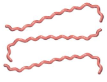
Spirochete

Budding & Appendaged

Flamentous

Single Bacillus

Diplobacilli

Streptobacilli

Coccobacillus

Diplococci
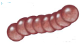
Streptococci

Tetrad

Sarcinae

Staphylococci
Viruses
Acellular, obligate intracellular parasites
contains a single type of nucleic acid surrounded by capsid
In viruses, the capsid determines…
the shape of the virus
Bacteriophages
viruses that infect bacteria
the most abundant type of virus
bacteriophage
Phage Therapy
use of bacteriophages as potential therapeutic agents for treating bacterial infectious disease
Microscopy
the use of light or electrons to magnify objects
Magnification
the process of enlarging an object in appearance only
Resolution (resolving power)
the ability to distinguish two points as distinct and separate
Light Microscopy
use of light (photons) to observe a specimen
Types of Light Microscopes
brightfieqld microscopy
phase-contrast microscopy
fluorescence microscopy
confocal laster scanning microscopy
Electron microscopy
uses elections instead of light to image cells and structures. Electomagnets function as lenses and can be fitted with a camera for taking photographs
Types of election microscopy
transmission electron microscopy
scanning electron microscopy
Brightfield microscopy
specimens are visualized due to differences in contrast between specimen and surroundings
Staining in Brightfeild Microscopy
if specimen is not naturally pigmented, can kill cells and distorts structures
Phase-Contrast Microscopy
uses a compound light microscope with special condenser and objectives, results in dark image on a light background (no staining). Used to see living cells
Flourescence microscopy
used to visualize specimens that fluoresce, either naturally or artificially with fluorescent dyes. Used in clinical diagnostic microbiology and microbial ecology for enumerating bacteria
Immunofluorescence
important for rapid detection of pathogens in clinical specimens [STAT+]
Confocal Laser Scanning Microscopy
uses a computerized florescent microscope coupled with a laser source. Computers focus the laser on single layers of the specimen. The images of the layers can then be complied to produced a 3-D image. Uses staining or requires inherent florescence
Electron micrographs
Images taken with an electron microscope fitted with a camera
Transmission electron microscopy (TEM)
much greater resolving power than light microscopes. the specimen in thinly cut and stained and sectioned, electrons pass through specimen and are scattered. Generates 2D image with details of internal structures (cannot use live specimens)
Scanning Electron Microscopy (SEM)
requires the specimen be coated with a thin heavy metal film. An electron beam scans the specimen, scattered electrons are collected and projected to produce a 3D image. Very high magnification and can view surface structures of cells & viruses (cannot be live)
Staining
increases contrast of specimen for brightfieqld microscopy. Contains dyes that bind to specific cellular material
Basic Dyes
are positively charged, bind strongly to negative charged cell components (cell surfaces, nucleic acids)
Steps of Staining cells for microscopic observation
spread culture in thin film over slide, dry in air
pass slide through flame to heat fix, then flood slide with stain and dry
place drop of oil on slide and examine under 100x objective lens
Simple Stain
uses only one dye to stain cells
Differential Stain
used two different coloured dyes (stains and counterstains) to distinguish between different types of bacteria
Special Stain
used to stain specific structures
Gram Stain
used to distinguish bacteria that differ in their cell wall structures (purple = ?, Pink = ?
Steps of Gram Stain
Flood the heat fixed smear with crystal violet (primary stain) for 1 min
add iodine (mordent) solution for 1 min
decolonize with alcohol for 20 sec
counterstain with safranin for 1-2 min
Gram Positive Vs Gram Negative
Acid Fast Stain
a differential stain used to identify bacteria from the genus Mycobacterium.
It classifies bacteria as either acid-fast or non-acid-fast
How Acid-Fast Stain works
Mycobacterium spp. have waxy lips in their cell wall called mycotic acids
the acid-fast stain uses special stain and procedure to stain mycobacterial cells
once stained, cells resist decolorization by acid-alcohol, and therefore are acid-fast and will appear red. Other bacteria decolourized and are counterstained blue
Steps of Acid-Fast Stain
apply carbolfuchsin, heat for 5 min, rinse
apply acid-acohol (decolourizer) 2× 15 sec
apply methylene blue 1 min (counterstain), rinse
Endospore Stain
a special stain;
endospores are highly resistant, dormant structures produced by some bacteria. The cannot be stained by any other method
Steps to Endospore Stain
apply malachite green (primary stain) heat 5min, then rinse 30 sec
apply safranin (counterstain) for 1 min, rinse, then blot dry
endospores will appear green, rest of cells as pink