Lower GI system & Minimally Invasive studies
 Call Kai
 Learn
Learn Practice Test
 Spaced Repetition
 Match
Match Flashcards
 Knowt Play
1/91

There's no tags or description
Looks like no tags are added yet.
Name | Mastery | Learn | Test | Matching | Spaced | Call with Kai | Chat |
|---|
No analytics yet
Send a link to your students to track their progress
92 Terms
what is the duodenum
“C-loop”
8-10 inches long
hystero should be scheduled approximately __ days after onset of menstruation
10
what is always given to a patient for a hystero that is ordered by the OBGYN
antibiotics
what are the muscles in the rotator cuff:
teres minor
infraspinatus
supraspinatus
subscapularis
what is the jejunum
feathery appearance. second section of the small bowel
what is the illeum
last portion of the small bowel, folowed by the large intestine
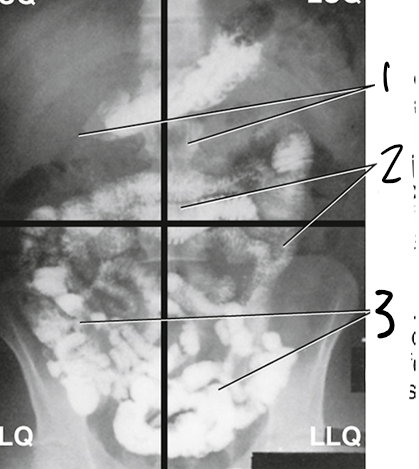
what is 1?
duodenum
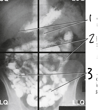
what is 2?
jejunum

what is 3?
ileum
what is villi?
series of fingerlike projections in the SB mucosa assisting in digestion and absorption
what is terminal ileum
most distal portion of the ileum
what is the ileocecal valve
one way spincter located between ileum and cecm to prevent reflux of fecal matter from large instestine back into small intestine
preparation for small bowel
NPO 8 hours prior
RH NPO after midnight
is a scout necessary for a small bowel study
yes
Small bowel series info
follows barium from the stomach to entrance of large intestine
timing is important
images of the small bowel are teken approx. every 15-30 min
RH 20 min
must be time annotated
small bowel completion
study is complete when the barium passes through ileocecal valve
barium typically enters first part of the large intestine in about 2-3 hours
when the barium reaches the cecum, the RA will spot fluoro over this area
focus will be at the terminal ileum entering into colon
technologist responsibilities of tech responsibilities
take scout abdomen and show RA/radiologst
give thin barium to patient
take the over hear stomach images, monitoring time of images, continum to take SM thru images per RA/Rad
assist pt and RA during fluoro portion of the study
if a pt cannot swallow the contrast or has pyloric difficulties, but small bowel needs to be evaluated you would do…
enteroclysis
what is enteroclysis
instertion of a tube through the nose or mouth into the patient’s stomach and into the beginning of the small bowel
stops at end of duodenum (duodenojejunal flexure)

what landmark is 1?
L4, L5 and iliac crest
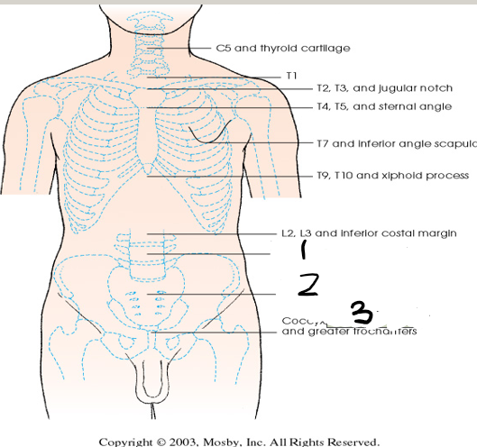
what landmark is 2?
SI and ASIS

what landmark is 3?
pubic symphysis

what is 1?
right colic flexure

what is 2?
ascending colon
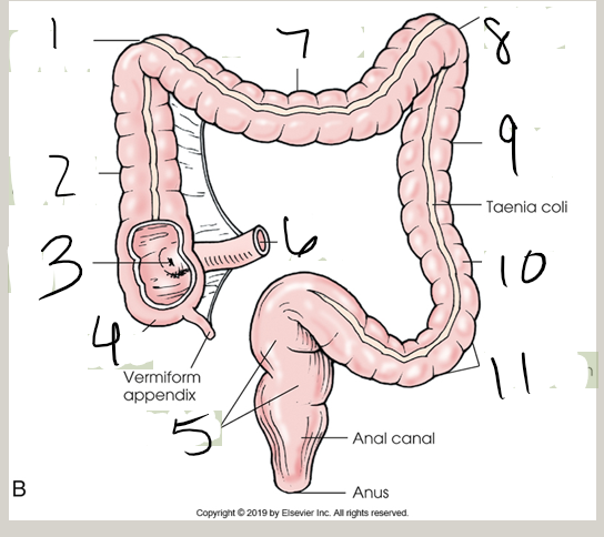
what is 3?
ileocecal valve

what is 4?
cecum
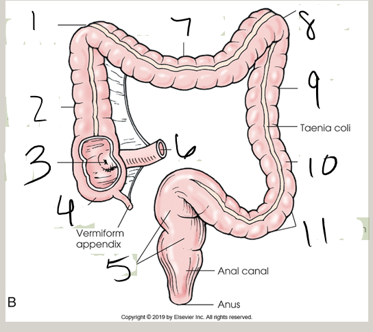
what is 5?
rectum

what is 6?
ileum

what is 7?
transverse colon

what is 8?
left colic flexure

what is 9?
descending colon

what is 10?
haustra

what is 11?
sigmoid colon
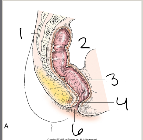
what is 1?
sacrum
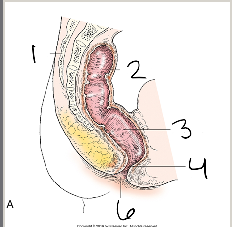
what is 2?
rectum

what is 3?
rectal ampulla

what is 4?
anal canal

what is 6?
anus
on a lateral view of the large intestine what parts are anterior
transverse and most of the sigmoid colon

what patient position is the circle?
supine

what patient position is the rectangle?
prone
what is the patient prep for BE
clear liquids only day prior to and morning of exam
no solid foods should be consumed
why are scout images important for BEs?
RA- still looking to see if patient is cleaned out, and if there are any obvious abnormalities
Techs- look at the gas pattern as well, how the intestine lay will determine whether or not you need to take images portrait, landscape or both
contrast needed for a single BE
2 bottle Vanilla SilQ HD mixed with 222mL of water
contrast needed for double BE
3 bottle vanilla SilQ, each mixed with 222 mL water
2 botles of Visacasure honey to each bottle of vanilla SilQ
never hang a BE bag more than _____ above tabletop
24”-30”
prior to insertion of the tip for a BE
explain to the patient what you’re doing
make sure the barium is clearned all the way to the enema tip
GLOVES! Lubricant!
have the patient in sims position
instering the enema tip
lift upper buttock to see the rectal area
instert tip on exhalation
slide tip back to front
inserting anteriorly and superiorly
no more than 4 inches
Who blows up the balloon for a BE
the RA does under fluoro guidance
tech will clamp tubing with hemostats and remove the square sponge
what are the minimally invasive procedures
arthrogram
myelogram
hysterosalpigograms
types of needle insertions:
joint: intraarticular
spine: intrathecal
what is an arthrogram
minimally invasive procedure using live imaging and a contrast material, such as dye, water, air, or a combo, to take images to visualize a joint
in conjuction with CT and MRI
shoulder, knee, hip, wrist, elbow
what type of contrast is used for pneumoarthrography
using gas as a contrast
what type of contrst is used for opaque arthrography
uses water-soluble iodinated contrast
what type of contrast is used for double contrast arthrography
using both (air/gas and contrast)
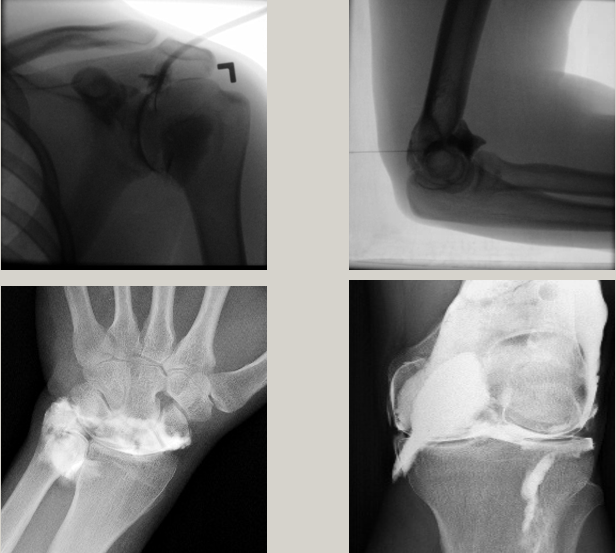
what are these images of?
arthrograms
post procedure by RA/RAD images per protocol (shoulder)
neutral
external
internal
axillary

where does the bowel sit in hypersthenic patients
periperally

where does the bowel sit in sthenic patients
right flexure lower

in hyposthenic patient where does the bowel sit
both flexures drop

in an asthenic patient where does the bowel sit
all low in abdomen
what is a myelogram
radiographic study of the spinal cord and its nerve root branches utilizing a contrast medium
what is the spinal cord
slender elongated structure consisting of an inner and outer section
extends from medulla oblongata to approx L1-L2 disk space
for myelograms what body parts is Omnipaque 300 used for
cervical
complete
for myelograms what body parts is Omnipaque 240 used for
thoracic
lumbar
what is a hysterosalpingogram
procedure to visualize uterus nd fallopian tubes with the use of water-soluble contrast through a uterine cannula
OBGYN with RA/RAD
what is a hysterosalpingogram for
determine size, shape and position of uterus and tubes
conception reasons
polyps, submucosa tumor, fistula tracts
successfulness of surgery to prevent conception
what is unicornuate uterus
abnormally shaped uterus with only one uterine tube
congenital abnormality
increased difficulty getting pregnant and may have issues while pregnant

what is this a picture of
unicornuate uterus

what is this an example of
bicornuate uterus
what is a bicornuate uterus
heart shaped uterus; paired uterine horns extending to uterine tubes
congenital; Mullerian dicts not fusing in uterine cavity while developing in womb
typically doesnt cause issues getting pregnant, but may have issues while pregnant
what is uterus didelphys
rare congenital condiiton where a person develops two uteruses and cervixes
increased risk of miscarriage or pre-term birthS

what is this an image of?
uterus didelphys
SIDE NOTE
STEP BY STEP PROCEDURES ARE NOT IN HERE, LOOK AT POWERPOINT
spinal nerves
31 pairs of spinal nerves
each pair arises from 2 roots nerves transmitted through IV and sacral foramina
what is the conum medullaris
pointed lower extremity of spinal cord
L1-L2
what is the cauda equina
spinal nerves below termination of cord
extend inferiorly through vertebral canal
what are the meninges
protective membrane that enclose the brain and spinal cord
where is the pia mater
closest to brain/spinal cord
where is the arachnoid
central layer
separated from the pia mater by the subarachnoid space
what is located in the subarachnoid space
CSF
myelogram injections are into the
subarachnoid space
where is the dura mater
outermost layer
what is the dura mater
strong, fibroid covering of brain and spinal cord
what is CSF
clear, colorless
surrounds and cushions the brain and spinal cord
where is CSF found
located in ventricles

what is 1?
conus medularis

what is 2?
cauda equina

what is 1?
dura mater

what is 2?
arachnoid
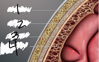
what is 3?
pia mater

what is 4?
subarachnoid space