Lecture 16- Functional Retina
1/38
There's no tags or description
Looks like no tags are added yet.
Name | Mastery | Learn | Test | Matching | Spaced |
|---|
No study sessions yet.
39 Terms
Functional retina
-Extension of brain
-Multiple layers
-Retina analyzes data
-Encodes data into neural signal
-Data transmitted to higher visual centers
retinal layers (anterior to posterior)
1. Inner limiting membrane
2. nerve fiber layer
3. ganglion cell layer
4. inner plexiform player
5. Inner nuclear layer
6. outer plexiform layer
7. outer nuclear layer
8. External limiting membrane
9. Outer Segment
10. Retinal pigment epithelium
What layer are photoreceptor cells found?
What is the direction of the flow of information in the retinal layers?
Why is the retina organizes in "backward direction?
-Photoreceptor dependence on RPE
-Protects the retina from blood-borne pathogens
-Light not absorbed by photoreceptor is all absorbed by RPE
What would happen is the retinal layers were reversed?
If the orders were reversed that photoreceptors were the front layer then RPE would absorb all the incoming light.
Receptive fields of ganglion cells
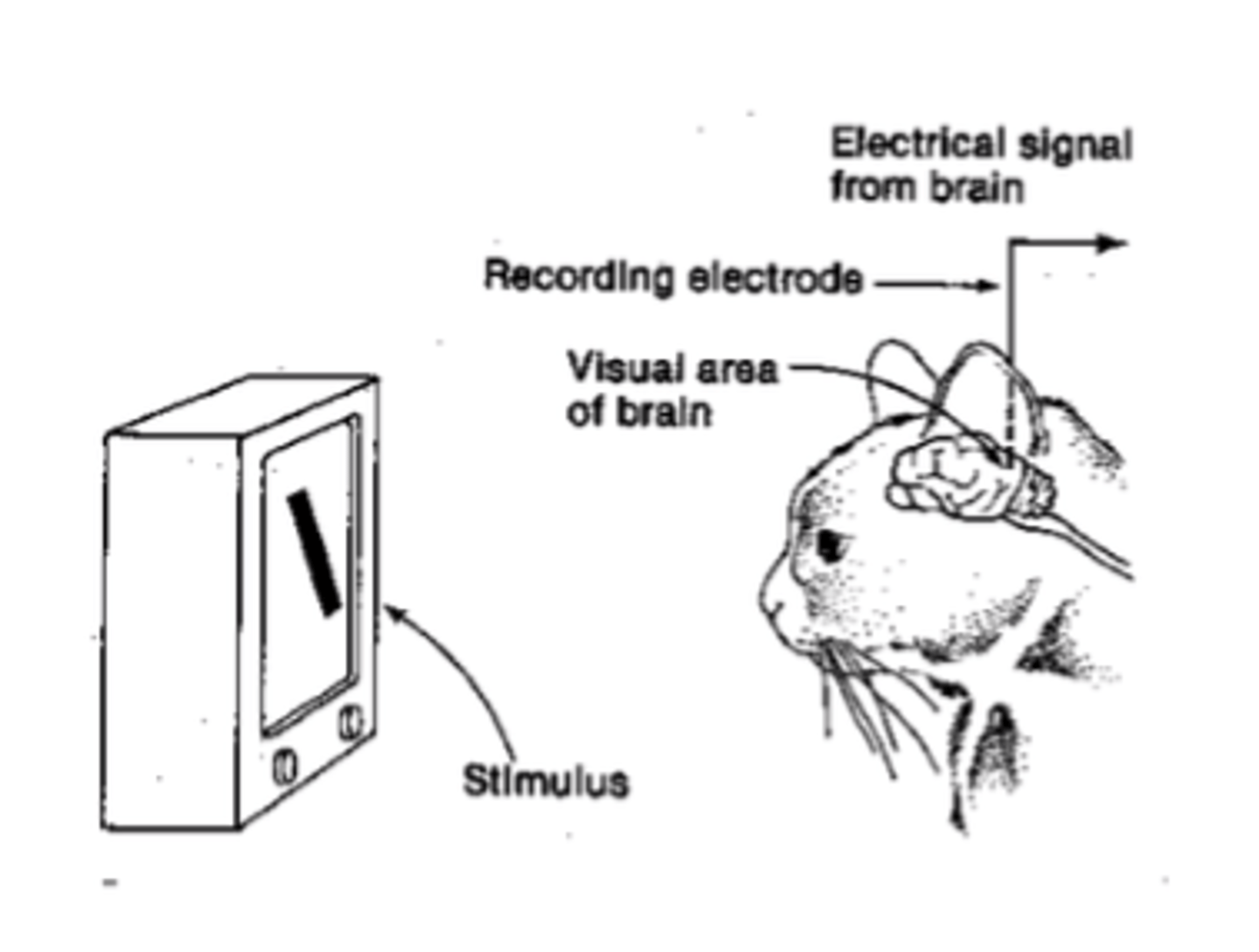
-electrophysiology used to study receptive fields
-fovea aligned with point on screen
-microelectrode placed in extracellular fluid next to ganglion cell
-action potentials read
-Spot do light elicits response from cell
Receptive field
region of space in which stimuli will affect the cell's activity
-Influence may be positive (by increasing in membrane depolarization or action potential frequency)
-influence may be negative
A small light located within the center of a receptive field that is on center/ off surround causes an ______ in the frequency of action potentials
increase
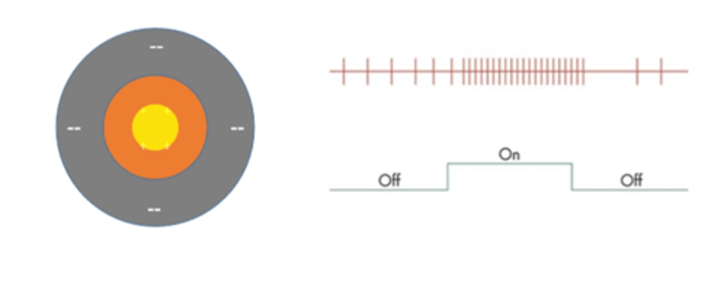
A small light positioned in the surround of a on center/ off surround receptive field produced a _________ in the frequency of action potentials
reduction

If the experiment is repeated with a larger spot of light, there is an increase in the frequency of action potentials due to ___________ ______________ that occurs within the receptive field's center.
spatial summation
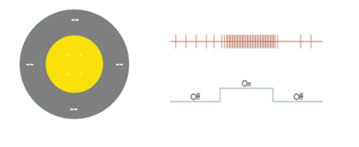
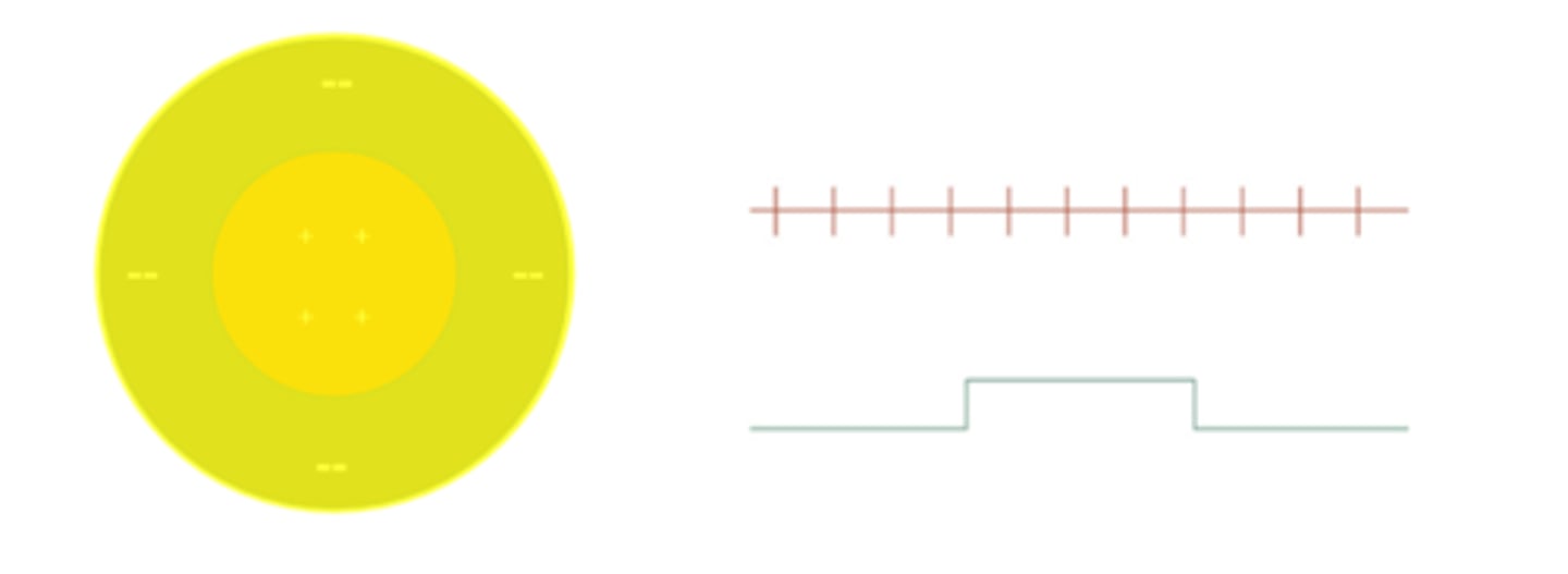
Ganglion cell response to a stimulus that fills the entire receptive field is about
the same as if there were no stimulus
-Spatially antagonistic ganglion cells do not response well to diffuse illumination
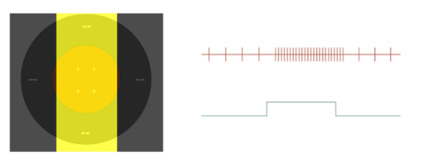
Sine-wave gratings are another strong stimulus for
ganglion cells
-Bright bar falls on the excitatory center =Increased frequency of action potentials
-Dark bands fall on inhibitory surround = Also increases the frequency of action potentials
-Spatial grating vigorously excites cells
Cat experiment
-Record extracellularly from an on-center, off-surround ganglion cell in a cat retina
-Shine a very small light onto the center of the cell's receptive field
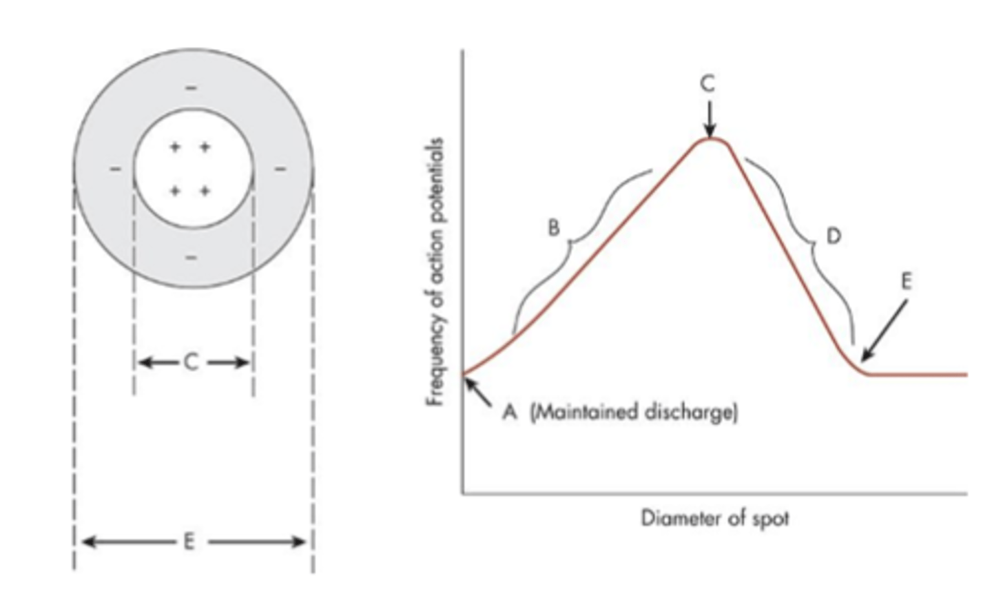
-Elicits an increase in the frequency of action potentials (as compared with the maintained discharge).
-If the experiment is repeated with a slightly larger spot of light, there is an increase in the frequency of action potentials due to spatial summation that occurs within the receptive field's center.
Cat experiment results
Plot the frequency of action potentials as a function of the diameter of the spot of light
-As the diameter of the spot increases, the cell's firing rate increases (section B) until the entire receptive field center is covered with light, and the response is at its maximum (point C).
-Any further increase in stimulus diameter produces a decrease in the firing rate (section D) because the stimulus now falls on the inhibitory surround, which is antagonistic to the excitatory center.
-Eventually, a point is reached where further increases in the stimulus diameter have no effect on the cell's response. This point, indicated by E, represents the termination of the cell's receptive field.
Which type of retinal neurons generate action potentials?
Amacrine cells and ganglion cells.
What type of potentials do all other retinal neurons generate?
Graded potentials.
What is the response of bipolar and ganglion cells to a spot and annulus?
They elicit responses of opposite signs.
Why do the spot and annulus elicit opposite responses in bipolar and ganglion cells?
The spot falls on the receptive field center, while the annulus falls on its antagonistic surround.
Photoreceptor
-Photoreceptors respond to light with a slow hyperpolarizing response
-Rods, detecting dim light, respond to relatively slow changes.
-Cones, detecting bright light, response to rapid light fluctuations.
-Rods and cones respond to light directly over them. So its receptive fields are very narrow.
Horizontal cell
-First synaptic level in the retina
-Horizontal cells receive input from many cones
-So, its receptive field is large.
-Consequently, H cells respond to light over a large area.
-H cells modulate the photoreceptor signal under different lighting conditions, which allows the signaling to become less sensitive in bright light and more sensitive in dim light.
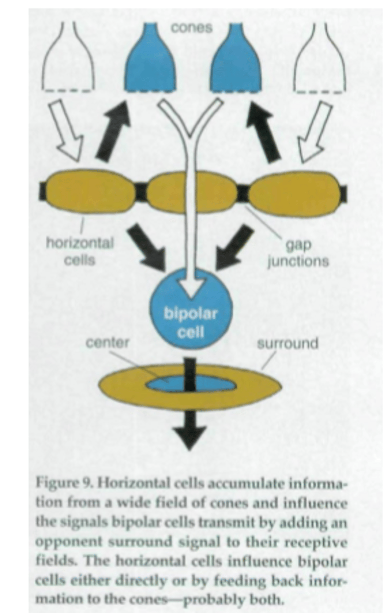
-Horizontal cells shapes the receptive field of bipolar cells
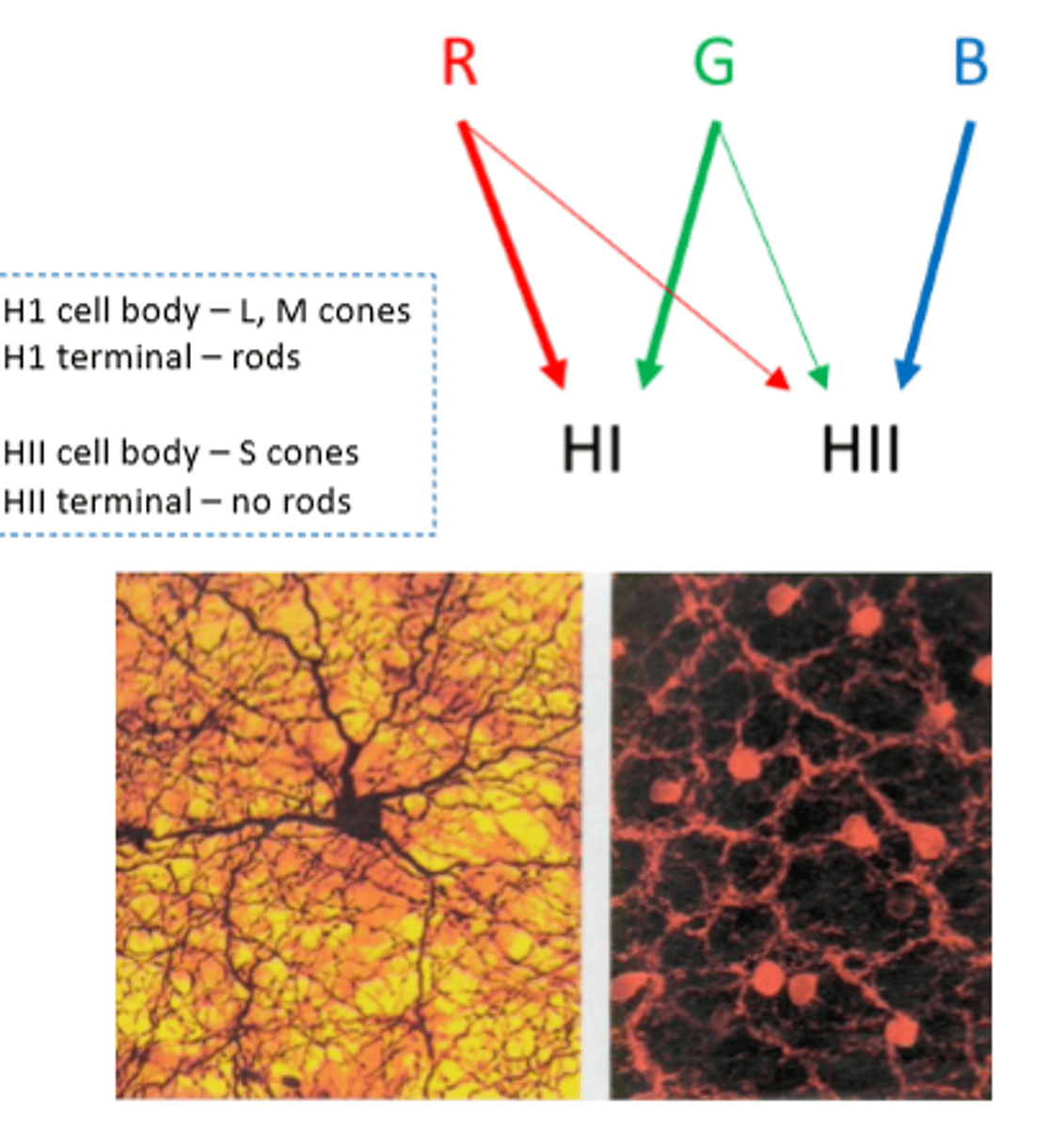
HI horizontal cells
receive an excitatory input from L- and M-cones, but lack a significant input from S-cones.
HII horizontal cells
excited by all three cone types, with the S-cones providing a particularly strong input
HI and HII horizontal cell types
each form an electrically couples network with receptive field diameters much larger than the dendritic field size of a single horizontal cell, effectively summing input from hundreds of cones.
Bipolar cell (off-center)
-Bipolar cells receives input from handful of cones
-Thus, it has a medium size receptive field
-Horizontal cells add an opponent signal giving bipolar cell its 'center -surround' organization
-If the bipolar center signals 'ON' then H cells will signal 'OFF', and vice versa.
What are the two types of amacrine cells?
Small field and Wide-field amacrine cells
Where do small field amacrine cells spread in the inner plexiform layer (IPL)?
They spread vertically across several strata in the IPL, which has approximately 5 strata.
How do small field amacrine cells interact with bipolar cells and ganglion cells?
Small field cells collect signals from about 30 rod-connected bipolar cells and synapse with both ON and OFF ganglion cells.
How do wide field amacrine cells differ in structure compared to small field cells?
Wide field cells stretch horizontally across the IPL for several hundred microns but typically confine to one strata.
How many bipolar and ganglion cells do wide field amacrine cells interact with?
Wide field amacrine cells interact with hundreds of bipolar cells and many ganglion cells.
What is one function of amacrine cells in vision?
They allow the perception of very dim light.
How do amacrine cells affect the perception of boundaries in vision?
Amacrine cells sharpen the boundary between center and surround even further than H cells do.
What is the structure of the receptive field of ganglion cells?
The receptive field is organized as concentric circles.
When are ON center ganglion cells activated?
When a spot of light falls in the center of their receptive field.
When are ON center ganglion cells deactivated?
When light falls on the periphery of their receptive field.
When are OFF center ganglion cells activated?
When a spot of light falls in the periphery of their receptive field.
When are OFF center ganglion cells deactivated?
When light falls on the center of their receptive field.
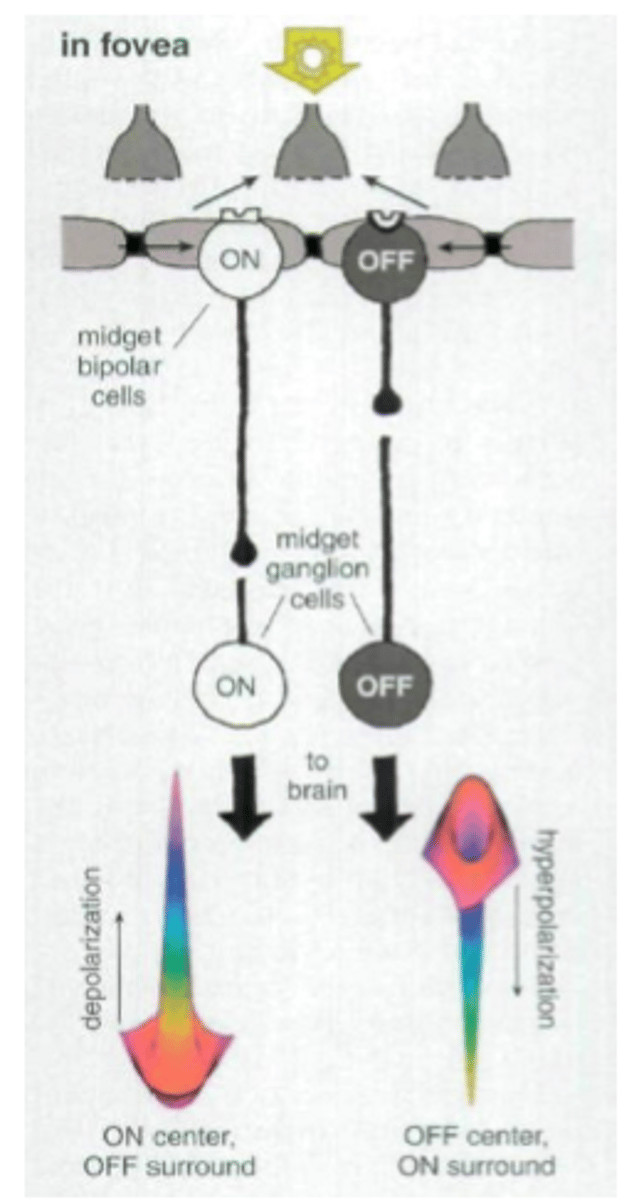
Ganglion cells in the fovea
Ganglion cells in fovea are connected to bipolar cells in one-to-one ratio.
-And, this info comes from a single cone. Thus relaying a point to point image from fovea to brain.
-The message that goes to the brain delivered from ganglion cells carry both spatial and spectral information of the finest resolution.
What if there are no retinal layers, that info passed on from photoreceptors to the brain?
1. The resulting image would probably be course-grained and blurry
2. The processing in retina layers defines precise edges to images and allows us to focus on find details.