Jim Exam 4 Neuropathology
 Call Kai
 Learn
Learn Practice Test
 Spaced Repetition
 Match
Match Flashcards
 Knowt Play
1/256

There's no tags or description
Looks like no tags are added yet.
Name | Mastery | Learn | Test | Matching | Spaced | Call with Kai | Chat |
|---|
No analytics yet
Send a link to your students to track their progress
257 Terms
Corpus callosum
What is this structure?
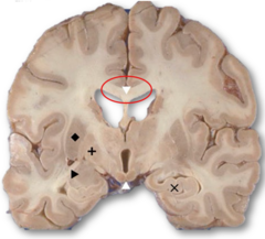
Caudate
What is this structure?
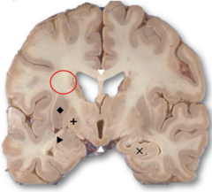
Putamen
What is this structure?
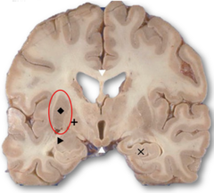
Amydgala
What is this structure?
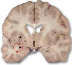
Globus pallidus
What is this structure?
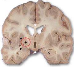
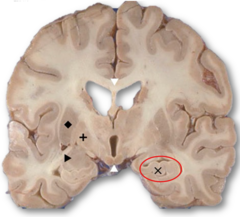
Hippocampus
What is this structure?
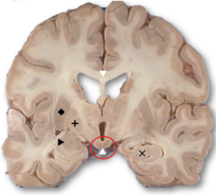
Mammillary bodies
What are these structures?
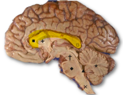
Corpus callosum
What is this structure?
Midbrain
What is this structure?
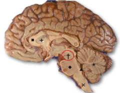
Pons
What structure is this?
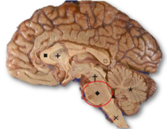
Medulla
What structure is this?
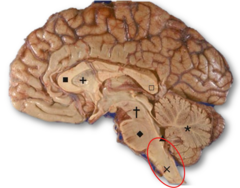
Cerebellum
What structure is this?
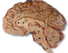
4th Ventricle
What structure is this?
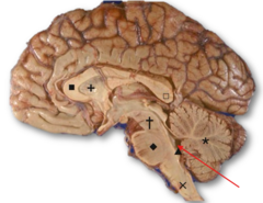
Corpus collosum
What structure is a large, thick band of nerve fibers that connects the two hemispheres of the brain?
Hippocampus
What is a crucial part of the limbic system located in the temporal lobe and functions in short-term and long-term memory?
Mamillary bodies
What are the paired brainstem nuclei on the posteroinferior aspect of the hypothalamus that play a role in memory recollection?
Basal ganglia
What is the group of nuclei in the central area of the brain that receives input from the cortex and functions to control conscious and proprioceptive movements?
Cerebellar Purkinje cells
What is the unique population of neurons in the cerebellum that function in regulating and coordinating motor movements?
Complete or partial absence of the corpus callosum
What is agenesis of the corpus callosum?
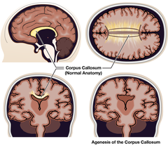
Yes, anterior commissure can assume interhemispheric tasks due to plasticity
Can agenesis of the corpus callosum be asymptomatic and why?
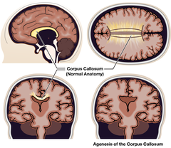
It is displaced downward
What happens to the cingulate gyrus in callosal agenesis?
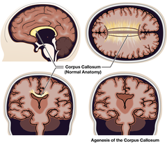
Increased volume of cerebrospinal fluid within the ventricular system
What is hydrocephalus?
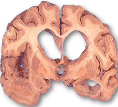
In the ventricles, not the brain parenchyma (fluid in the parenchyma is cerebral edema
Where is the excess fluid in hydrocephalus located?
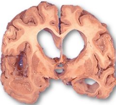
Headache, behavioral change, lethargy, delayed development
What are common clinical signs of hydrocephalus?
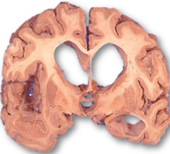
Ventricular dilation upstream from a blockage of CSF outflow
What is noncommunicating hydrocephalus?
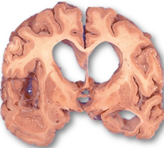
Congenital malformations, tumors, or certain infections
What causes noncommunicating hydrocephalus?
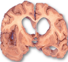
CSF accumulation from impaired absorption or excess production affecting all ventricles
What is communicating hydrocephalus?
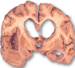
Ventricular enlargement secondary to loss of brain parenchyma from atrophy
What is hydrocephalus ex vacuo?
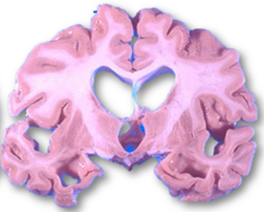
Accumulation of fluid within the brain parenchyma
What is cerebral edema?
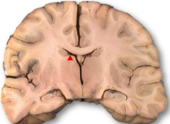
Inflammation, ischemia, toxic injury, or space-filling lesions like tumors
What are typical causes of cerebral edema?
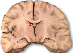
Increased vascular permeability
What drives cerebral edema formation at the vascular level?
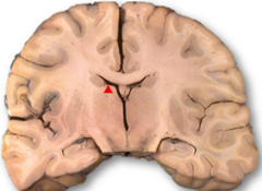
Swollen flattened gyri, narrowed sulci, compressed ventricles
What are gross features of generalized cerebral edema?
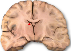
Brain herniation
What is a major risk of severe cerebral edema?
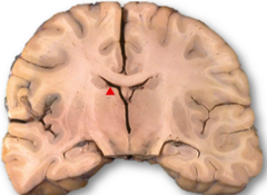
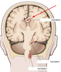
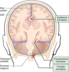
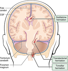
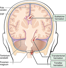
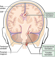
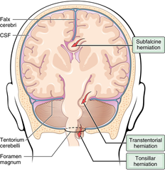
Subfalcine herniation
What kind of herniation is this?
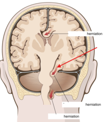
Transtentorial herniation
What kind of herniation is this?
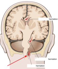
Tonsillar herniation
What kind of herniation is this?
Cingulate gyrus pushed under the falx due to unilateral hemispheric expansion
What is subfalcine herniation?
Anterior cerebral artery
Which artery can be compressed in subfalcine herniation?
Medial temporal lobe forced against the tentorial edge
What is transtentorial (uncal) herniation?
Oculomotor nerve (CN III) with ipsilateral blown pupil due to impaired ocular reflexes
Which cranial nerve is compressed in transtentorial/uncal herniation and what is the sign?
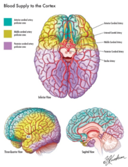
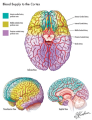
Posterior cerebral artery causing ischemia of primary visual cortex
Which artery may be compressed in transtentorial/ uncal herniation and what results?
Compression of the contralateral cerebral peduncle against the tentorium
How can uncal herniation produce ipsilateral hemiparesis?
Loss of consciousness
What can occur when the brainstem is compressed in transtentorial herniation?
Duret hemorrhages
With which hemorrhage is transtentorial/uncal herniation classically associated?
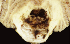
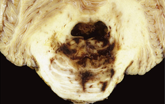
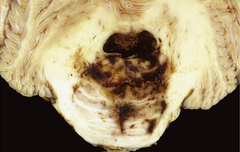
Linear or flame-shaped hemorrhages in midbrain and pons from downward displacement in uncal herniation
What is a Duret hemorrhage?
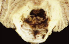
Tearing of basilar artery branches entering the pons along the midline
What vessel disruption underlies Duret hemorrhage?
Poor, indicating severe brainstem injury
What is the prognosis with Duret hemorrhages?
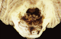
Altered consciousness, respiratory irregularities, autonomic instability
What symptoms suggest brainstem involvement in Duret hemorrhage?
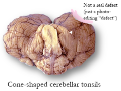
Cerebellar tonsils displaced through the foramen magnum
What is tonsillar herniation?
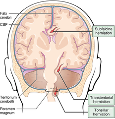
Brainstem compression causing respiratory and cardiac failure
What are life-threatening consequences of tonsillar herniation?
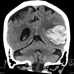
1) Trauma with contusions
2) Hemorrhagic transformation of ischemic infarct
3) Cerebral amyloid angiopathy
4) Hypertension
5) Tumors (primary or metastatic)
What are common etiologies of intraparenchymal hemorrhage?
Selective involvement of gyri
What are the features of intraparenchymal hemorrhage due to trauma (contusions)?
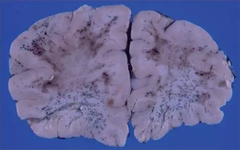
Petechial hemorrhages in a previously ischemic territory due to reperfusion injury
What are the features of intraparenchymal hemorrhage due to ischemia (hemorrhagic conversion of ischemic infarct)?
Lobar hemorrhages involving subcortical white matter with possible extension into subarachnoid space
Due to amyloid deposition making vessel walls fragile
What are the features of intraparenchymal hemorrhage due to cerebral amyloid angiopathy?
Hemorrhage is centered in the deep white matter, thalamus, basal ganglia, brainstem with possible intraventricular extension
What are the features of intraparenchymal hemorrhage due to hypertension?
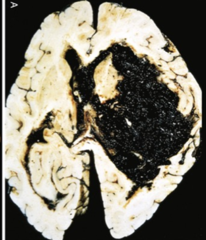
Minute aneurysms / microaneurysms in the brain that occur in small penetrating vessels that can rupture and cause intraparenchymal hemorrhage
What are Charcot-Bouchard aneurysms?
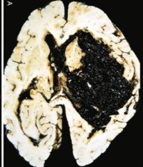
High-grade gliomas and certain metastases
Which tumors commonly bleed within brain parenchyma?
1) Acute hypertensive encephalopathy
2) Intraparenchymal/lobar hemorrhage
3) Arteriosclerosis
4) Charcot-Bouchard aneurysms
5) Rupture of berry aneurysms
6) Lacunar infarcts
7) Predilection for vessels involving the basal ganglia
8) Atherosclerosis
9) Subarachnoid hemorrhage
10) Cerebral edema
11) Petechiae and necrosis of arterioles in the gray and white matter
List CNS findings associated with systemic hypertension
Trauma or rupture of vascular abnormalities such as saccular aneurysm or AVM
What are common causes of subarachnoid hemorrhage?
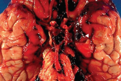
Sudden severe headache ("thunderclap
headache) and rapid neurologic decline with risk of vasospasm from blood in CSF
What clinical presentation suggests aneurysmal subarachnoid hemorrhage?
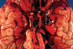
Venous bleeding from torn bridging veins often after minor trauma with slow progression
What is the usual mechanism of subdural hematoma?
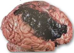
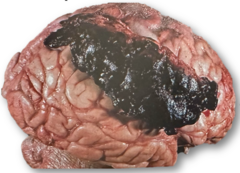
Early clot draws in water and enlarges into a mass-like lesion
Why can a subdural hematoma enlarge over time?
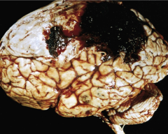
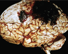
Laceration of the middle meningeal artery associated with skull fracture (usually due to trauma)
What causes an epidural hematoma?
Brief lucid interval followed by rapidly evolving neurologic deterioration
What is the classic course of an epidural hematoma?
Amyloid-β derived from APP proteolysis
What amyloid species is usually deposited in cerebral amyloid angiopathy (CAA)?
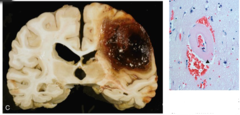
Cortical and leptomeningeal vessels
Which vessels are primarily involved in CAA?
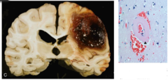
CAA is cortical and leptomeningeal whereas arteriosclerosis predominates in deep structures like basal ganglia
How does CAA differ from hypertensive arteriosclerosis in distribution?
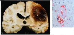
Makes vessel walls rigid and fragile increasing hemorrhage risk
How does amyloid deposition affect vessel integrity in CAA?
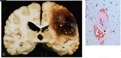
Lobar hemorrhages and microhemorrhages in cortex
What hemorrhage pattern is typical of CAA?
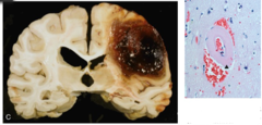
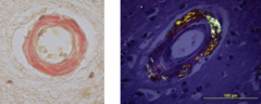
Congo red with apple-green birefringence under polarized light
What stain highlights amyloid in CAA?
Hemorrhagic parenchymal injury with edema from rapid tissue displacement
What is a cerebral contusion?
Orbitofrontal surfaces and temporal tips over rough skull base
Where are contusions most common and why?
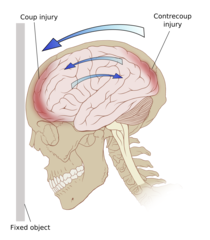
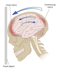
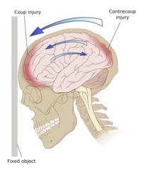
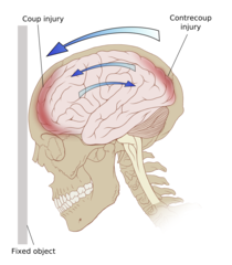
Contusion at the site of head impact
What is a coup injury?
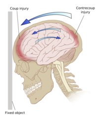
Contusion opposite the site of impact due to rebound
What is a contrecoup injury?
Wedge-shaped reddish purple lesion widest at point of impact
What are gross features of an acute contusion?
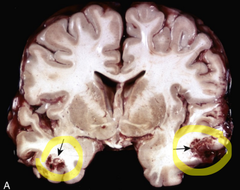
Neuronal injury and hemorrhage
What are microscopic features of an acute contusion?
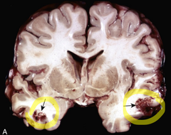
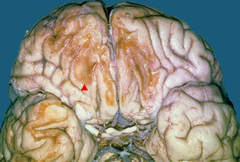
Yellow brown discoloration from hemosiderin with firmer tissue due to fibrosis
What are gross features of a remote contusion?
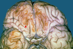
Hemosiderin deposition with neuronal loss and chronic inflammation with lymphocytes and macrophages
What are microscopic features of a remote contusion?
Eye opening verbal response and motor response
What does the Glasgow Coma Scale assess?
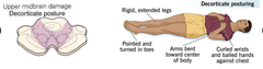
Upper midbrain damage with flexed arms and extended legs
What does decorticate posturing indicate anatomically?
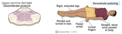
Upper pontine damage with extended rigid arms and legs
What does decerebrate posturing indicate anatomically?
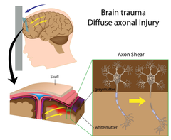
Widespread white matter tract injury from shearing between brain regions
What is diffuse axonal injury?
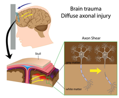
Coma due to widespread disconnection
What is a clinical consequence of diffuse axonal injury?
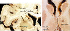
Small cavitary infarct due to occlusion of a single penetrating artery
What is a lacunar infarct?
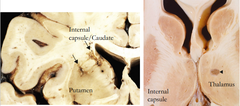
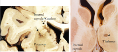
Basal ganglia, thalamus, internal capsule, deep white matter, and pons
Where do lacunar infarcts most commonly occur?
Yes they can be clinically silent or produce focal deficits
Can lacunar infarcts be asymptomatic?
Global brain dysfunction from malignant hypertension with raised intracranial pressure
What is acute hypertensive encephalopathy?
Headache confusion vomiting seizures and possible coma
What are clinical features of acute hypertensive encephalopathy?
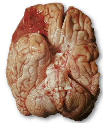
Embolism thrombosis and global ischemia
What are the main mechanisms leading to cerebral infarct?
Post-MI mural thrombus, atrial fibrillation, and valvular disease
What are common cardiac sources of cerebral emboli?
Via paradoxical embolism through a right-to-left shunt
How can a DVT cause a brain embolus?
Middle cerebral artery
Which artery is most frequently occluded by emboli?
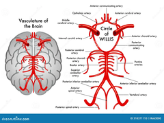
Carotid bifurcation, origin of MCA, and ends of basilar artery
Where do thrombotic occlusions most often form in the cerebrovasculature?
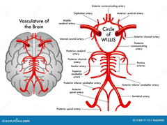
Severe hypotension in cardiac arrest or shock
What causes global cerebral ischemia without focal occlusion?
Infarct in border-zone regions most distal to arterial supply during global hypoperfusion
What is a watershed infarct?
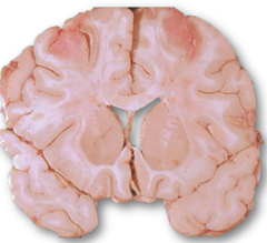
Red neurons with cytoplasmic eosinophilia and edema followed by neutrophil infiltration
What are early histologic changes after acute infarct?
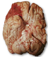
Liquefactive necrosis resolving to a cystic cavity with macrophage cleanup
What is the end stage of infarct evolution after weeks?
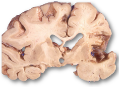
Penetrating arterioles develop atherosclerosis and occlude
How does hypertension lead to lacunar infarcts pathologically?
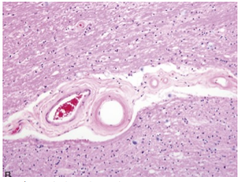
Sensory disturbances
What deficits can lacunar infarcts in the thalamus cause?
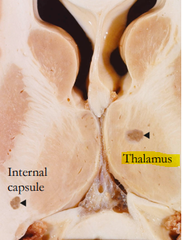
Internal capsule
What deep white matter tract is critical for motor and sensory transmission, and is often affected by lacunar infarcts?