Mandible and temporomandibular joint
1/35
There's no tags or description
Looks like no tags are added yet.
Name | Mastery | Learn | Test | Matching | Spaced |
|---|
No study sessions yet.
36 Terms
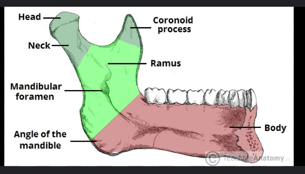
Mandible
The mandible consists of a horizontal part called the body and the vertical part called the ramus
Body of Manible (External surface)
The external surface is made up:
Mental foramen
Alveolar process
Alveolar yolk
Mental protuberance.
Mental foramen
Mental foramen- there are two mental foramina. The mental nerve and vessels exit through it.

Alveolar process
Alveolar process- this is the tooth-bearing area of the mandible.
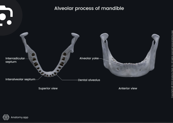
Alveolar yolk
These are a series of eminences corresponding to the position of the roots of the teeth.
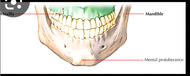
Mental protuberance
This forms the prominence of the chin.
Internal surface of body of mandible
Mylohoid line
Submandibular fossa
Mental spine
Sublingual fossa
Digastric fossa
Mylohyoid line
The attachment of the mylohoid muscle
Submandibular fossa
Inferior to the mylohoid line for the submandibular gland

Mental spine
Found at the midline

Sublingual fossa
Found on either sides of the mental spine for the sublingual gland
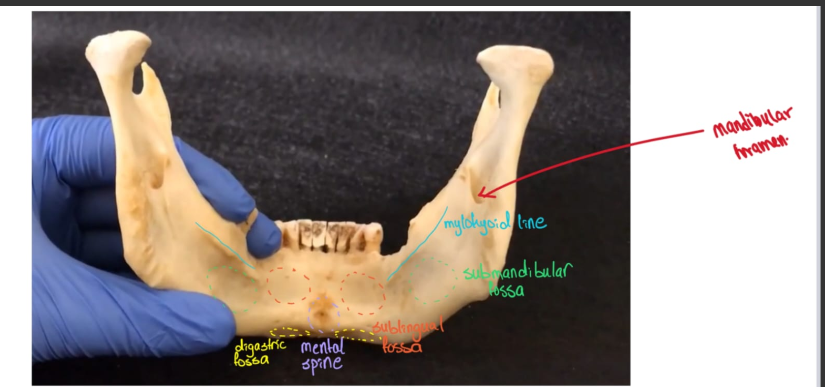
Digastric fossa
Found at the base of the mandible for the attachment of the anterior belly of the digastric muscle
External surface of ramus of mandible
Angle of Mandible
Massteric tuberosity
Coronoid process
Oblique line
Condylar process
Mandible notch
Angle of mandible
Junction between the body and the ramus of the mandible.
For the attachment of the masseter muscle and the stylomandibular ligament.
Masseteric tuberosity
For the attachment of the massester muscle
Coronoid process
for the attachment of the temporalis muscle.
Mandibular notch
Betwenn the coronoid and teh condylar process.
Internal surface of the ramus of the mandible
Condylar process
Neck of the mandible
Mandiblar foramen
Lingula
Mylohoid groove
Ptyergoid tuberosity,
Condylar process
This consists of the head of the mandible and the pterygoid fovea inferior to it.
The ptergoid fovea is the place of attachment of the lateral ptergoid muscle.


Neck of mandible

Mandibular foramen
paired, superior to the mandibular angle.
Content: Inferior alveolar vessels and nerve enter the mandibular canal through it. These supply the lower part of the teeth
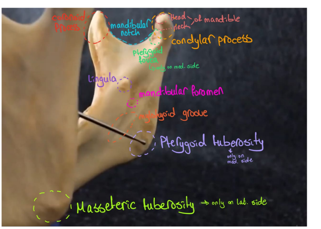
Lingula
for the attachment of the sphenomandibular ligament
Mylohoid groove
For the attachment of the mylohoid muscle
Pterygoid tuberosity
For the attachment of the medial pterygoid muscle
Temporomandibular joint
Type of joint: It is a paired, modified condyloid type of synovial joint.
Articular surface: The head of the mandible with the mandibular fossa of the temporal bone. There is an articular disc between the joint cavity.
Joint Capsule: is loose
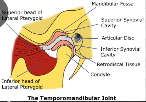
Articular disc
Articular disc: This divides the joint into 2 separate compartments this creates separate superior and inferior articular cavities, which are lined by 2 synovial membranes:
Superior synovial membrane: lines the fibrous layer of the capsule superior to the articular disc.
Inferior synovial membrane: inferior to the articular disc.
Ligaments
Lateral ligament:
Stylomandibular ligament:
Sphenomandibular ligament:
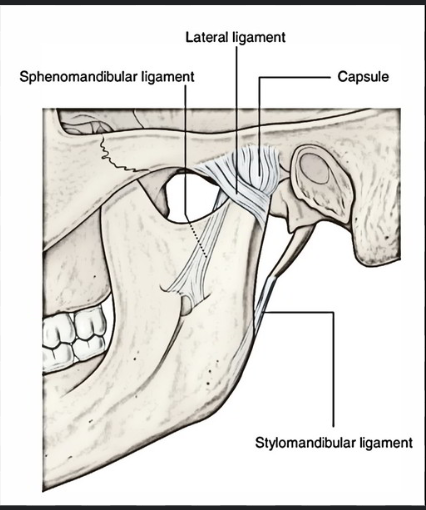
Lateral ligament
This ligament runs from the articular tubercle and attaches to the neck of the mandible
Acts to prevent posterior dislocation.
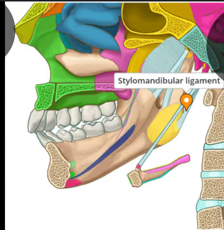
Stylomandibular ligament
This ligament runs from the styloid process to the angle of the mandible
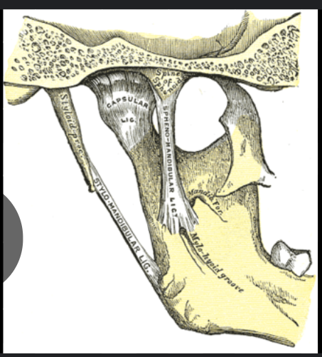
Sphenomandibular ligament
This runs from the spine of the sphenoid to the lingula of the mandible.
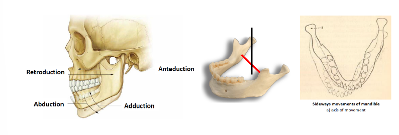
Movements:
Elevation: close mouth
Depression: open mouth
Protrusion: chin goes forward
Retrusion: chin goes backwards
Lateral movements: chewing
What muscles produce the TMJ movemnts
These are called the muscles of Mastication:
Temporalis
Masseter muscle
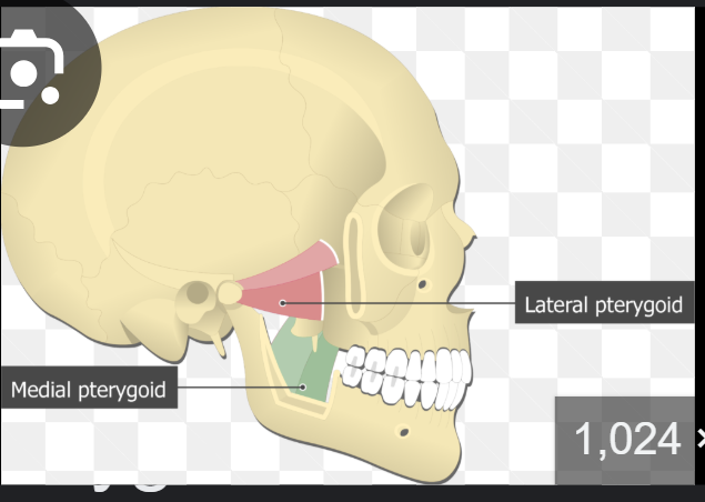
Medial and lateral pteygoid muscle
Which muscle is the only opener
the lateral ptergoid muscle is the only opener,
The other muscles close the jaw.
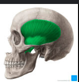
What muscle fills up the temoporal fossa entirely?
Temporalis muscle
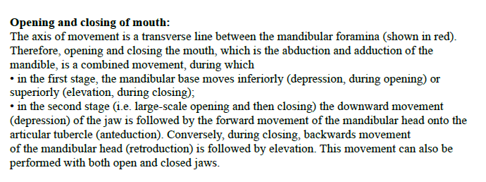
Opening and closing of the mouth
The axis of movement is a transverse line between the manibulae foramina.
Therefore, opening and closing of the mouth which is the abduction and adduction of the mandible is a combined movement during which:
In the first stage, the mandibular base moves inferiorly (depression during opening) or superiorly (elevation during closing).
In the second stage, the downward movement of the jaw is followed by the forward movement of the mandibular head onto the articular tubercle.
Coversely, during closing, the backward movement of the mandibular head into the mandibular fossanand this is followed by elevation.
This movement can also be performed with bothe open and closed jaws.
Note: Anteduction is the same as protrusion/protraction
Retroduction is the same as retrusion/retraction

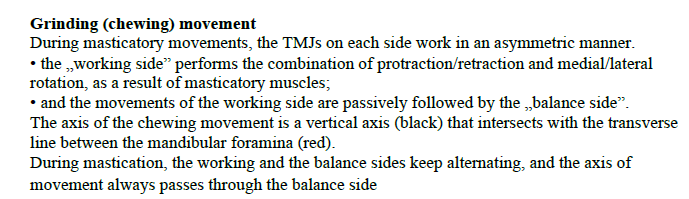
Grinding (chewing) movement
During masticatory movements, the TMJ on each side work in an asymmetric manner.
The working side performs the combination of protraction/retraction (protrusion/retrusion) and medial/lateral rotation as a result of the masticatory muscles.
And the movements of the working side are passively followed by the balance side.
The axis of the chewing movement is a vertical axis that intersects with the transverse line between the mandibular foramina.
During mastication, the working and balance sides keep alternating and the axis of the movement always passes through the balance side.
Note: Anteduction is the same as protrusion/protraction
Retroduction is the same as retrusion/retraction
