Pelvic Structures
 Call Kai
 Learn
Learn Practice Test
 Spaced Repetition
 Match
Match Flashcards
 Knowt Play
1/51

There's no tags or description
Looks like no tags are added yet.
Name | Mastery | Learn | Test | Matching | Spaced | Call with Kai | Chat |
|---|
No analytics yet
Send a link to your students to track their progress
52 Terms
The bony pelvis consists of the —
sacrum, coccyx, and innominate bones
The posterior border of the pelvic cavity is marked by the —
sacrum and coccyx
The innominate bones consist of the —
ilium, ischium, and pubic symphysis
The boundaries of the female pelvis are considered to be from the iliac crest to a group of muscles known as the —, located at the base of the pelvis.
pelvic diaphragm
The pelvis can further be divided into a true pelvis (lesser pelvis) and false pelvis (major pelvis) by an imaginary line known as the —
linea terminalis
The false pelvis is located more — than the true pelvis,
superiorly
The true pelvis contains the —
urinary bladder, small bowel, sigmoid colon, rectum, ovaries, fallopian tubes, and uterus.
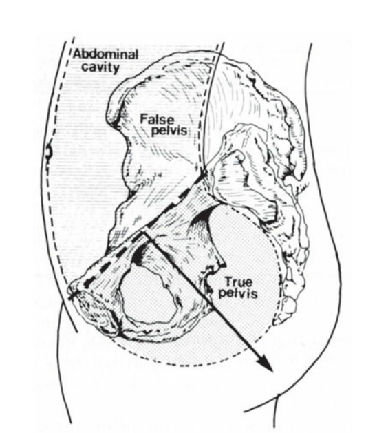
Relationship of the abdominal cavity to the true and false pelvis.
Muscles visualized on a sonogram include the
rectus abdominis muscles, the iliopsoas muscles, obturator internus muscles, piriformis muscles, and a group of muscles known as the pelvic diaphragm, which is composed of the levator ani and coccygeus muscles
The pelvic diaphragm muscles provide
support to the pelvic organs
The weakening of the pelvic diaphragm, which includes the levator ani muscles, can lead to
prolapse of the uterus.
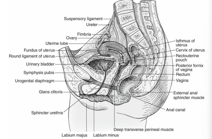
Organs of the female pelvis in the sagittal section.
The broad ligaments and suspensory ligament of the ovary are actually
double folds of peritoneum
In addition to providing support, the — of the ovary contains the ovarian artery, ovarian vein, lymphatics, and ovarian nerves.
suspensory ligament
cardinal ligaments house the
vasculature of the uterus
The vesicouterine pouch, or anterior cul-de-sac, is located —
anterior to the uterus and posterior to the urinary bladder
The rectouterine pouch, located between the —, may also be referred to as the posterior cul-de-sac or pouch of Douglas
rectum and uterus
— is considered the most dependent part of the peritoneal cavity, making it the most likely place for fluid to collect in the pelvis
The rectouterine pouch
Between the anterior wall of the urinary bladder and symphysis pubis lies the —
space of Retzius, or retropubic space
the space of Retzius, or retropubic space is an area that contains
extraperitoneal fat.
Rectus abdominis muscle (2) location
anterior
Iliopsoas muscles (2) location
Lateral and anterior to iliac crest
Obturator internus (2) location
Lateral to ovaries
Piriformis (2) location
Posterior
Pelvic diaphragm (levator ani and coccygeus muscles) location
Inferior near the vagina in transverse
The most dependent part of peritoneal cavity is the —, making it the most likely place for free fluid to collect in the pelvis.
posterior cul-desac or pouch of Douglas
The abdominal aorta supplies blood to —
the female genitalia.
The paired right and left uterine arteries are branches of the —
internal iliac arteries.
The paired right and left uterine arteries supply blood to the uterus, the fallopian tubes, and the ovaries, and course along the —
lateral aspect of the uterus within the folds of the broad ligaments
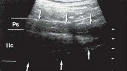
Sagittal scan of the psoas major (Ps) and the iliac muscle (Ilc).
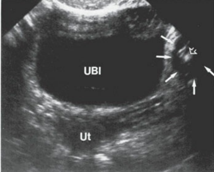
Transverse scan of the pelvis reveals the urinary bladder (UBl), uterus (Ut), and left iliopsoas muscle (arrows).
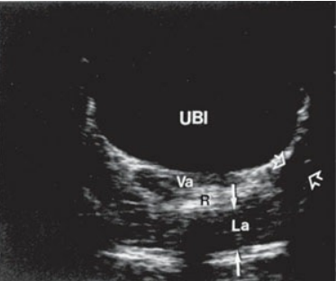
Transverse scan at the level of the vagina (Va), urinary bladder (UBl), and rectum (R) demonstrating the levator ani (La) muscle group (solid white arrows) and the obturator internus muscle (open arrows).
Branches of the uterine artery include the —, which may be visualized with Doppler interrogation along the lateral aspect of the myometrium
arcuate arteries
The arcuate vessels progress further within the uterus and eventually become the —, which supply blood to the deeper layers of the myometrium.
radial arteries
The radial arteries then divide into the —
straight arteries and spiral arteries
The spiral arteries are the tiny, coiled vessels that supply blood to the —
functional or decidual layer of the endometrium
The ovarian arteries originate from the —
lateral aspect of the abdominal aorta
Each ovary receives its nourishment from an —
ovarian artery and a branch of the uterine artery
broad ligament supports
uterus
tubes
ovaries
broad ligament location
Extends from the lateral aspect of the uterus to the side walls of the pelvis
round ligaments support
uterus (fundus)
round ligaments location
Extends from uterine cornua to labia majora between the folds of the broad ligaments
the suspensory ligament of the ovaries (infundibulopelvic) supports
ovaries and tubes
the suspensory ligament of the ovaries (infundibulopelvic) location
Extends from the ovaries to the pelvic side walls
ovarian ligaments support
ovaries
ovarian ligaments location
Extends from ovary to lateral surface of the uterus
cardinal ligament supports
cervix
caerdinal ligament location
Extends from the lateral surface of the cervix to the lateral fornix of vagina
uterosacral ligament supports
uterus
uterosacral ligament location
Extends from uterus to sacrum
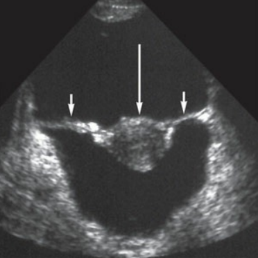
Broad ligament outlined by ascites. The bilateral broad ligaments (short arrows), outlined by ascites, are clearly noted in this transverse image. The longer arrow is identifying the uterus.
All venous structures mirror their arterial counterparts with the exception of the left ovarian vein, which instead of returning blood to the inferior vena cava, drains directly into —
the left renal vein