lower extremity Muscles: Human Anatomy
 Call Kai
 Learn
Learn Practice Test
 Spaced Repetition
 Match
Match Flashcards
 Knowt Play
1/41

There's no tags or description
Looks like no tags are added yet.
Name | Mastery | Learn | Test | Matching | Spaced | Call with Kai | Chat |
|---|
No analytics yet
Send a link to your students to track their progress
42 Terms

Anterior View
Muscles
Name: Tensor fascia latae
Description: Helps keep knee extended during upright posture. Keeps pelvis level when opposite leg is raised
O: Anterior external lip of iliac crest and the ASIS
I: IT band which attaches to lateral condyle of the tibia
A: Assists in ABduction and medial rotation of the thigh
N: Superior Gluteal nerve
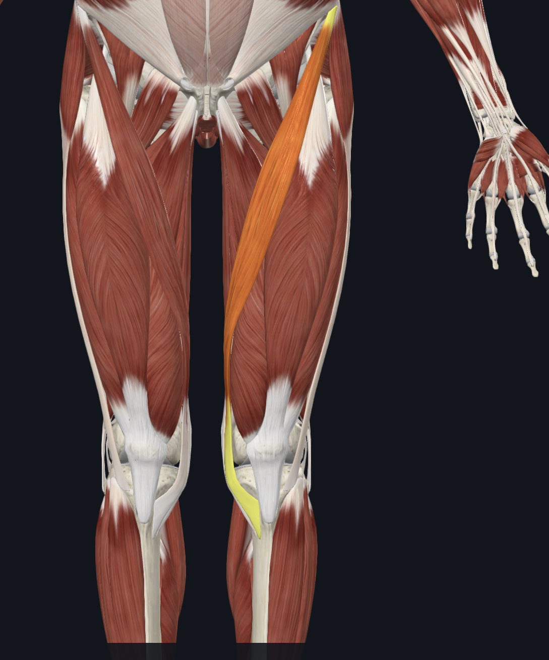
Anterior view
Muscles
Name: Sartorius
Description: Part of thigh. Most superficial muscle of ant. thigh. Seatbelt. Not affected by position of knee
O: ASIS
I: Pes anserine
A: main action is hip flexion; can produce ABduction and external rotation
N: Femoral Nerve

Anterior View
Muscles
Name: Rectus femoris
Description: Power as a hip flexor. Only portion of the quad that passes the hip and the knee. Optimal length for hip flexor is when knee is flexed
O: AIIS
I: Common tendon to tibial tuberosity
A: assists illiopsoas in hip flexion and extends the knee with the quad.
N: Femoral Nerve
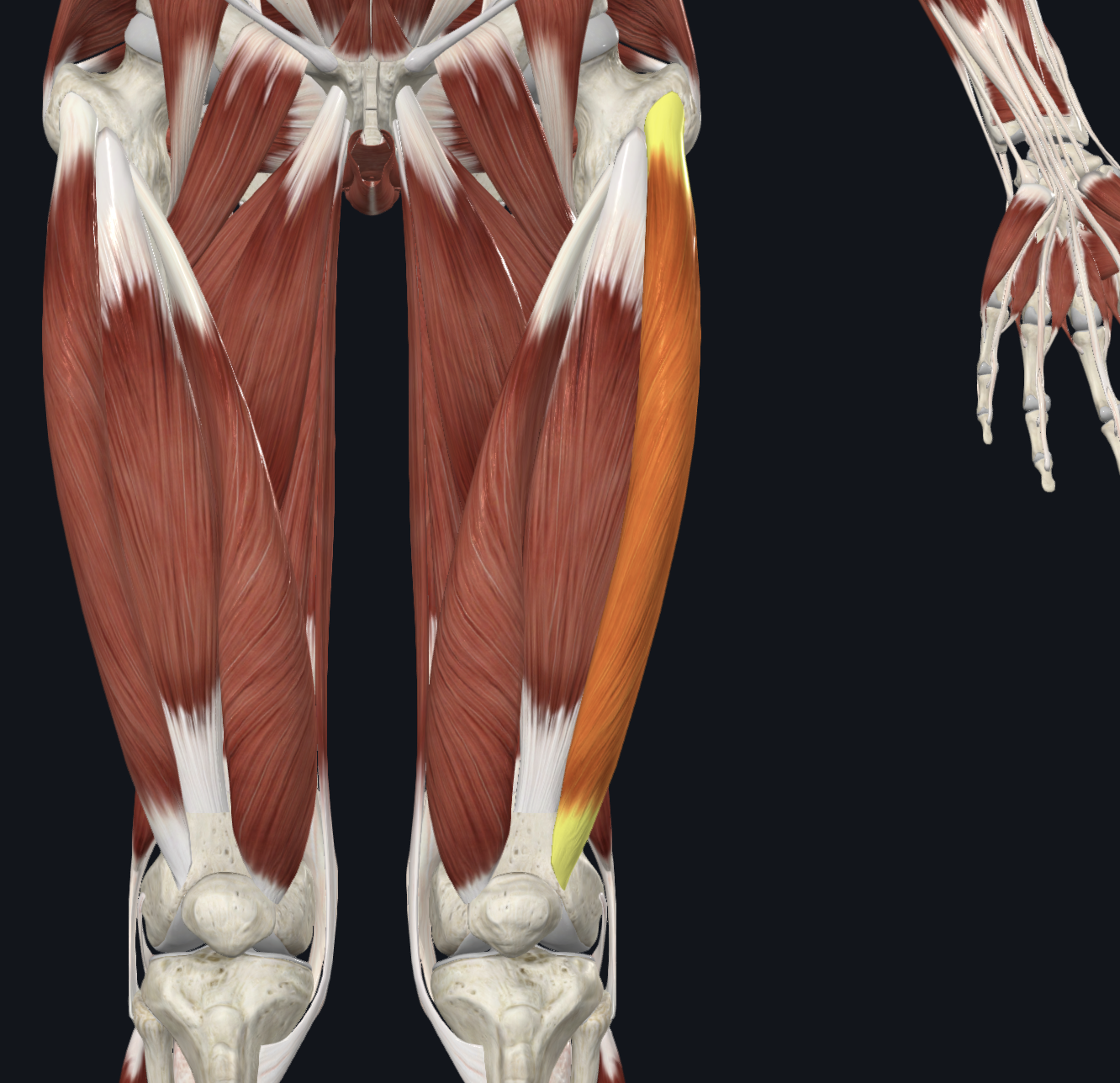
Anterior view
Muscles
Name: vastus lateralis
Description:
O: Greater trochanter and lateral lip of linea aspera
I: Common tendon to tibial tuberosity
A: extends knee
N: femoral nerve

Anterior view
Muscles
Name: vastus medialis
Description:
O: Medial lip of linea aspera
I: Common tendon to tibial tuberosity
A: extends knee
N: femoral nerve
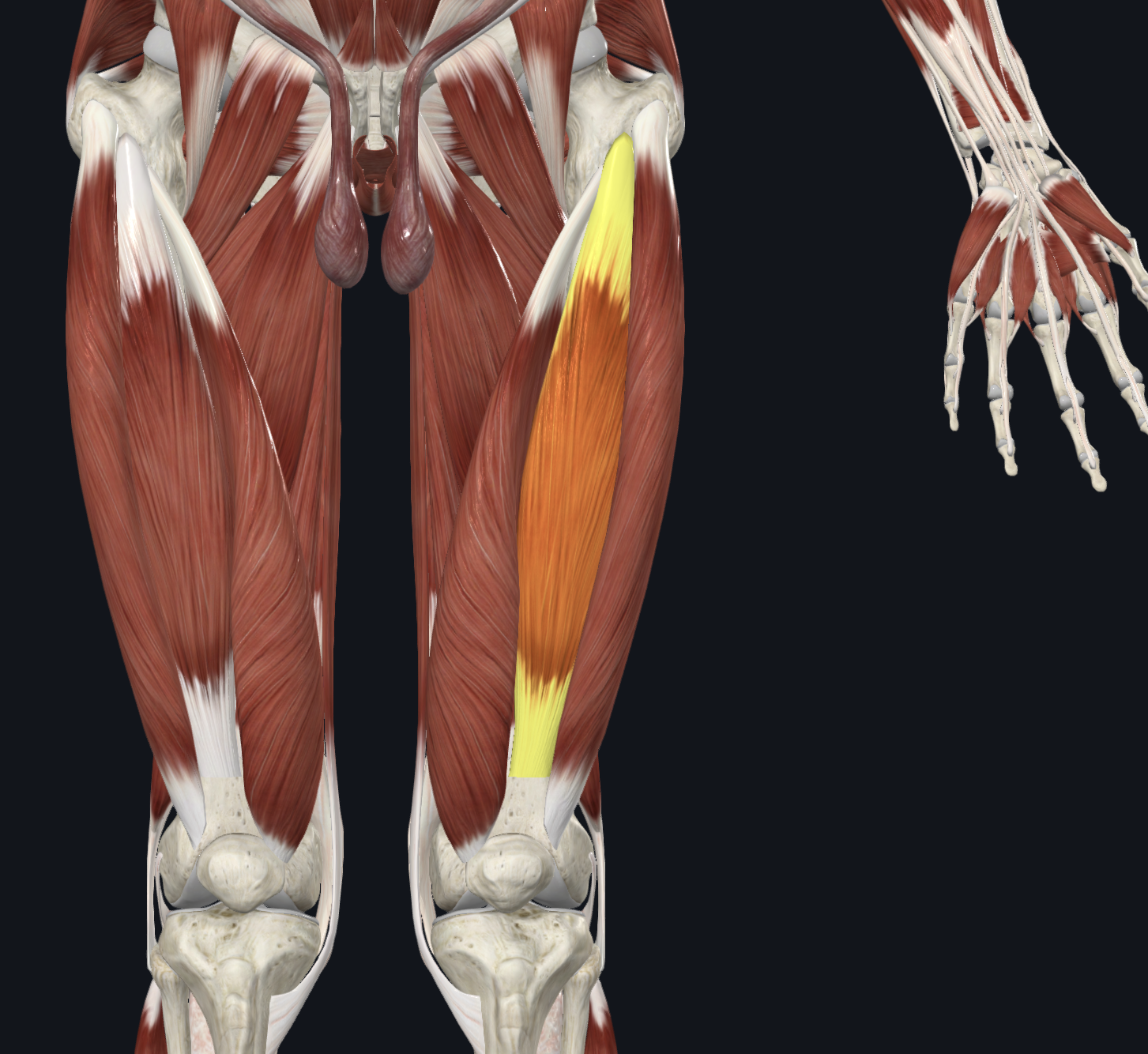
Anterior view
Muscles
Name: vastus Intermedius
Description:
O: anterior and lateral surface of the femur
I: Common tendon to tibial tuberosity
A: extends knee
N: femoral nerve
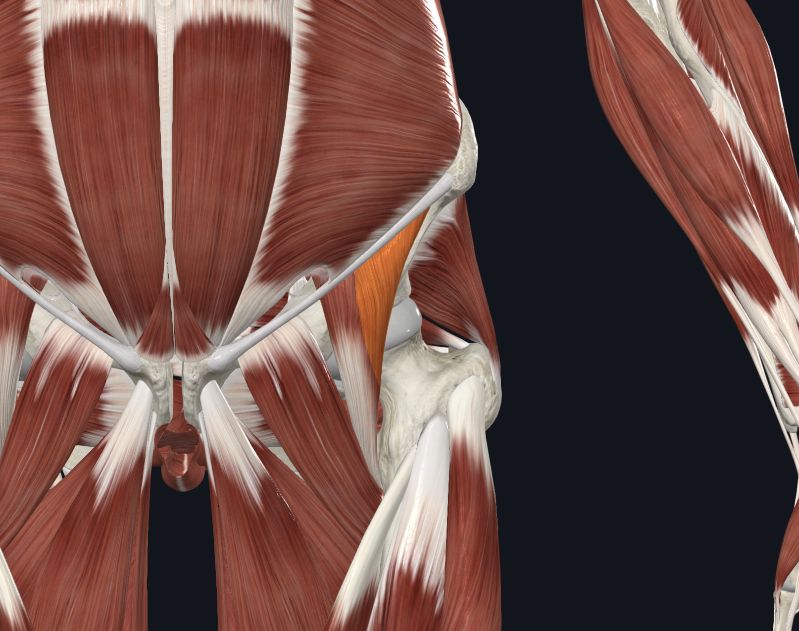
Anterior view
Muscles
Name: Illiacus part of the iliopsoas
Description:
O: Iliac fossa, illiac crest, ala of sacrum
I: lesser trochanter of the femur
A: hip flexion
N: femoral nerve
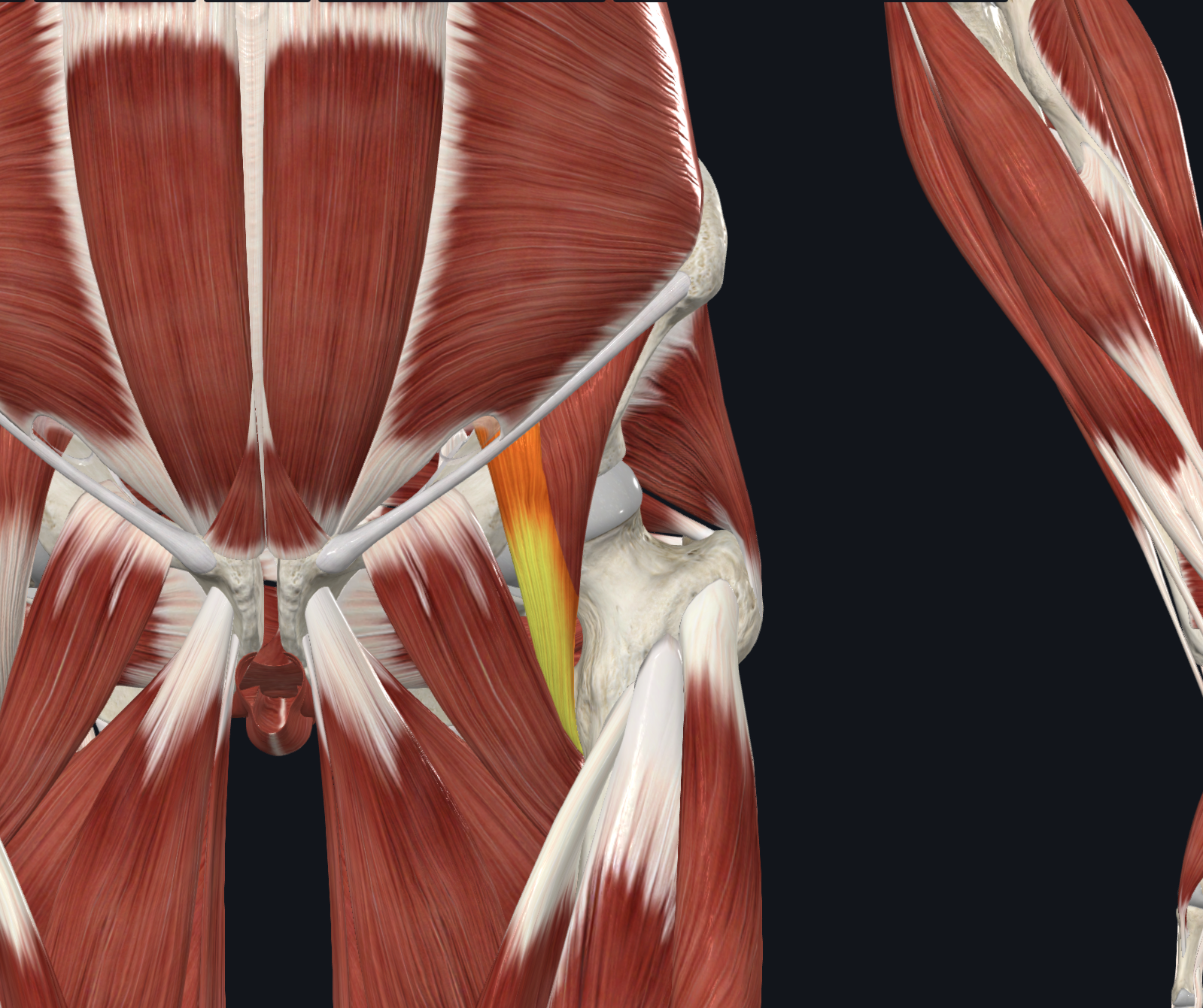
Anterior view
Muscles
Name: Psoas Major part of the iliopsoas
Description:
O: Transverse processes of lumbar vertebrae T12-L-5
I: lesser trochanter of the femur
A: hip flexion for lateral rotation
N: Anterior rami of the lumbar nerves L1, L2, L3

Anterior view
Muscles
Name: Pectineus
Description:
O: Superior ramus of pubis
I: Pectineal line of femur inferior to lesser trochanter
A: adduction and flexion in the thigh> Assists with medial rotation
N: femoral nerve

Anterior view
Muscles
Name: Adductor Longus
Description:
O: Body of pubis inferior to crest
I: Middle third of linea aspera
A: Adduction and flexion in the thigh. Assists with medial rotation
N: Obturator Nerve
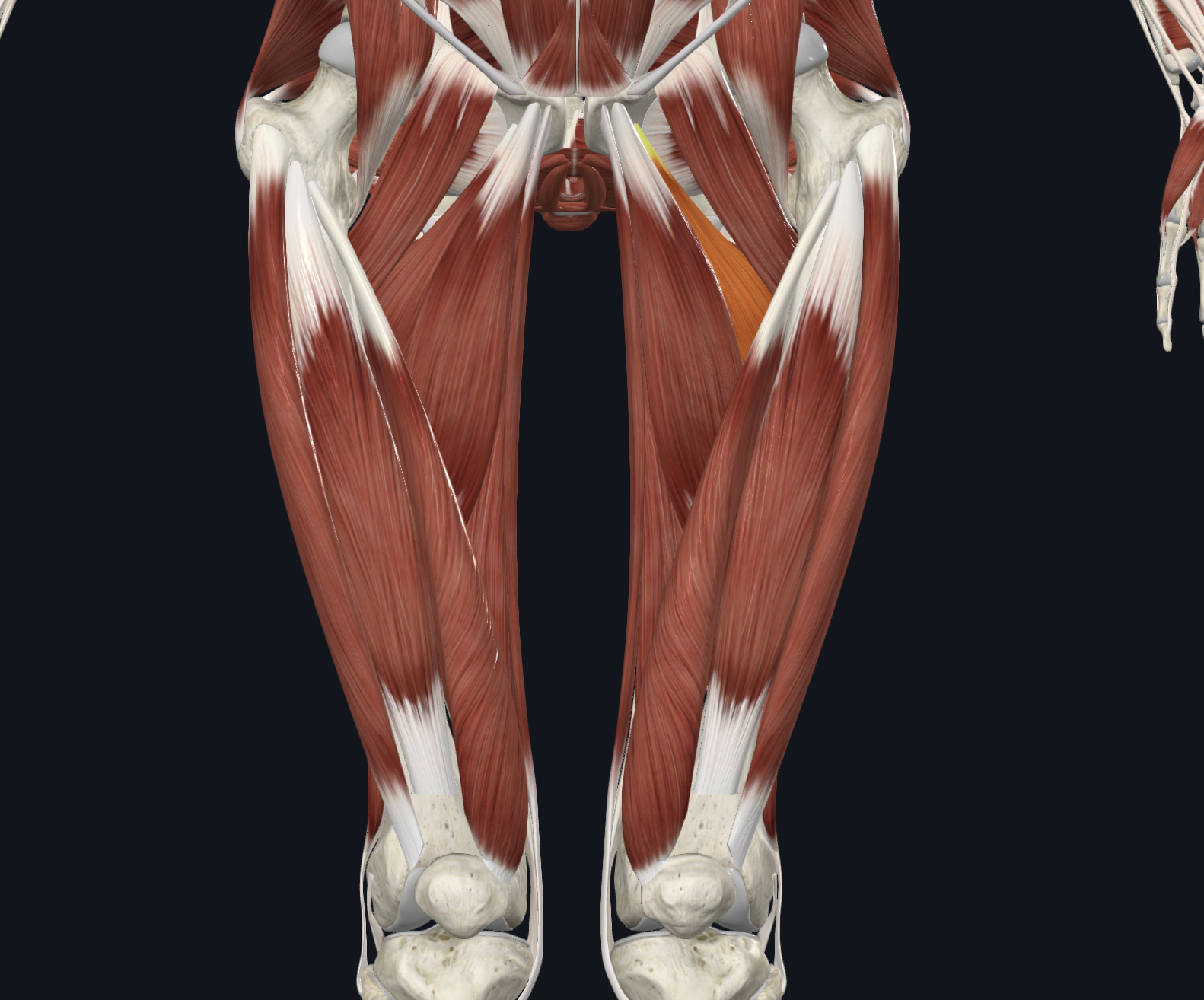
Anterior view
Muscles
Name: Adductor brevis
Description:
O: Body and inferior ramus of pubis
I: Pectineal line of femur, proximal linea aspera
A: Adduction and flexion in the thigh. Assists with medial rotation
N: Obturator Nerve

Anterior view
Muscles
Name: Adductor magnus
Description:
O: Inferior ramus of pubis nad ramus of ischium
I: Adductor part: Gluteal tuberosity, linea aspera, and medial supracondylar line.
Hamstring part: Adductor tubercle
A: Adduction and flexion in the thigh. Assists with medial rotation
N: Adductor part: Obturator Nerve
Hamstring part: tibial portion of the Sciatic Nerve

Anterior view
Muscles
Name: Gracilis
Description:
O: Body and inferior ramus of pubis
I: Pes Anserine
A: Adduction and flexion in the thigh. Assists with medial rotation
N: Adductor part: Obturator Nerve
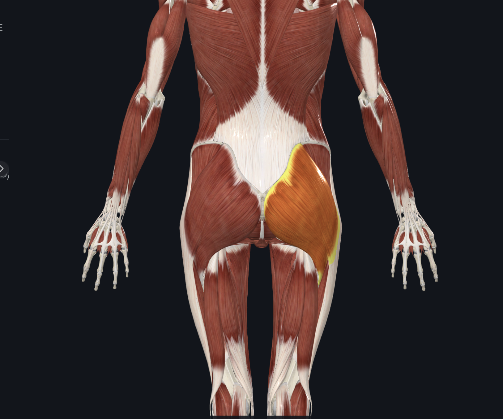
Posterior view
Muscles
Name: Gluteus maximus
Description:
O: Posterior ilium and sacrum and associated ligaments
I: Superior fibers insert into ITB inferior fibers into gluteal tuberosity on the femur
A: Hip extension, external rotation
N: inferior gluteal nerve

Posterior view
Muscles
Name: Gluteus medius
Description:
O: external surface of ilium
I: Greater trochanter of femur
A: hip abduction
N: Superior gluteal nerve
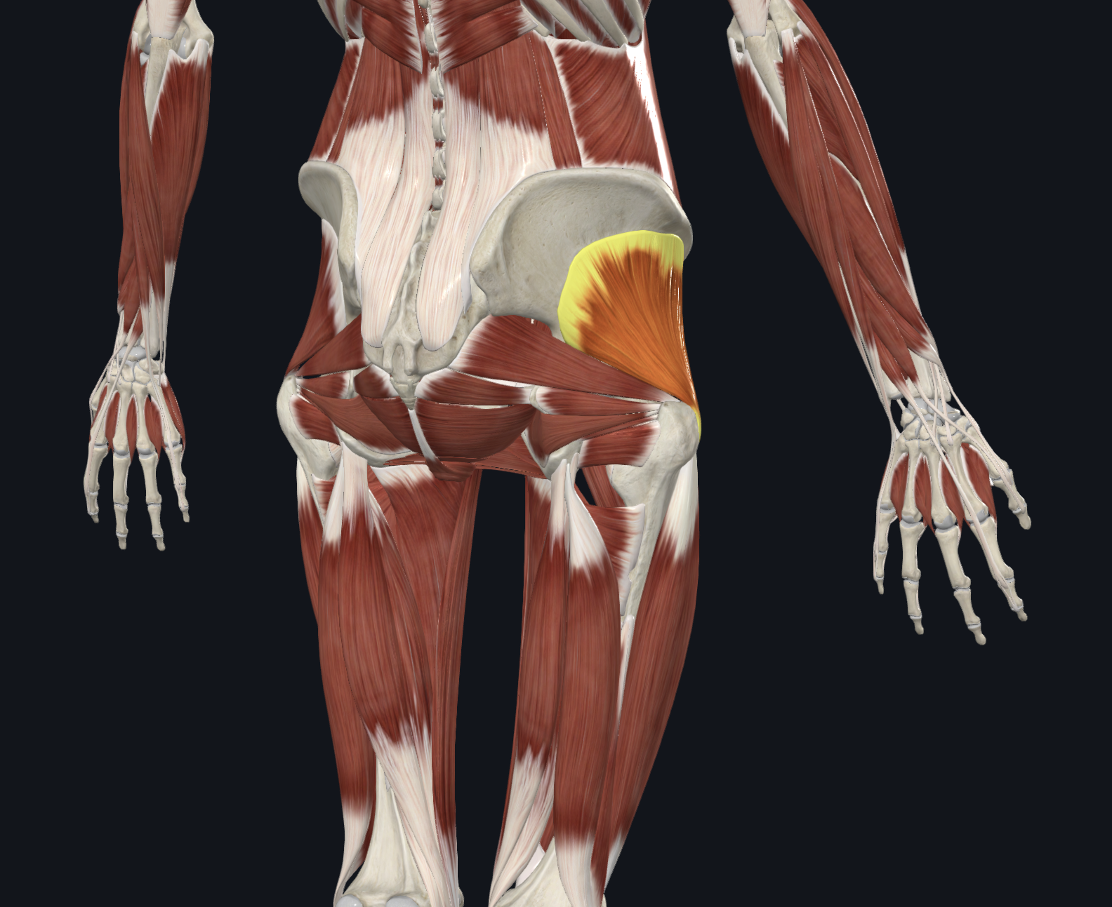
Posterior view
Muscles
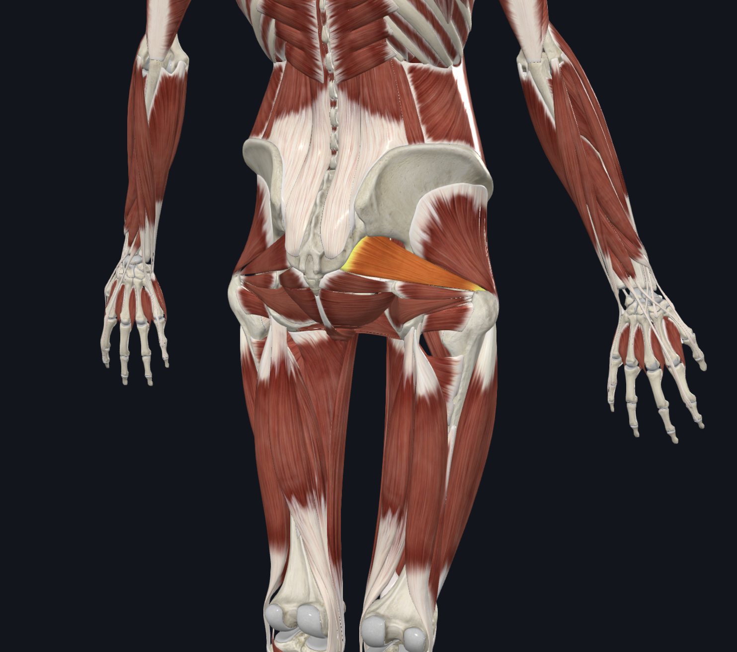
Name: Gluteus minimus
Description:
O: external surface of ilium
I: Greater trochanter of femur
A: hip abduction
N: Superior gluteal nerve
Posterior view
Muscles
Name: Piriformis
Description:
O: Anterior sacrum
I: Greater trochanter of femur
A: Laterally rotates extended thigh, abducts a flexed thigh
N: Nerve to Piriformis

Posterior view
Muscles
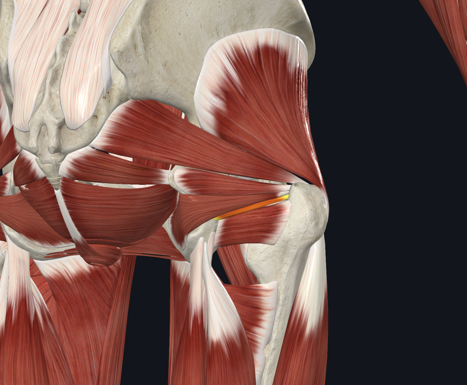
Name: Superior gemellus
Description:
O: ischial spine
I: Greater trochanter of femur
A: Laterally rotates thigh, abducts thigh when flexed
N: Nerve to Obturator internus
Posterior view
Muscles
Name: Inferior gemellus
Description:
O: ischial tuberosity
I: Greater trochanter of femur
A: Laterally rotates thigh
N: Nerve to Quadratus Femoris
Posterior view
Muscles
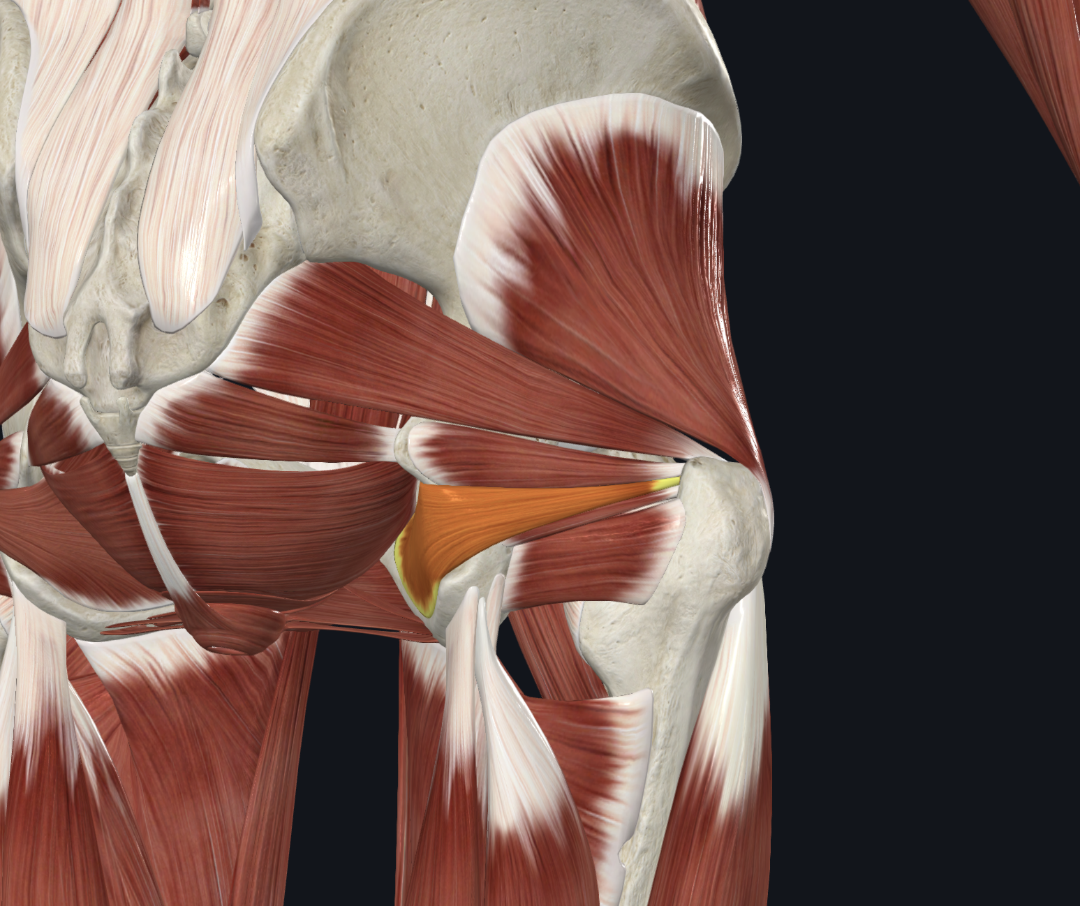
Name: Obturator internus
Description:
O: Posterior margins of obturator foramen, obturator membrane
I: Greater trochanter of femur
A: Laterally rotates thigh, abducts thigh when flexed
N: Nerve to obturator internus
Posterior view
Muscles
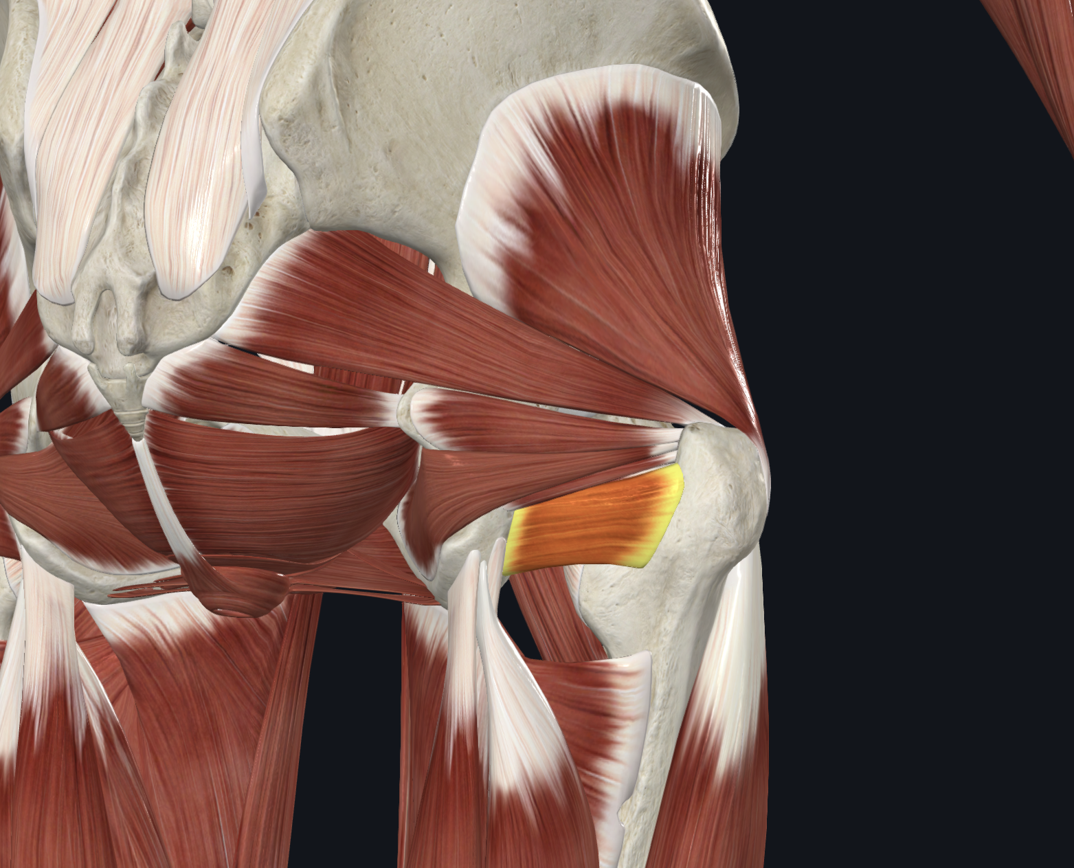
Name: Quadratus femoris
Description:
O: Lateral aspect of ischial tuberosity
I: Greater trochanter of femur
A: Laterally rotates thigh
N: Nerve to Quadratus Femoris

Posterior view
Muscles
Name: Biceps femoris Long and short head
Description:
O: Long head- ischial tuberosity
O: Short head-lateral linea aspera
I: fibular head
A: flexes knee and extends hip
N: Long head- tibial nerve
N: Short head-comon peroneal

Posterior view
Muscles
Name: Semitendinosus
Description:
O: ischial tuberosity
I: pes anserine
A: flexes knee and extends hip
N: sciatic nerve

Posterior view
Muscles
Name: Semimembranosus
Description:
O: ischial tuberosity
I: Medial condyle of the tibia
A: flexes knee and extends hip
N: sciatic nerve

Posterior view
Muscles
Name: adductor magnus
Description:
O: ischial tuberosity
I: Medial condyle of the tibia
A: flexes knee and extends hip
N: sciatic nerve

Transverse view
Name: Pes Anserine
Descripton: Attatchment site for semitendinosus, sartorius and gracilis

Posterior view
Muscles
Name: Popliteus
Description:
O: Anterior lateral condyle of femur
I: posterior surface of tibia near soleal line
A: internal rotation of knee joing, unlocks extended knee, weak knee flexor
N: tibial nerve

Posterior view
Muscles
Name: Gastrocnemius
Description:
O: Posterior parts of medial and lateral femoral condyles
I: achilles tendon
A: Primary ankle plantarflexor, secndary knee flexor
N: tibial nerve

Posterior view
Muscles
Name: Plantaris
Description:
O: distal end of lateral supracondylar line
I: posterior calcaneus
A: works with gastrocnemius to plantarflex the ankle and assist in flexion of the knee
N: tibial nerve

Posterior view
Muscles
Name: Soleus
Description:
O: Posterior surfaces of the head of the fibula and soleal line
I: posterior calcaneus
A: Plantarflexor of ankle
N: tibial nerve

Posterior view
Muscles
Name: Calcaneal (achilles) tendon
Description:
O: Posterior surfaces of the head of the fibula and soleal line
I: posterior calcaneus
A: Plantarflexor of ankle
N: tibial nerve

Posterior view
Muscles
Name: Flexor hallucis longus
Description:
O: Posterior surface of distal 2/3 of fibula and interosseus membrane
I: Base of distal phalanx of great toe (plantar surface)
A: flexes the great toe
N: tibial nerve

Posterior view
Muscles
Name: Flexor digitorum longus
Description:
O: Middle 3/5 of posterior surface of body of tibia
I: Base of distal phalanges of digits 2-5
A: Flexes proximal and distal interphalangeal and metatarsophalangeal joints of digits 2-5, assists with plantarflexion
N: tibial nerve

Posterior view
Muscles
Name: Tibialis posterior
Description:
O: Most of interosseus membrane, lateral portion of posteiror surface of tibia, proximal 2/3 of medial surface of fibula
I: Navicular tuberosity, cunieforms, cuboid and the bases of the 2-4 metatarsals
A: plantarflexes and inverts the ankle
N: tibial nerve

sagittal View
Muscles
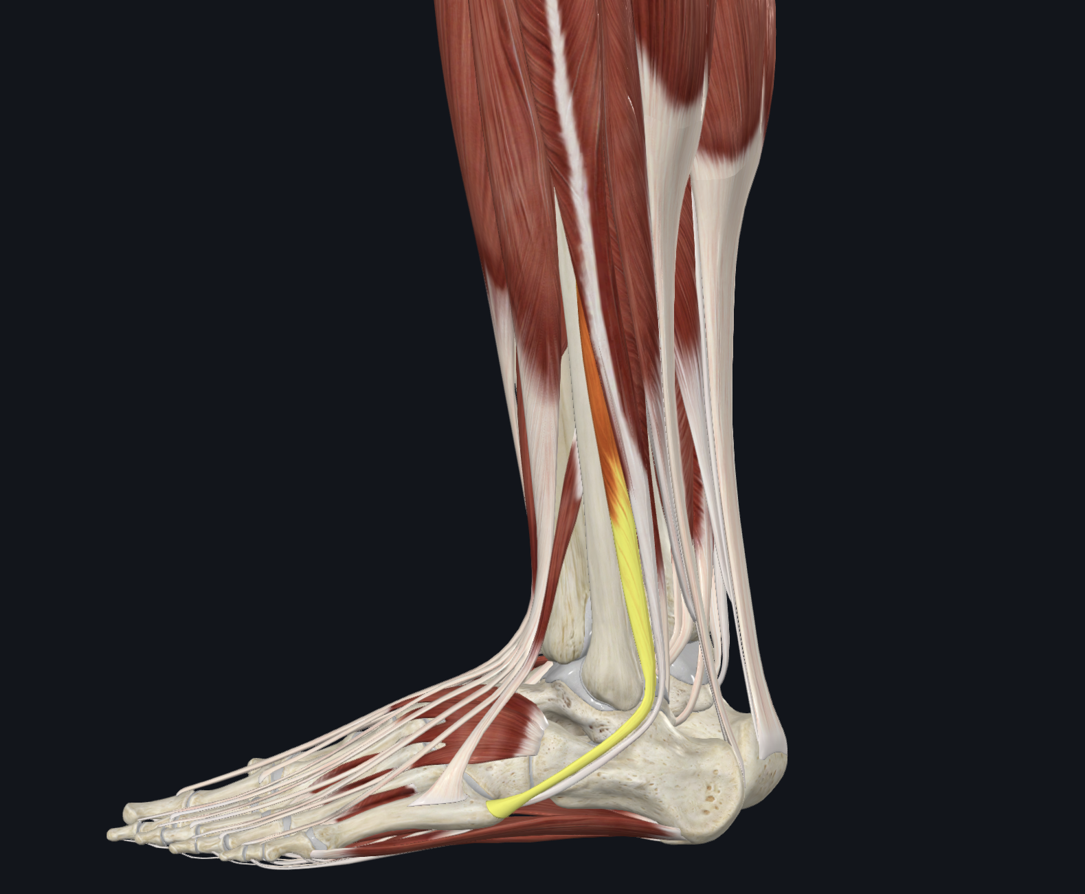
Name: Peroneus Longus
Description:
O: Head and proximal 2/3 of lateral surface of fibula
I: Lateral side of the base of 1st metatarsal and of medial cuneiform
A: everts foot, assists in plantarflexion, depresses 1st metatarsal
N: superficial peroneal
sagittal View
Muscles
Name: Peroneus Brevis
Description:
O:distal 2/3 of the lateral surface of the fibula
I: Tuberosity of the 5th metatarsal
A: Everts foot and assists in plantarflexion
N: Superficial peroneal

Anterior view
Muscles
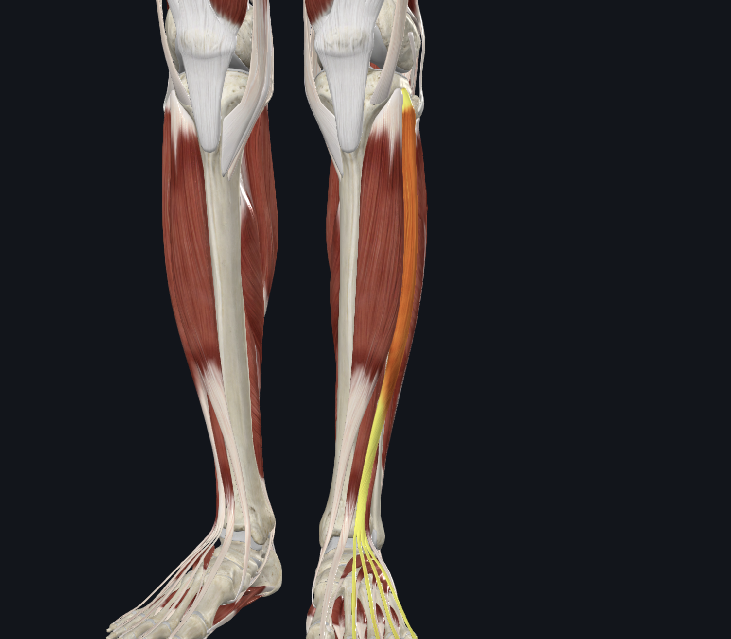
Name: Tibialis anterior
Description:
O: Lateral condyle and proximal ½ of lateral surface of tibia
I: medial and plantar surface of medial cuneiform and base of 1st metatarsal
A: Dorsiflexes and inverts the ankle
N: Deep peroneal
Anterior view
Muscles
Name: Extensor digitorum longus
Description:
O: Lateral condyle of tibia, proximal ¾ of anterior surface of the body of fibula and the interosseus membrane
I: by four tendons to the digits 2-5 attached to base of middle and distal phalanges
A: Extends metatarsophalangeal joins and assists extending the interphalangeal joints of digits 2-5, dorsiflexes the ankle
N: Deep peroneal

Anterior view
Muscles
Name: peroneus tertius
Description:
O: Distal 1/3 of anterior surface of the fibula
I: dorsal surface, base of the 5th Metatarsal
A: elevates the base of the 5t metatarsal, dorsiflexes and everts the foot
N: Deep peroneal

Anterior view
Muscles
Name: Extensor hallucis longus
Description:
O: Middle two quarters of the anterior surface of the fibula
I: base of the distal phalanx of the great toe
A: extends the metatarsalphalangeal and interphalangeal joints of the great toe
N: Deep peroneal

transverse superior view
Muscles
Name: Extensor digitorum longus
Description:
O: Lateral condyle of tibia, proximal ¾ of anterior surface of the body of fibula and the interosseous membrane
I: By four tendons to the digits 2-5 attached to base of middle and distal phalanges
A: extends the metatarsophalangeal joints and assists in extending the interphalangeal joints of digits 2-5, dorsiflexes the ankle
N: Deep peroneal
anterior view
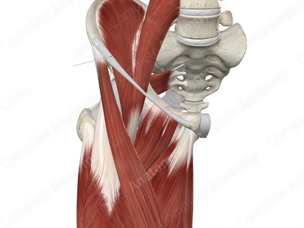
Name: inguinal ligament
Description: attaches to anterior superior iliac spine (ASIS) of the pelvis to the pubic tubercle of the pubic bone