muscoskeletal system
1/68
There's no tags or description
Looks like no tags are added yet.
Name | Mastery | Learn | Test | Matching | Spaced |
|---|
No study sessions yet.
69 Terms
Axial Skeleton
The part of the skeleton that includes the skull, vertebral column, and rib cage.
Appendicular Skeleton
The part of the skeleton that includes the limbs and girdles (shoulder and pelvic girdles).
Primary ossification center
The first location in a developing bone where ossification (bone formation) occurs.
Secondary ossification center
Locations in bones where ossification occurs after the primary center, usually in the epiphyses.
function of the skeleton
• Protection: Encloses internal organs

• CNS, Cardio, Resp, Repro...

• Support: Rigid structural framework

• Movement: Anchors skeletal muscle

• Mineral Storage: homeostasis

• Blood cell production: Red bone marrow

• Fat (energy storage): Yellow bone marrow
Blood Cell production and energy storage as a
function of the skeleton
Bone Marrow is a dynamic semi-solid tissue found within bone
Red Marrow: high number of haematopoietic stem cells (blood cell formation)
• Erythrocytes, monocytes, neutrophils, etc
• Located in spongy/trabecular bone, in ends of bones or flat bones
• Yellow Marrow: high number of adipocytes (fat cells) for energy/fat storage
> Located in medullary cavity of long bones
Collagen fibers
Organic substances produced by the body that provide strength and flexibility in bone.
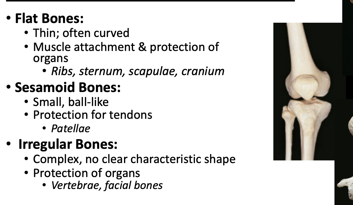
bone types
Describes bone shape and not length
Long bones in arms or legs
Short bones mainly in wrist and hands or ankle
Skull bones and ribs/sternum are flat
Sesamoid bones form from within tendon, eg is patella
Irregular is vertebra
Long bones
Bones that are longer than they are wide, such as those found in the arms and legs.
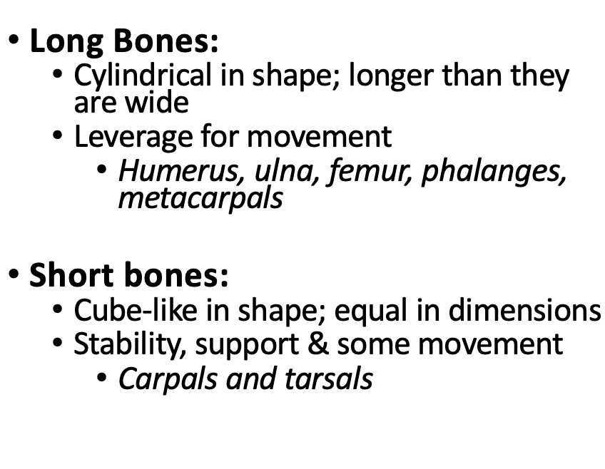
Short bones
Bones that are approximately equal in length and width, such as those in the wrist and ankle.
Irregular bones
Bones that have complex shapes, such as vertebrae.
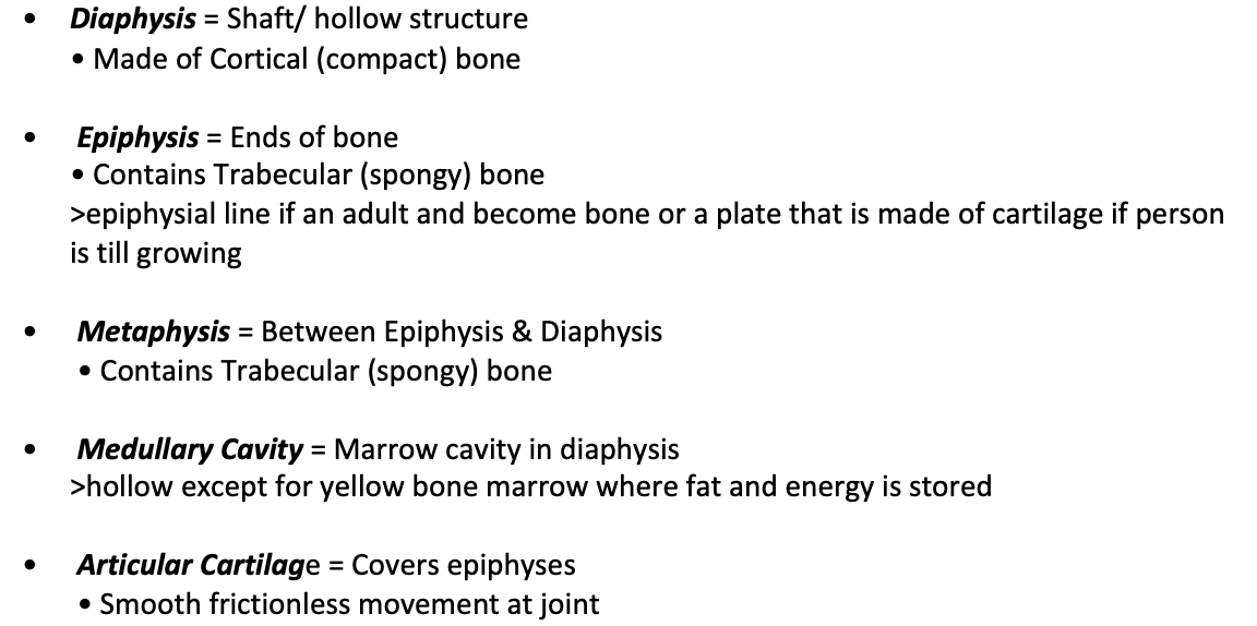
Cortical bone
Also known as compact bone, it's the dense outer layer of bone.
Prominent composition/type in diaphysis (shaft of long bone)
• Contains Osteons (how bone is built)
> Main unit of compact bone microstructure
• Strength in uniform direction due to being compact
Trabecular bone
Also known as spongy bone, it's characterized by a lattice-like structure.
Prominent composition/type in heads of long bone & other bone structures (flat, irregular, etc)
• Contains Trabeculae (bony struts)
• Strength in multiple directions bc of latticework and space In/ around it, has red bone marrow which is essential for the making of blood cells (haematopoiesis)
gross anatomy of the long bone
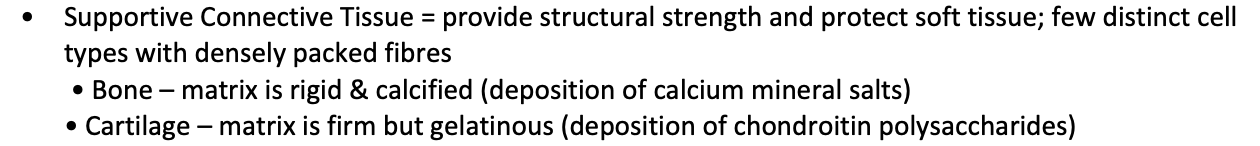
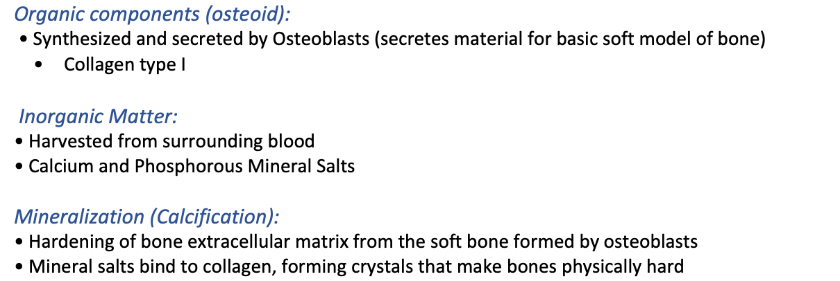
bone as a suppourtive connective tissue/ its composition
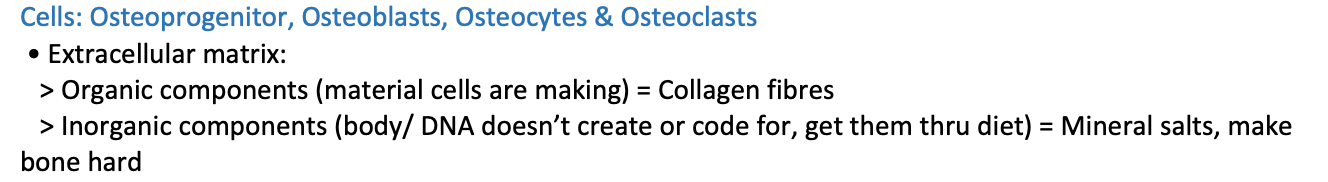
Bone extracellular matrix
=
Osteons
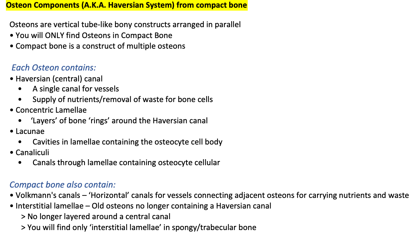
The structural unit of compact bone, consisting of a central Haversian canal surrounded by concentric lamellae.
Haversian canal
Central canal in an osteon that contains blood vessels and nerves.
Lacunae
Small cavities in bone tissue that contain osteocytes.
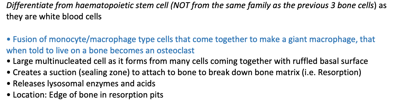
Osteoclast
Bone-resorbing cell responsible for breaking down bone tissue.
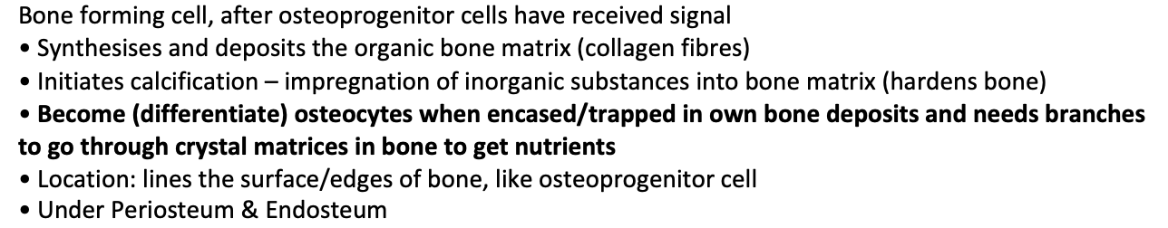
Osteoblast
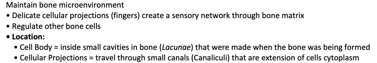
Osteocyte
Mature bone cell that maintains the bone matrix and communicates via canaliculi.
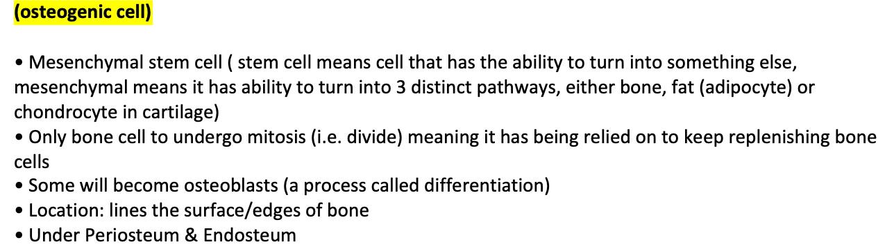
osteoprogenitor cells
osteon components
Osteon Components (A.K.A. Haversian System) from compact bone
Trabeculae
Structural units of spongy bone, consisting of thin struts that create a web-like matrix.
Trabeculae is found in spongey/ trabecular bone, in the heads of bones
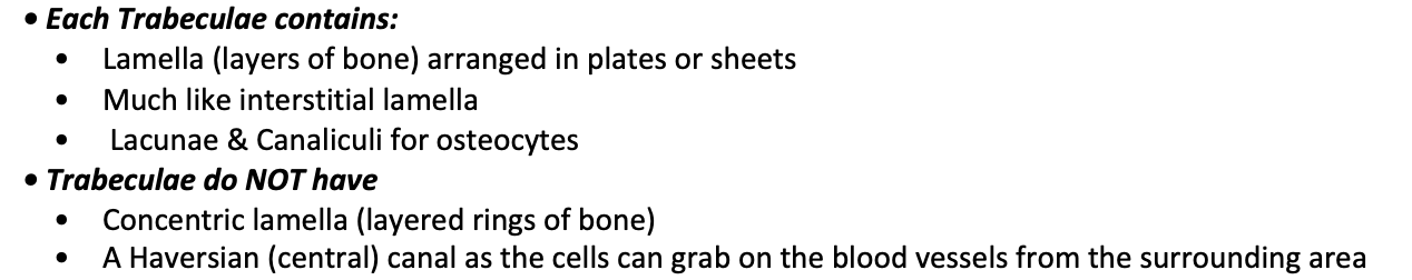
• Each Trabeculae contains:
Lamella (layers of bone) arranged in plates or sheets
Much like interstitial lamella
Lacunae & Canaliculi for osteocytes
• Trabeculae do NOT have
Concentric lamella (layered rings of bone)
A Haversian (central) canal as the cells can grab on the blood vessels from the surrounding area
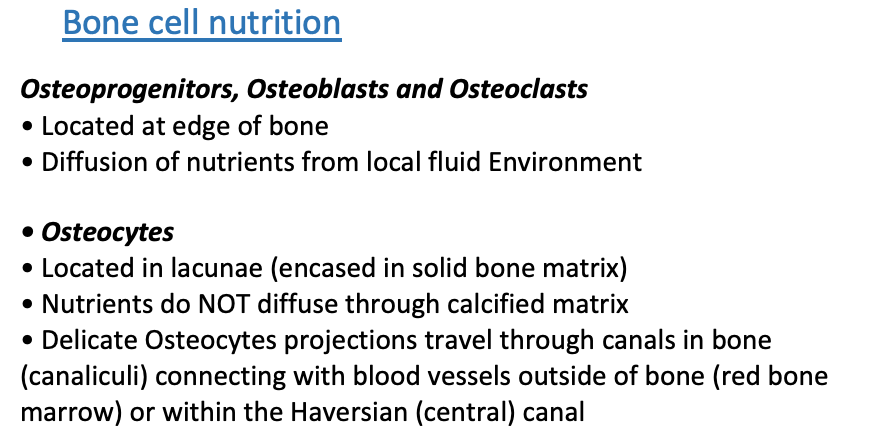
bone cell nutrition
Osteoprogenitors, Osteoblasts and Osteoclasts
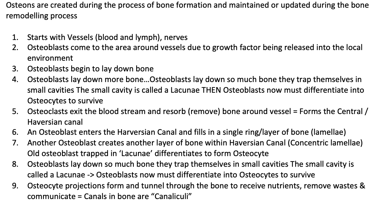
how do osteons forms
Osteons are created during the process of bone formation and maintained or updated during the bone remodelling process
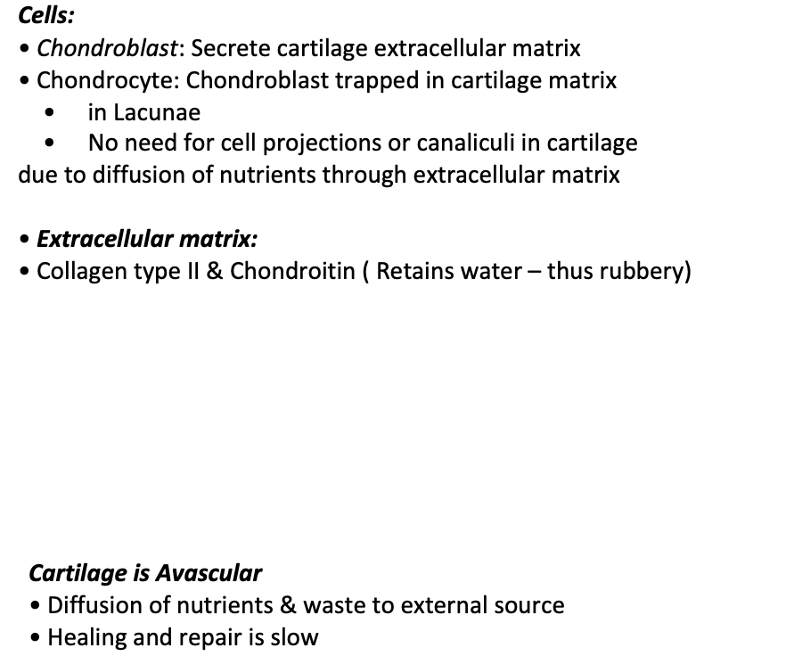
function of cartilage tissue
Maintains shape
Resist compression & absorbs shock
Provides smooth surface to minimise friction (articular cartilage in long bone does this and resists compression)
cartilage compositon and cells
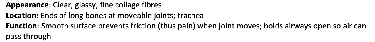
types of cartilage- hyaline
Appearance: Clear, glassy, fine collage fibres
Location: Ends of long bones at moveable joints; trachea
Function: Smooth surface prevents friction (thus pain) when joint moves; holds airways open so air can pass through
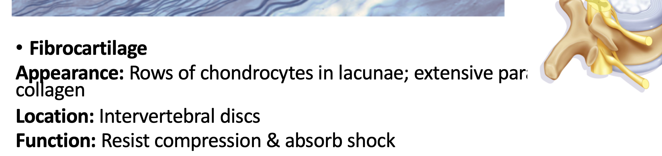
fibrocartilage
Very fibrous and with lots of collagen bundles, giving it more strength and resistance, so good for areas that need to maintain integrity
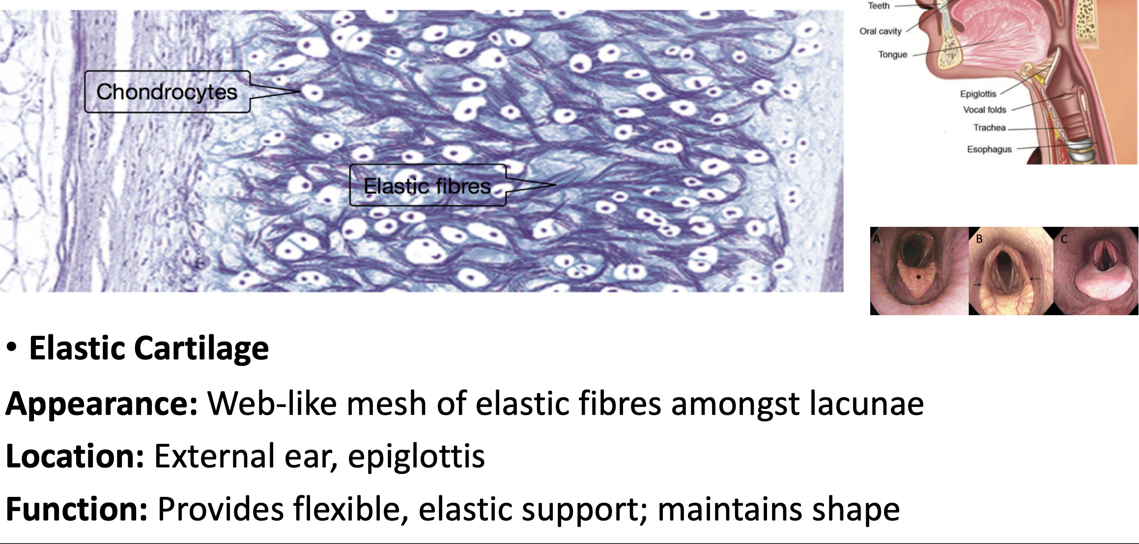
elastic cartilage
Lots of elastic fibres, giving web appearance, have some stretch and recoil back into shape
ossification
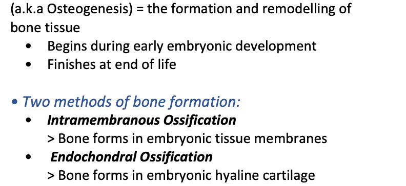
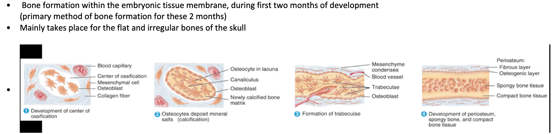
intramembranous ossification
Bone formation within the embryonic tissue membrane, during first two months of development (primary method of bone formation for these 2 months)
Mainly takes place for the flat and irregular bones of the skull
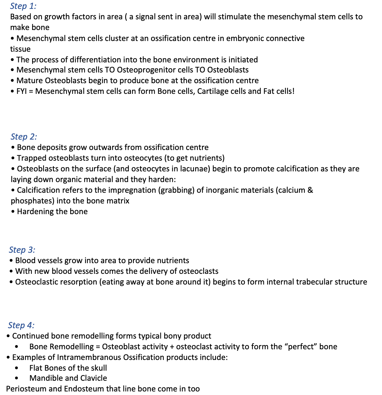
intramembranous ossification steps
Based on growth factors in area ( a signal sent in area)
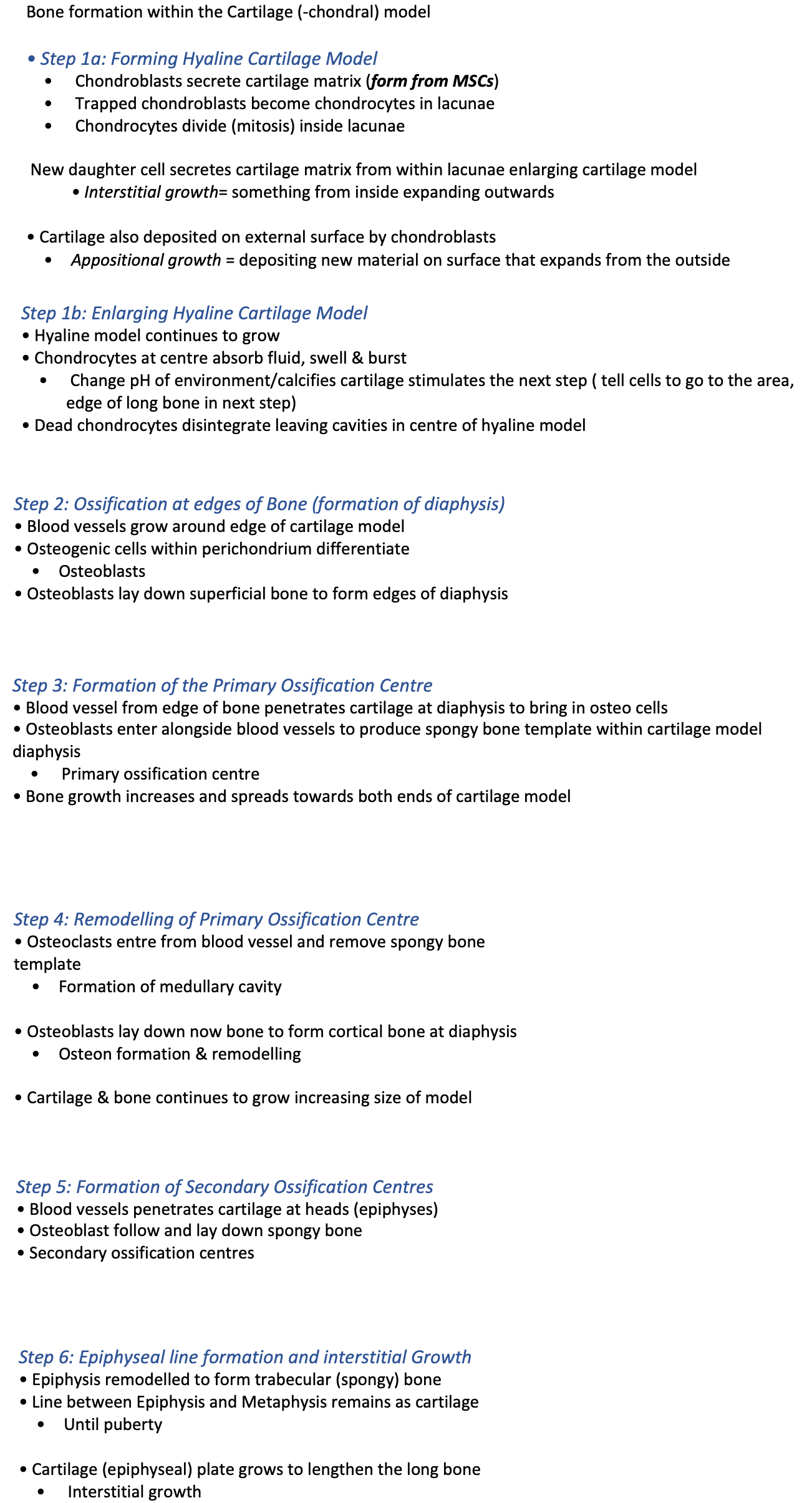
endochondral ossification
Bone formation within the Cartilage (-chondral) model
interstitial vs appositional growth
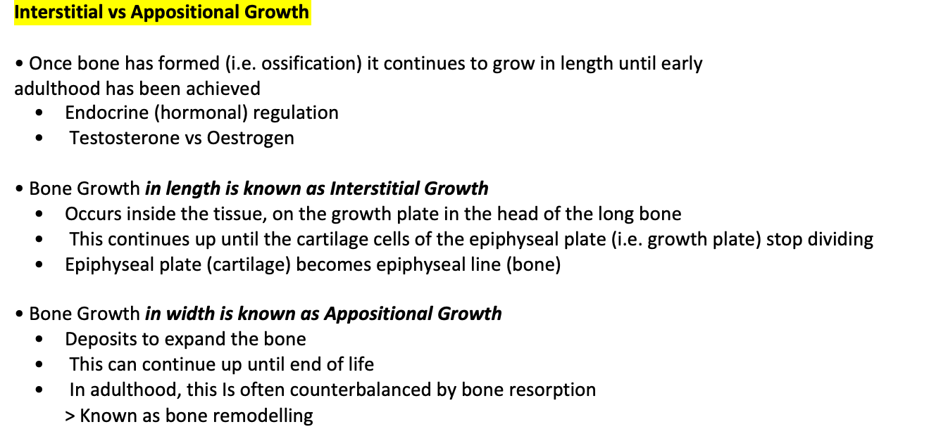
interstitial growth
(inside tissue at interstitial plate)
• Bone growth in length
• Occurs ‘within bone’
• Occurs at Epiphyseal Plate
• Chondrocytes divide increasing Hyaline cartilage from the inside
• Growth of cartilage pushing epiphysis further from metaphysis
• Hyaline cartilage gradually ossifies (Adulthood)
• Epiphyseal line
s
appositional growth
Bone growth in width, Occurs on ‘edges of bone’
• Bony structure is formed
• Osteogenic cells within periosteum differentiated to form osteoblasts
• Osteoblasts lay down new layers of bone (lamellae) on the edges of bone
• Osteoblasts trap themselves TURN INTO osteocytes

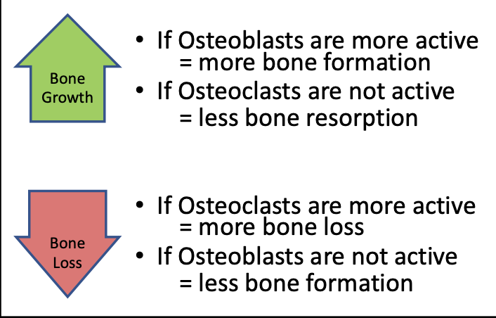
bone remodelling
Process of bone cells removing old bone and replacing with new bone
• Osteoclasts removing bone by reabsorbing it at equal rates of Osteoblasts forming new bone (important balance)
• Balance of bone removal and bone formation = repair/maintenance
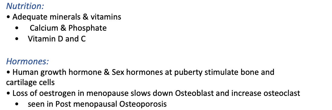
Breaking down of the bones releases minerals into the body
• Adeq
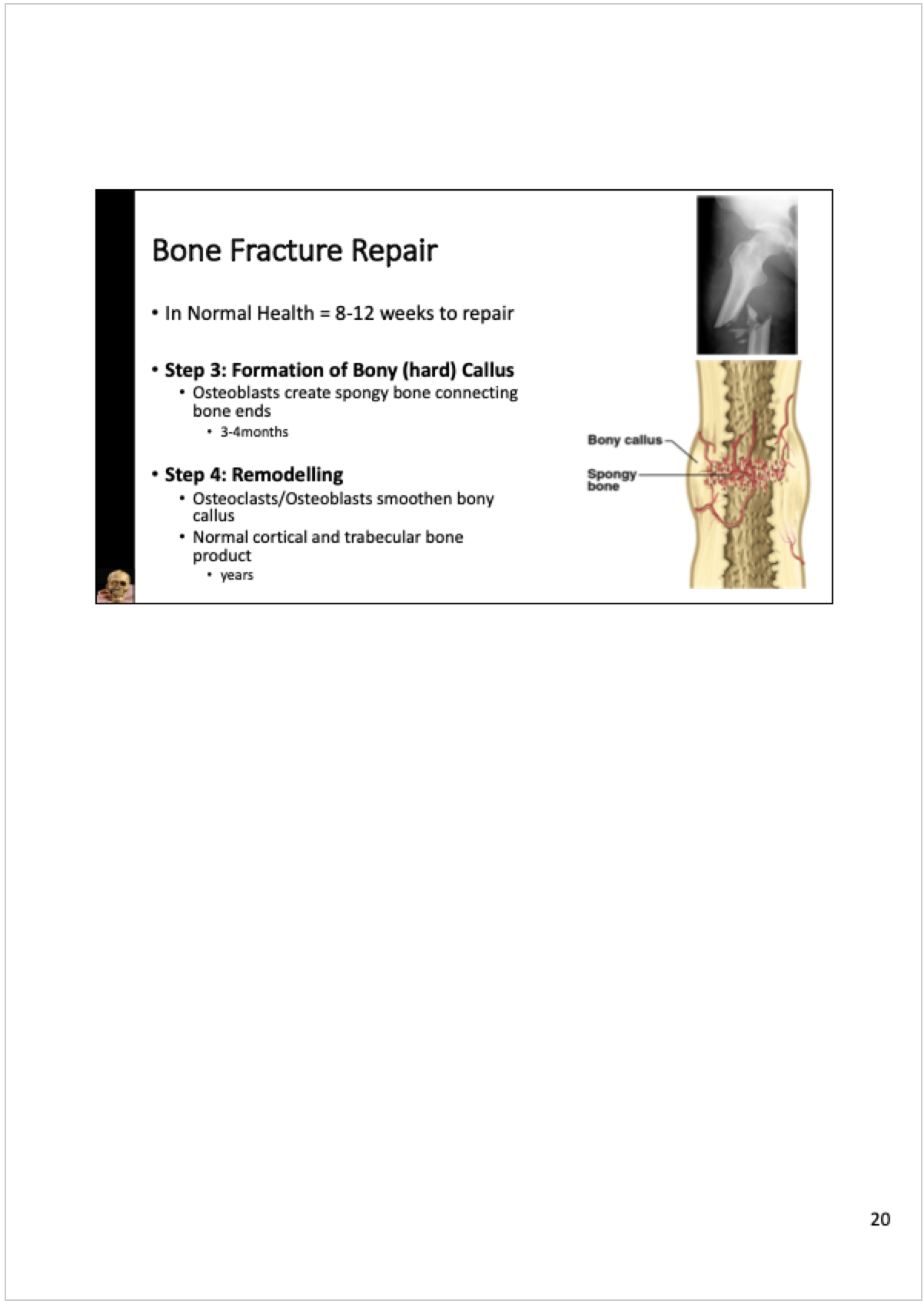
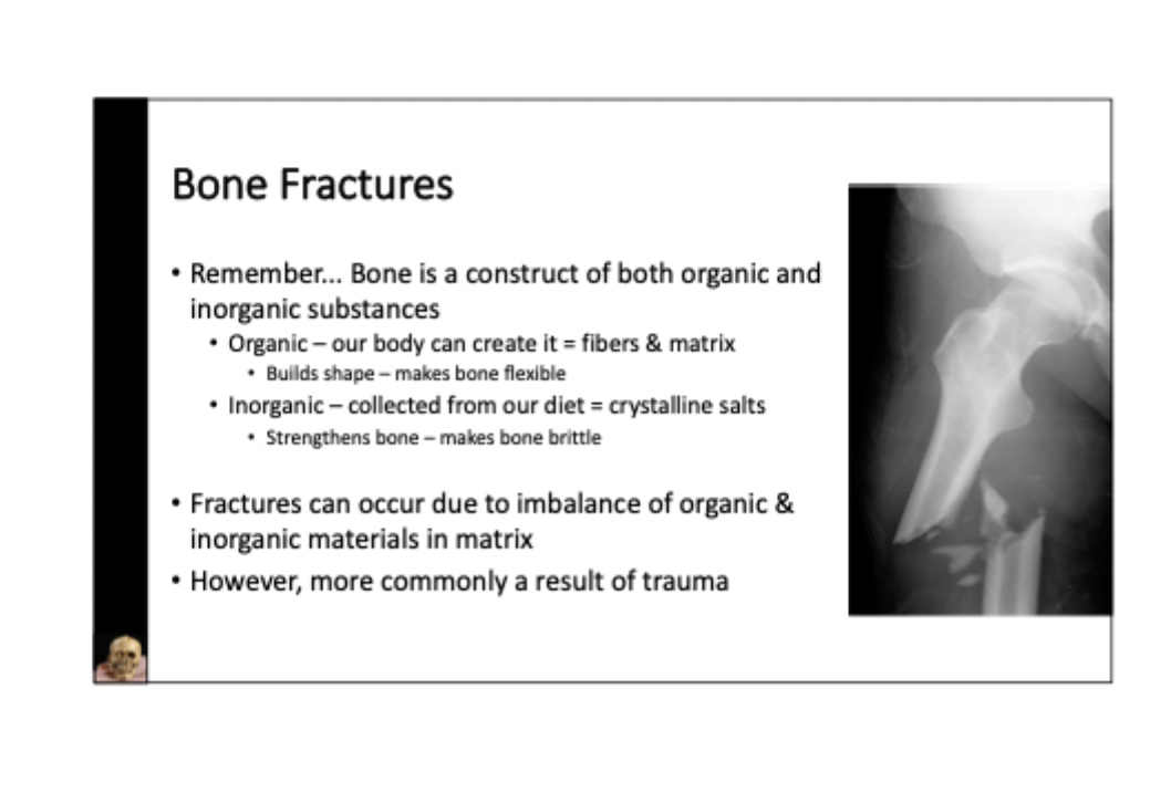
bone fracture and repair
what are joints
Joints (a.k.a. articulations) form where two bones are interconnected
• Can also be considered the point of contact between two structures
Where bone meets bone
Where cartilage meets bone
Where teeth meet bone
Classifying joints - structural and functional classifications
Functional Classifications: Refers to the range of motion at an articulation
Immobile (synarthrosis) = no movement
Partly mobile (amphiarthrosis) = little movement
Freely mobile (diarthrosis) = wide range of movement
• Structural Classifications: Refers to the components & features of the joint
Bony = complete fusion of two bones
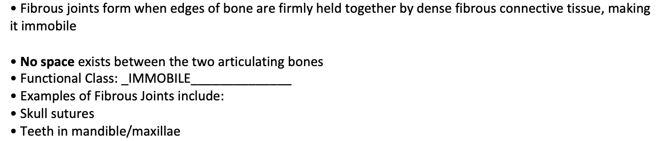
Fibrous = held together by dense collagen fibres
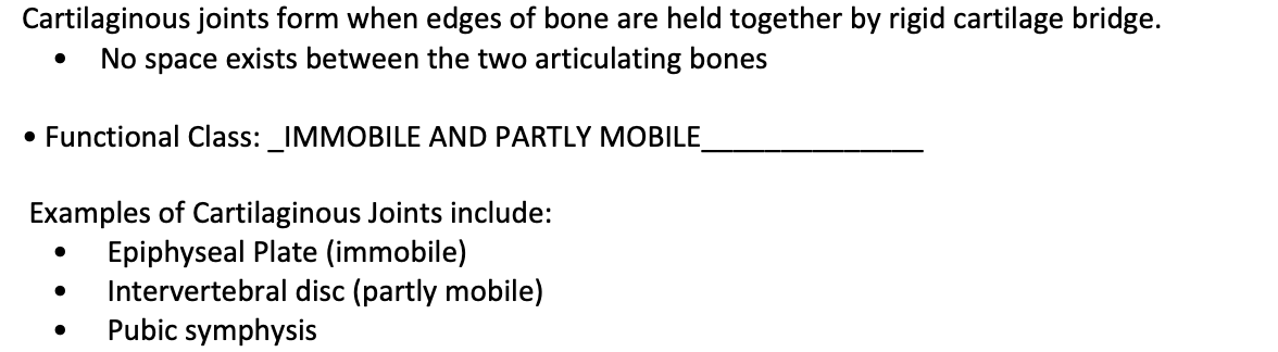
Cartilaginous = held together by cartilage
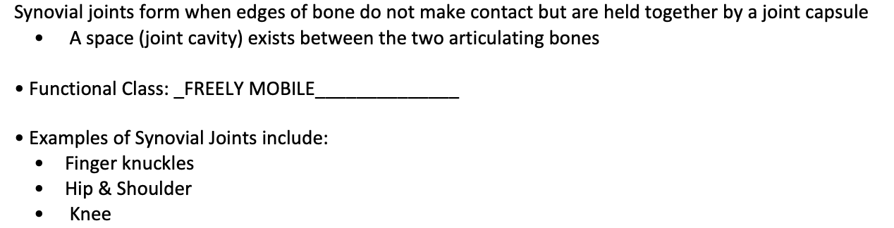
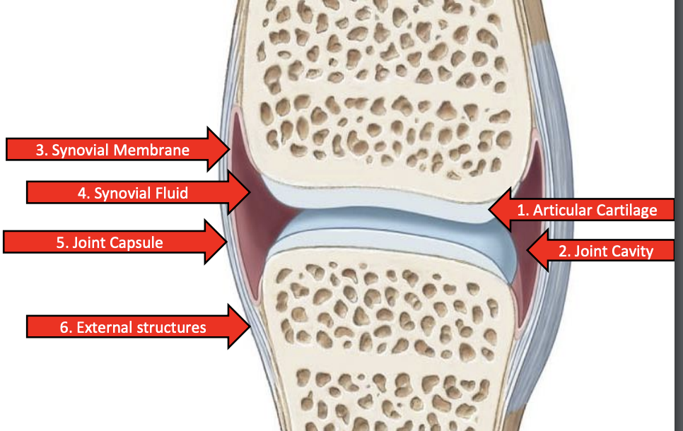
Synovial = contain a joint space held together by joint capsule
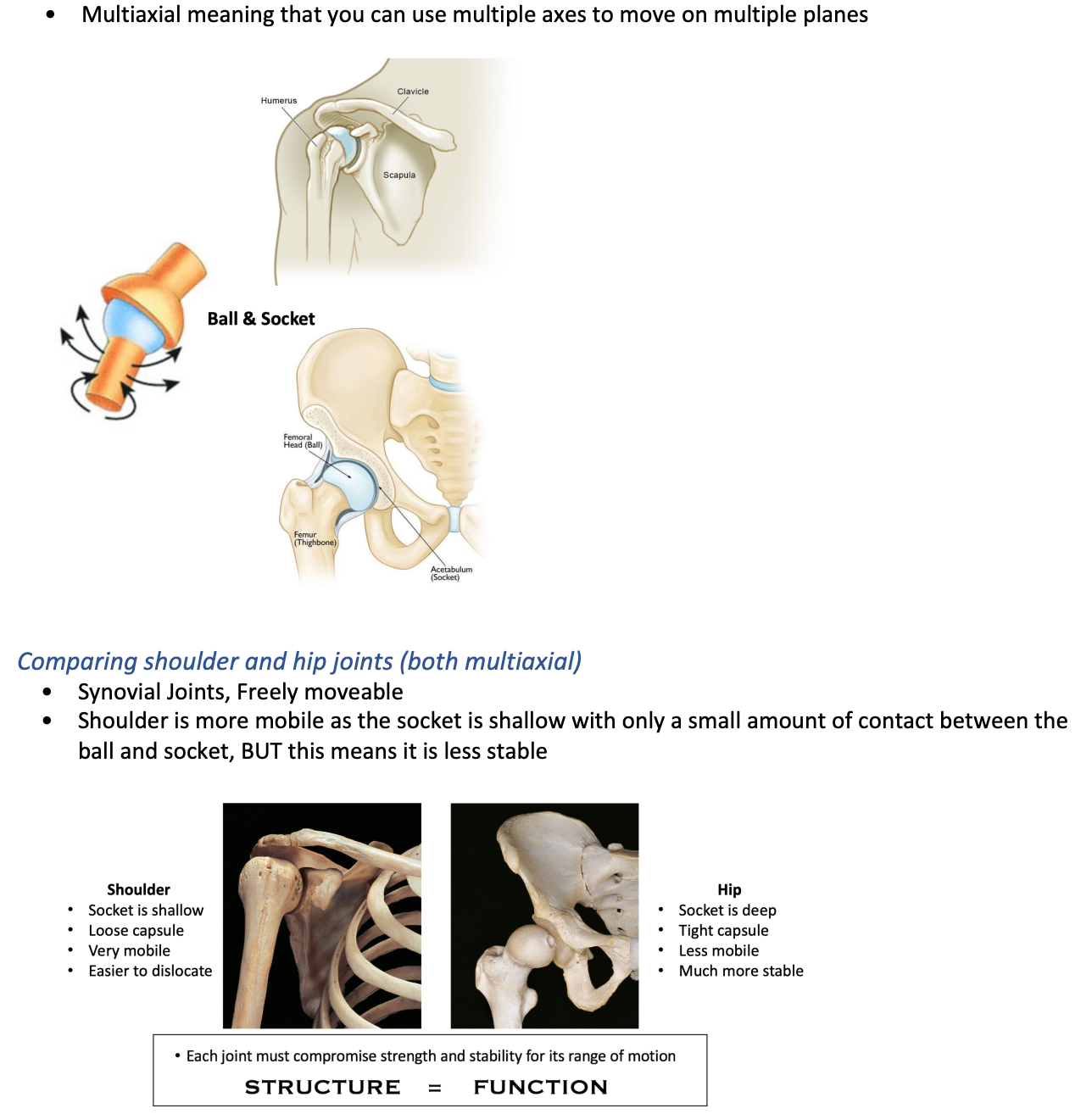
Each joint must compromise strength and stability for its range of motion
STRUCTURE = FUNCTION
structural classification of joints- bony joints
structural classification - fibrous joints


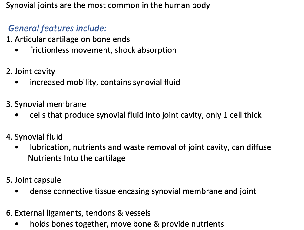
general features of synovial joints
Synovial joints are the most common in the human body
1.
the six types of synovial joints
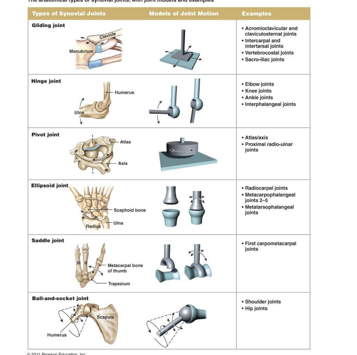
ball and socket joints
Multiaxial meaning that you can use multiple axes to move on multiple planes
hinge joints
Uniaxial meaning that it uses one axes to move on one plane
Meaning you can only go forwards and backwards so flexion and extension
Example is the ankle,
plane/ gliding joints
Nonaxial or multiaxial meaning that they slide/move across all plains depending on the environment, they have no specific movement, found in the feet e.g

saddle joints
Can do adduction, abduction, extension and flexion
Only in few places, including thumb
Biaxial meaning uses two axes to move on two planes
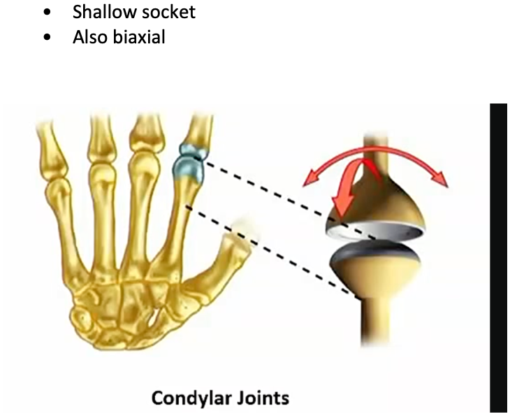
condylar joints
Shallow socket
Also biaxial
pivot joints
allow for rotation
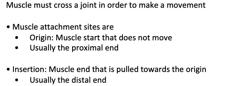
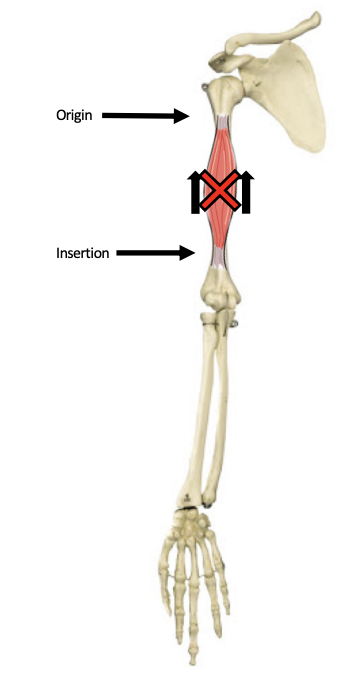
how do muscles allow for movement
Usually the distal end
movement at synovial joints
The joint must be from the moveable class for movements to occur
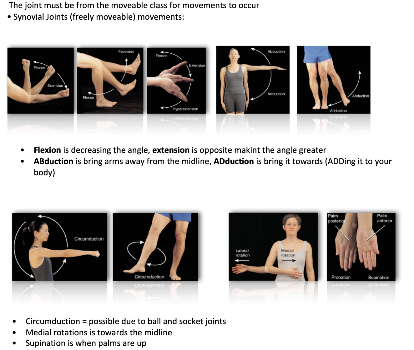
muscles function
Motion: walking, running, moves blood around the body, urine, muscles allow our chest muscles to work and for air to come in
2. Stabilising Body Position: posture/tension on skeleton frame
3. Regulation of Organ Volumes: sphincters, oesophageal is an example, keeps acid in stomach
4. Support of Soft Tissue: shield and weight support of organs, i.e the pelvis and the chest
5. Maintain body temperature: heat through contractions/shivering of muscles to keep us warm
6. Storage nutrients: Amino acid release from skeletal muscle breakdown
Co-ordinated action of muscle groups
Skeletal muscles function in groups to move a joint
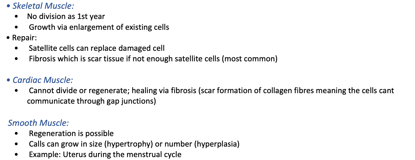
3 types of muscles and characteristics
Skeletal Muscle: Attaches to bone- moves things at the joints
2. Cardiac Muscle: Forms the heart
3. Smooth Muscle: Located within walls of hollow organs, like blood vessels, in intestines to contract and push food through
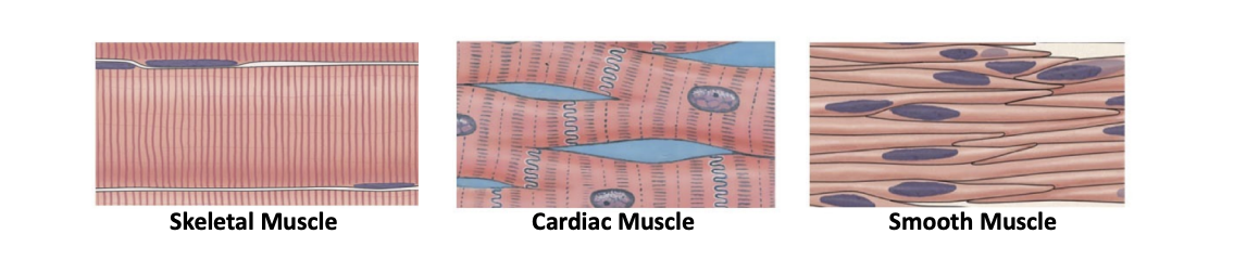
skeletal muscle
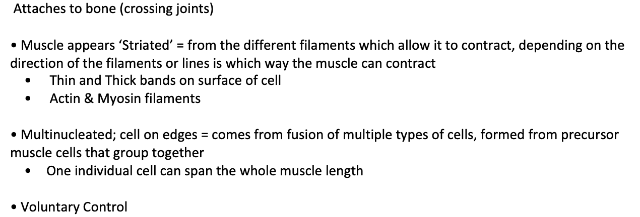
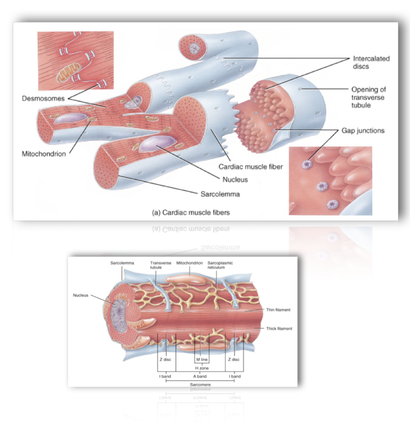
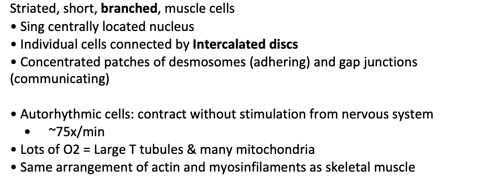
cardiac muscle
The Muscles of the Heart
• Muscle appears ‘Striated’ in one direction
• cells have one central nucleus and are branched to connect togethor
• Involuntary Control; Autorhythmic
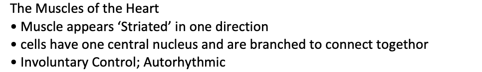
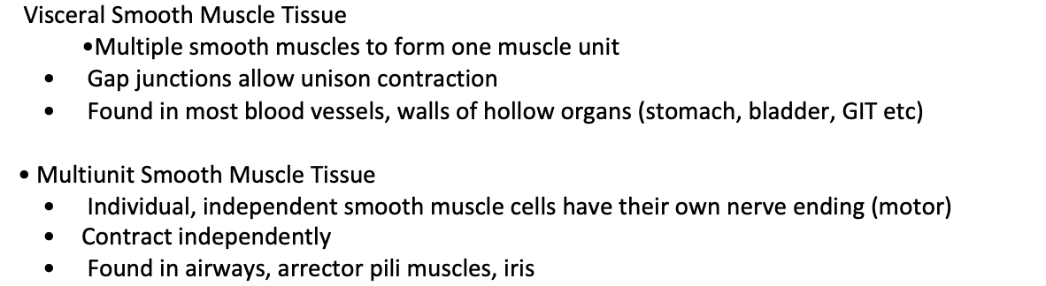
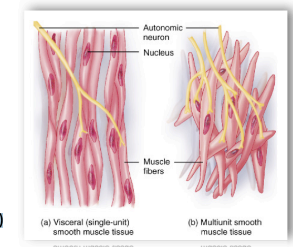
smooth muscle
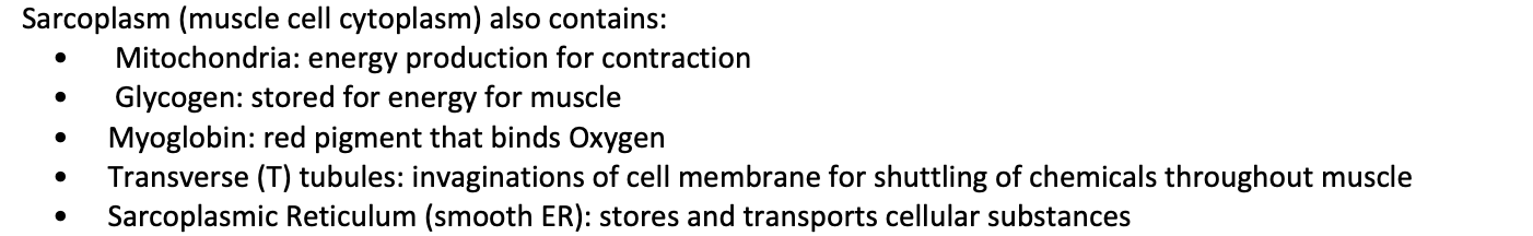
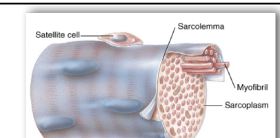
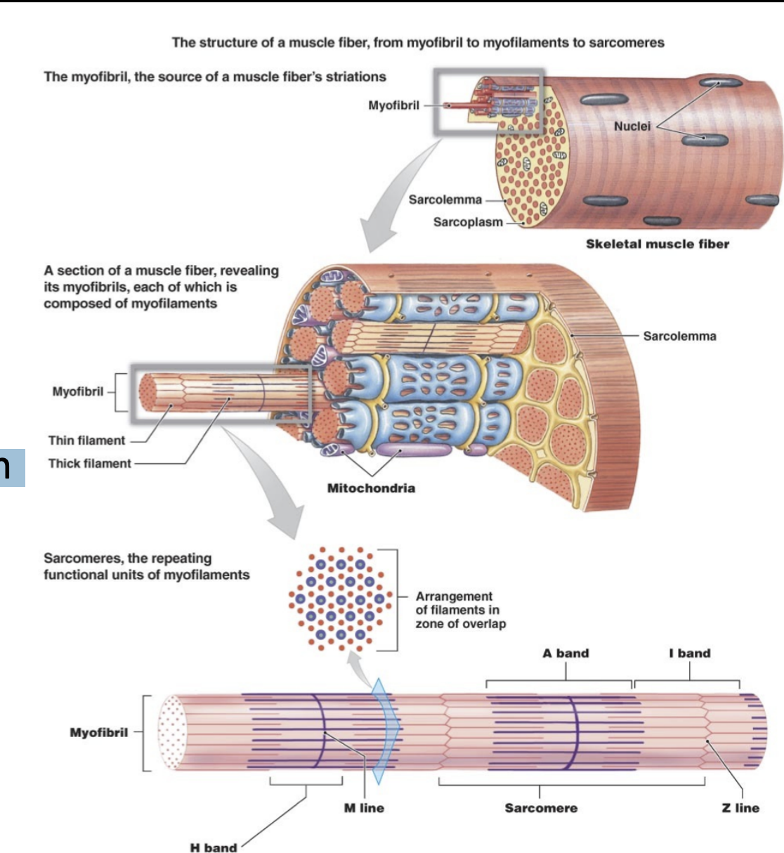
skeletal muscle cells
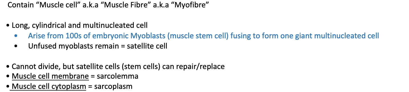
Sarcoplasm (muscle cell cytoplasm) also contains:
Mitochondria: energy production for contraction
Glycogen: stored for energy for muscle
Myoglobin: red pigment that binds Oxygen
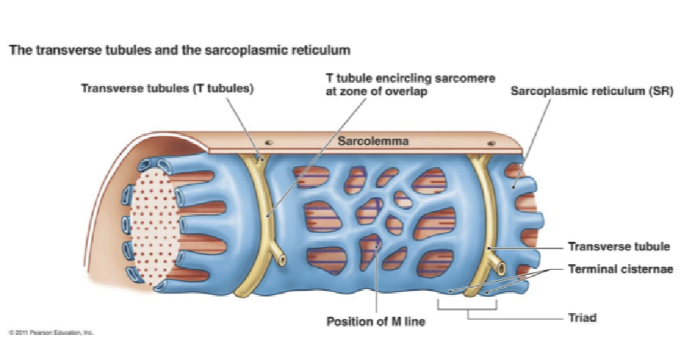
Transverse (T) tubules: invaginations of cell membrane for shuttling of chemicals throughout muscle
Sarcoplasmic Reticulum (smooth ER): stores and transports cellular substances
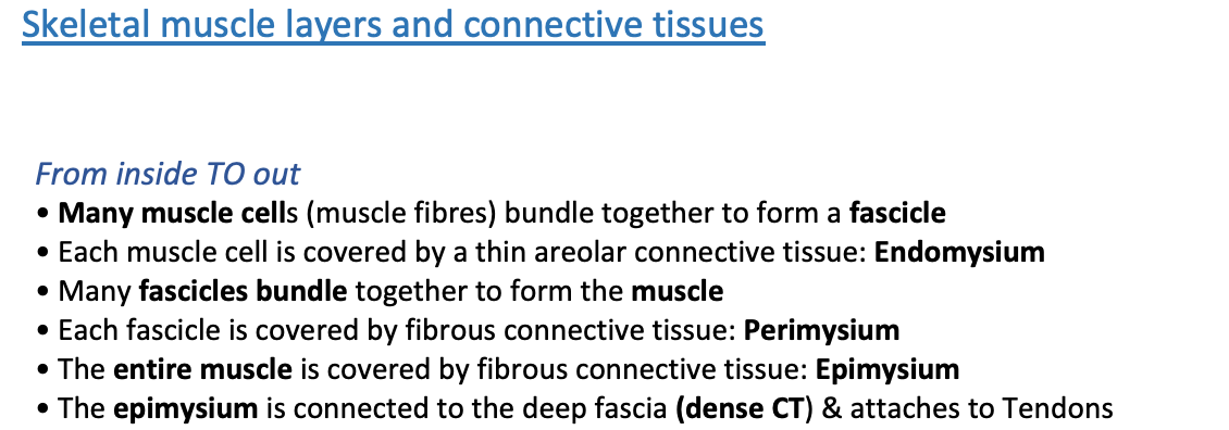
skeletal muscle layers
skeletal muscle contractions

cardiac muscle cells
smooth muscle
muscle regeneration