Image ID Skull, Orbits & Sinuses
1/107
There's no tags or description
Looks like no tags are added yet.
Name | Mastery | Learn | Test | Matching | Spaced | Call with Kai | Chat |
|---|
No analytics yet
Send a link to your students to track their progress
108 Terms
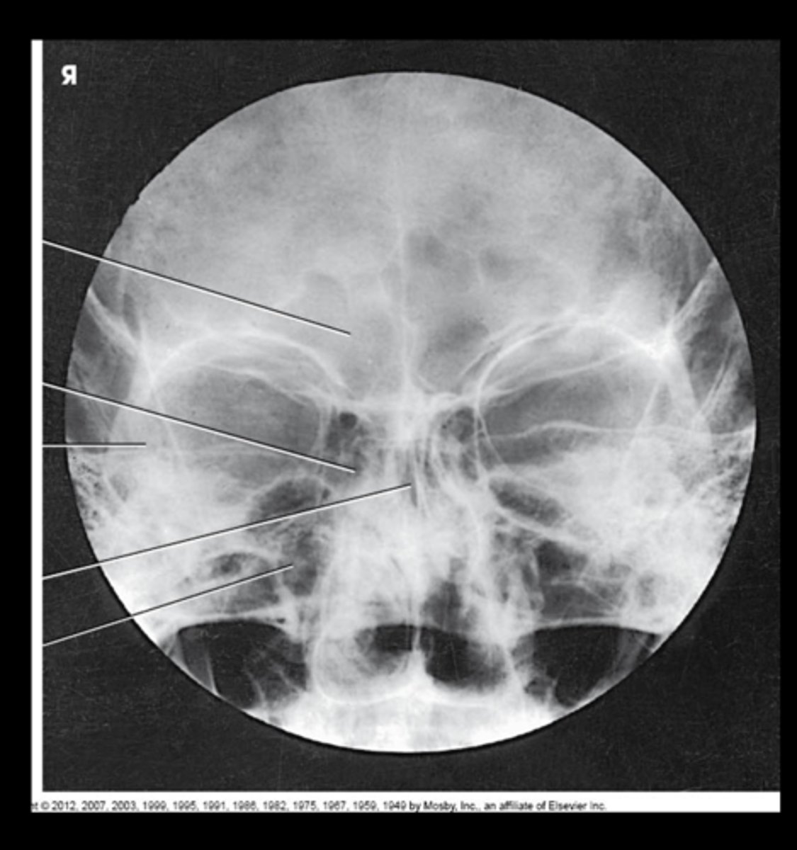
PA Axial Caldwell
What projection is this?

Frontal Bone
What is 1?
crista galli of ethmoid bone
What is 2?

Petrous Ridges of Temporal bone
What is 3?

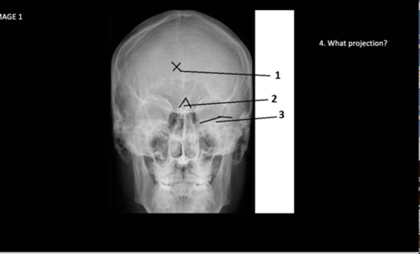
Bregma
What is 5?

Coronal Suture
What is 6?

Greater Wing of Sphenoid
What is 7?
Lesser Wing of Sphenoid
What is 8?

Orbital Plates of Frontal Bone
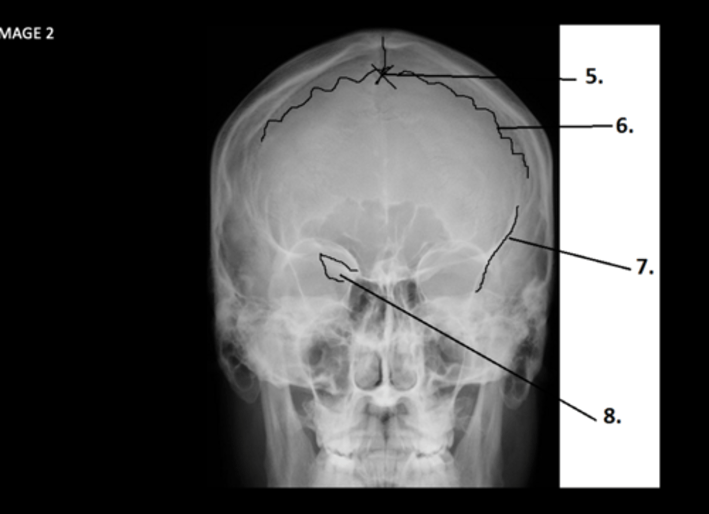
What is 9?

Posterior clinoid process of sphenoid bone
What is 10?

Dorsum sellae of sphenoid bone
What is 11?

Clivus of occipital bone
What is 12?

External auditory meatus EAM
What is 13?
OML (Orbitomeatal Line)
Identify the topographic line:
What is A?

IOML (Infraorbitomeatal line)
Identify the topographic line:
What is B?
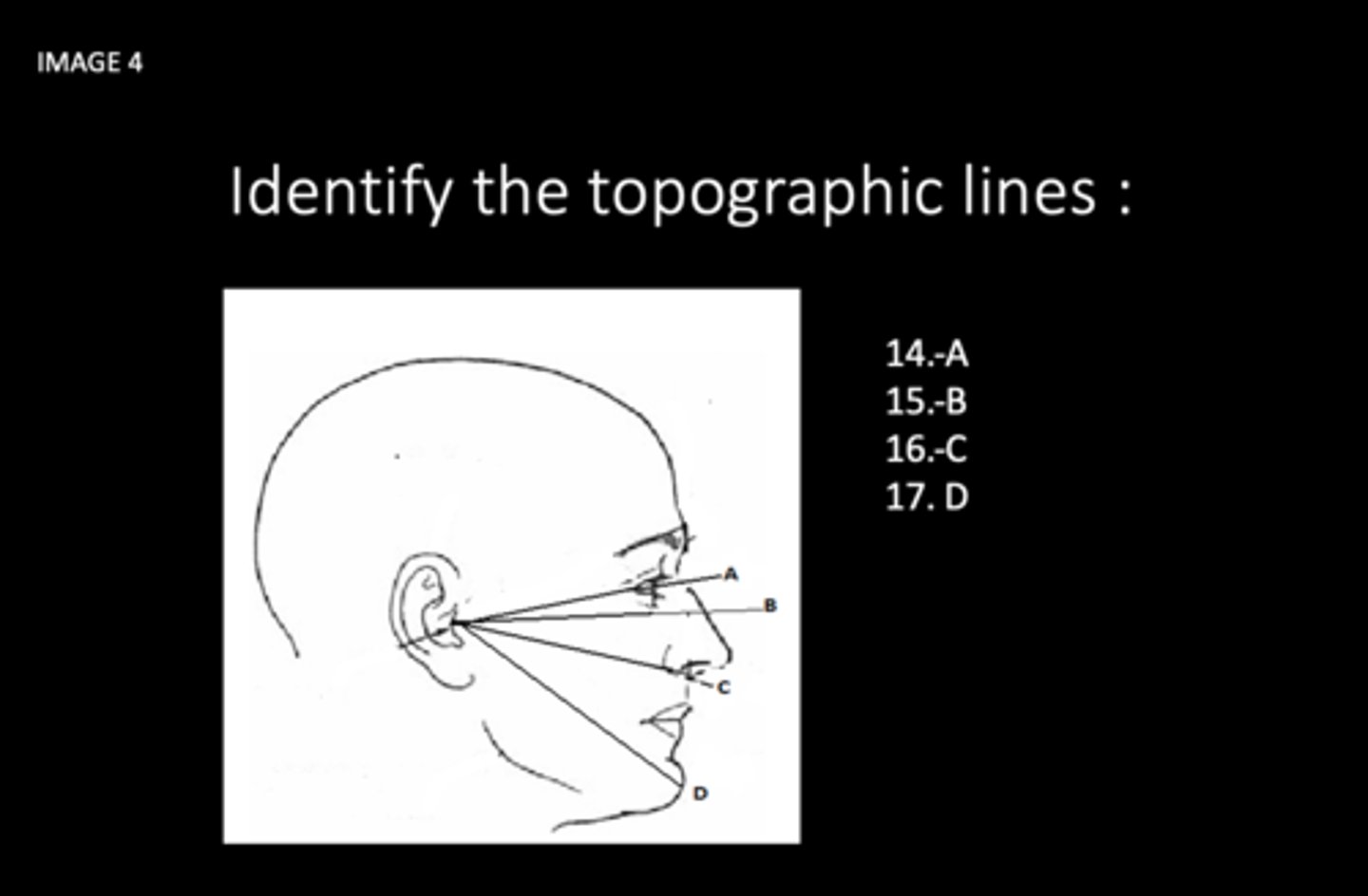
AML (acanthiomeatal line)
Identify the topographic line:
What is C?

MML (mentomeatal line)
Identify the topographic line:
What is D?

Glabella
Identify the topographic landmarks:
What is 18?

IPL (interpupillary line)
Identify the topographic landmarks:
What is 19?

Nasion
Identify the topographic landmarks:
What is 20?
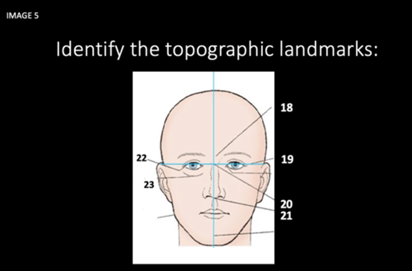
Acanthion
Identify the topographic landmarks:
What is 21?

Outer Canthus
Identify the topographic landmarks:
What is 22?

Infraorbital margine (IOM)
Identify the topographic landmarks:
What is 23?

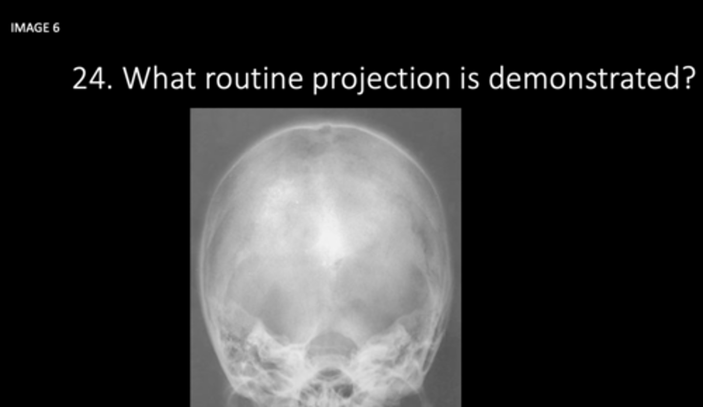
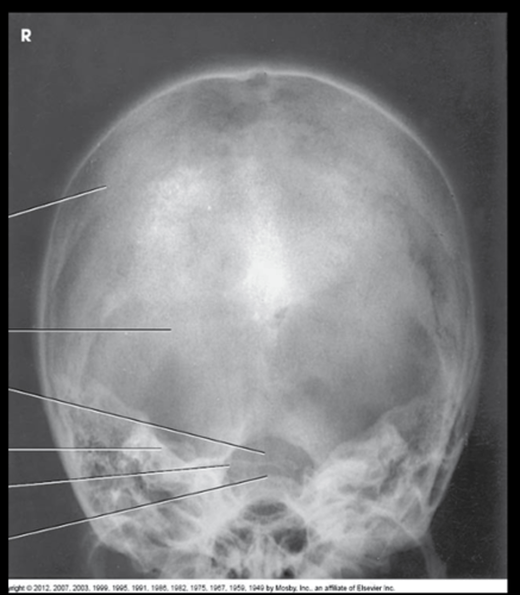
AP Axial Townes method
What projection is this?
petrous ridge of temporal bone
What is 25?
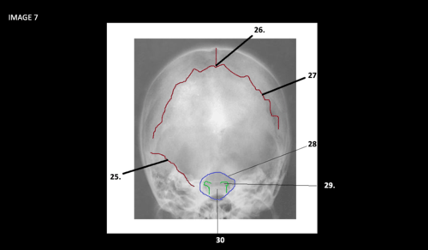
Lambda
What is 26?

Lambdoidal Suture
What is 27?

foramen magnum of occipital bone
What is 28?

posterior clinoid processes of sphenoid bone
What is 29?

dorsum sellae of sphenoid bone
What is 30?

Lateral Skull
What projection is this?
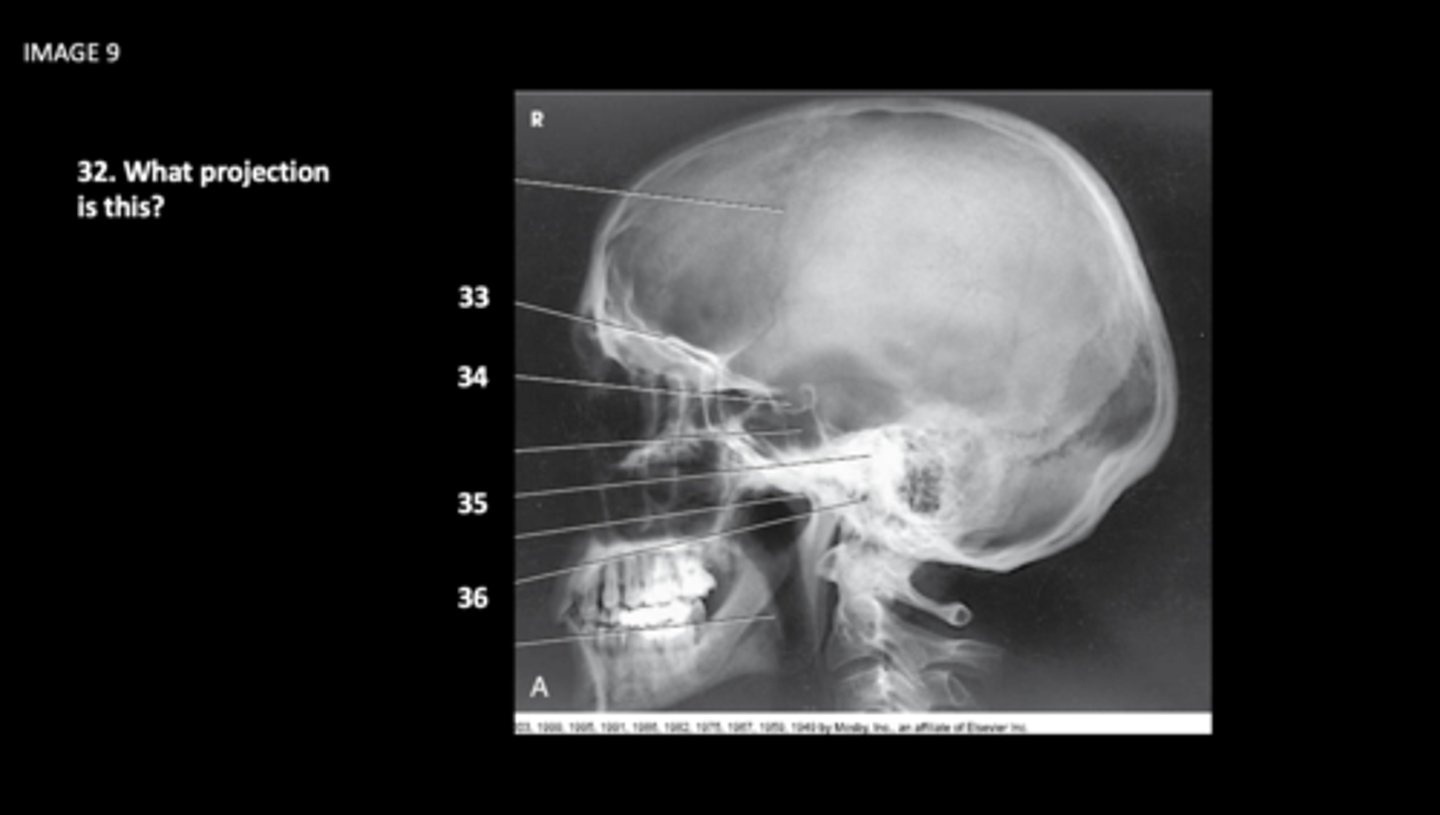
- Sella turcica should not be rotated, but should be seen in profile
-Superimposition of: orbital roofs, greater wings of sphenoid, mastoid regions, EAMS and the tempromandibular joints should be superimposed too.
What is the exam evaluation criteria?

Superimposed orbital roof
What is 33?

sella turcica of sphenoid bone
What is 34?

petrous portion of temporal bone
What is 35?

external acoustic meatus
What is 36?

dorsum sellae of sphenoid bone
What is 37?

Superior orbital margin
What is 38?

petrous ridge of temporal bone
What is 39?

infraorbital margin
What is 40?

crista galli of ethmoid bone
What is 41?
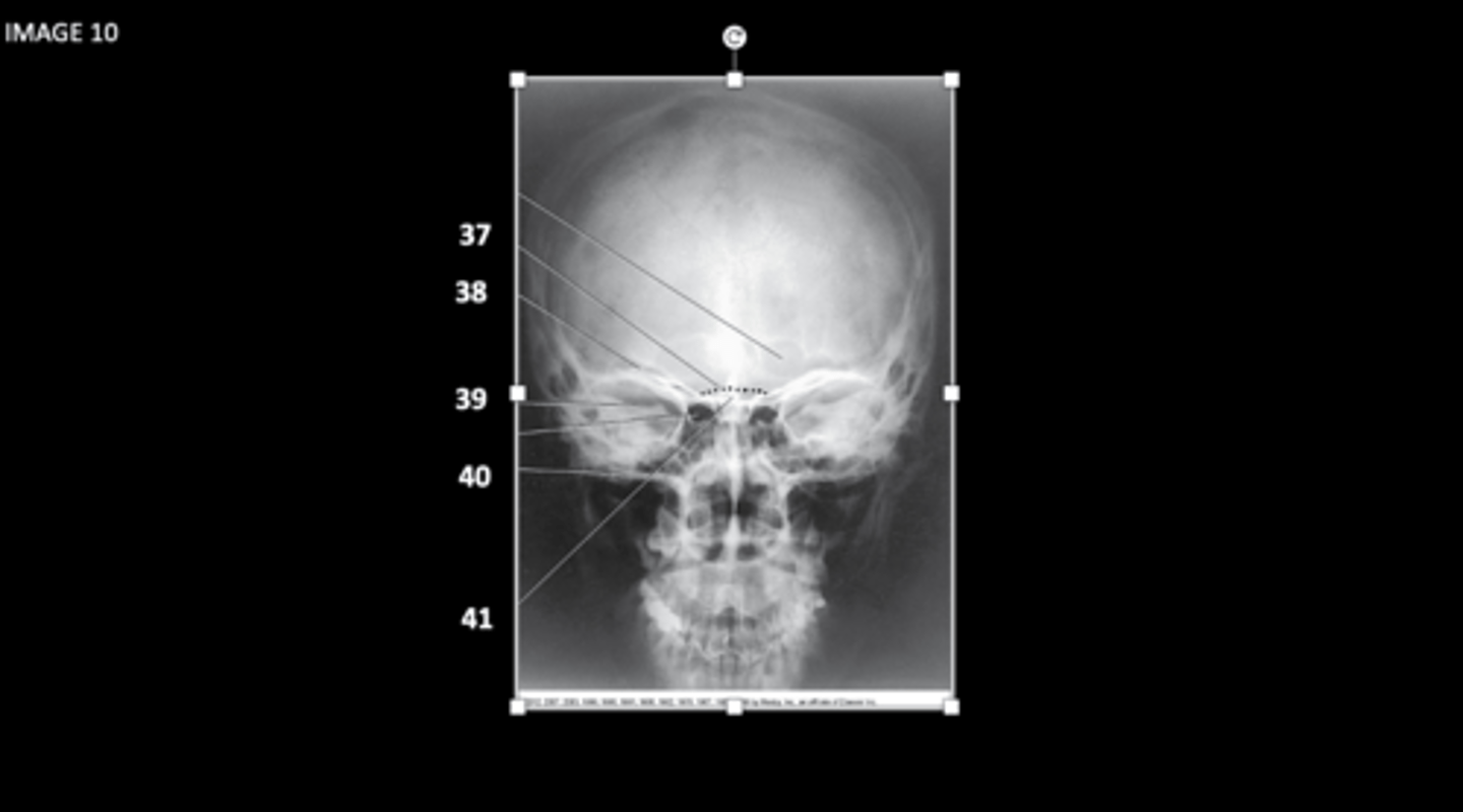
Modified Waters
What projection is this?

Petrous Ridges
What is 47?
PA Axial Caldwell Skull
What projection is this?

- The petrous bones should lie in the lower third of the orbits
- The distance between the lateral border of the skull to the lateral border of the orbit should be equal on both sides
What is the evaluation criteria for this image?
AP Axial Townes Method
What is this projection?

- the dorm sella and the posterior crinoid processes should be visualized within the magnum foramen
- the distance from the lateral border of the skull to the lateral margin of the foramen magnum should be equal on both sides
What is the evaluation criteria for this image?
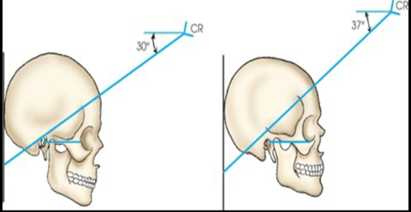
AP Axial Towne Method
- OML 30 degrees caudal
-IOML 37 degrees caudal
Which projection uses these angulations?
Superior orbital margin
What is 31?

Lateral orbital margin
What is 32?
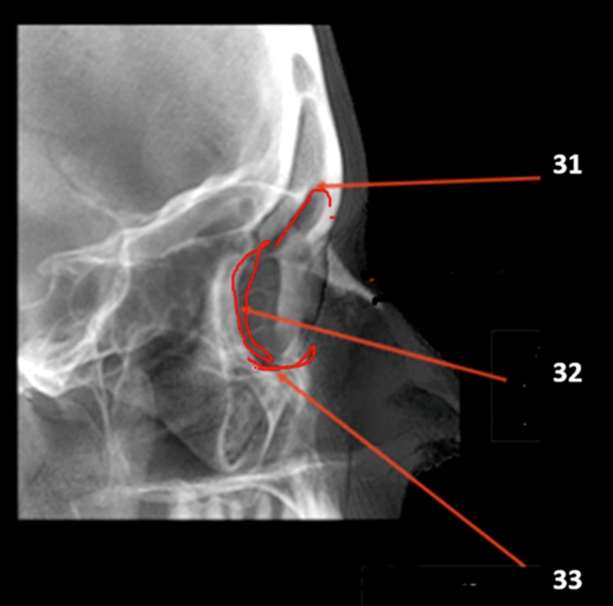
Inferior orbital margin
What is 33?

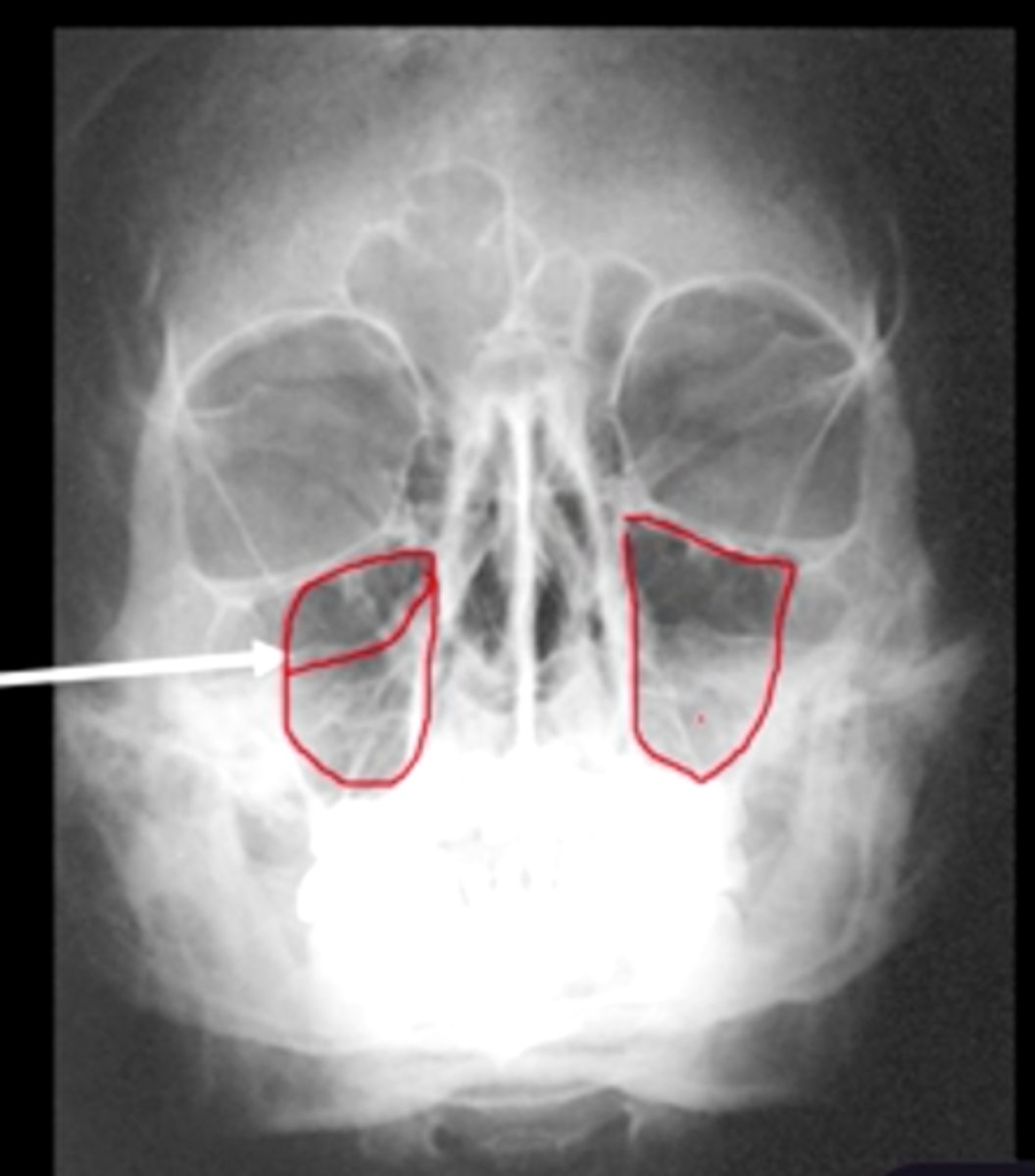
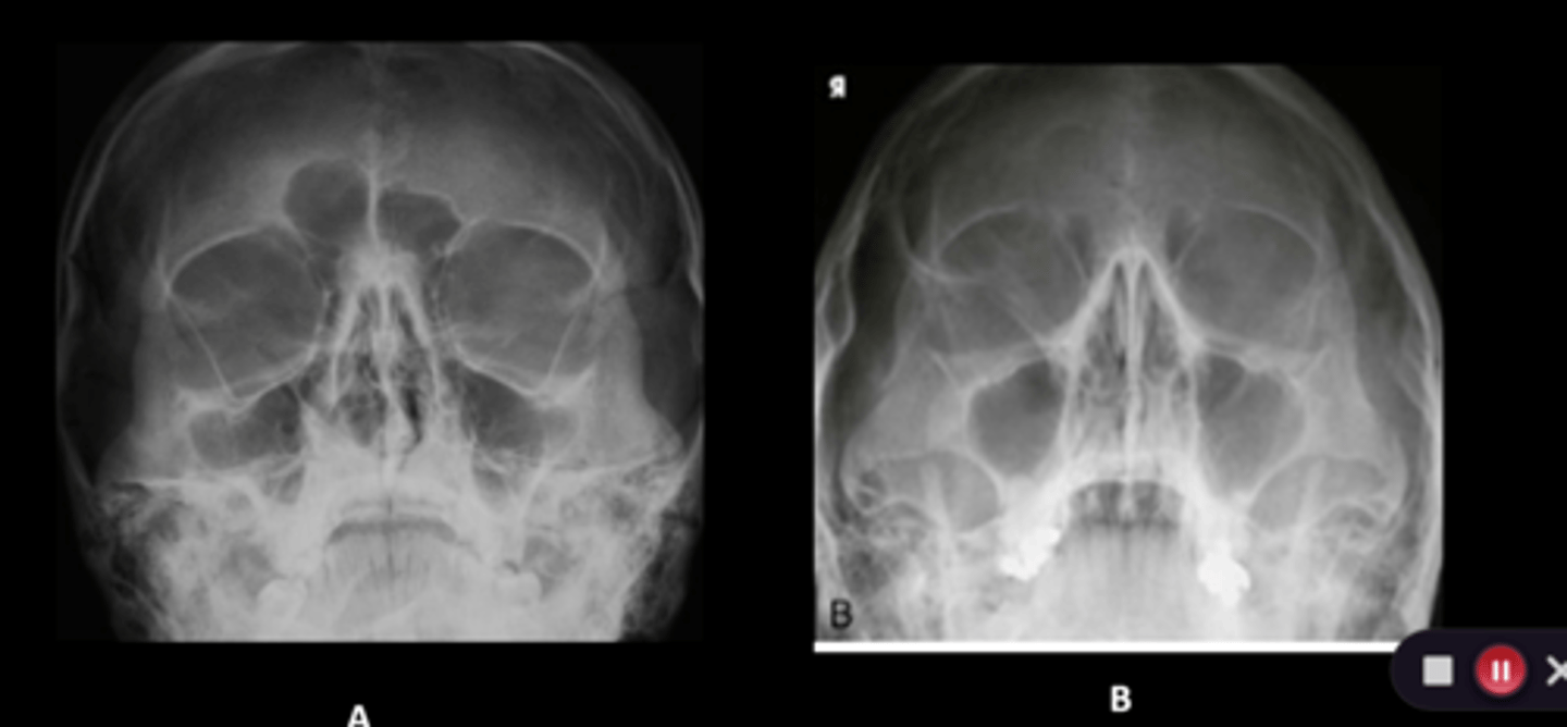
image A
the petrous ridges are projected below maxillary sinuses for the modified waters
Which image is the modified waters?
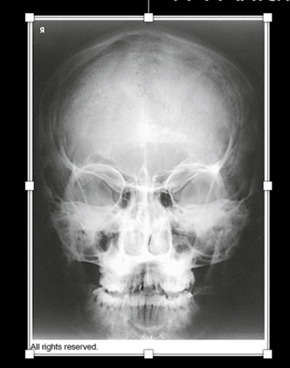
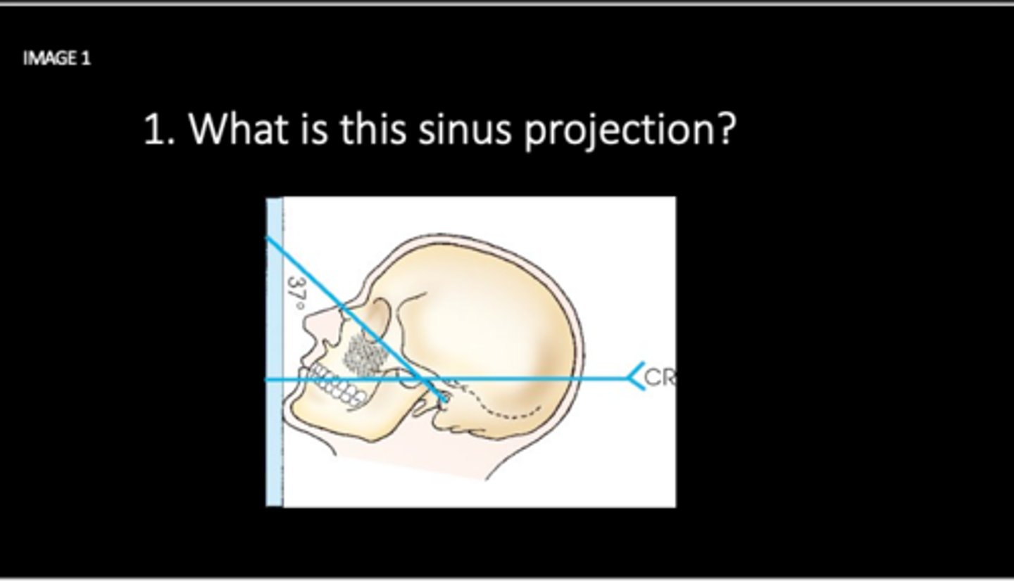
Waters: demonstrates maxillary sinuses.
What is this sinus projection?
Maxillary Sinus
What is A?

Frontal Sinus
What is B?

perpendicular plate of ethmoid bone
What is C?

petrous ridge of temporal bone
What is D?
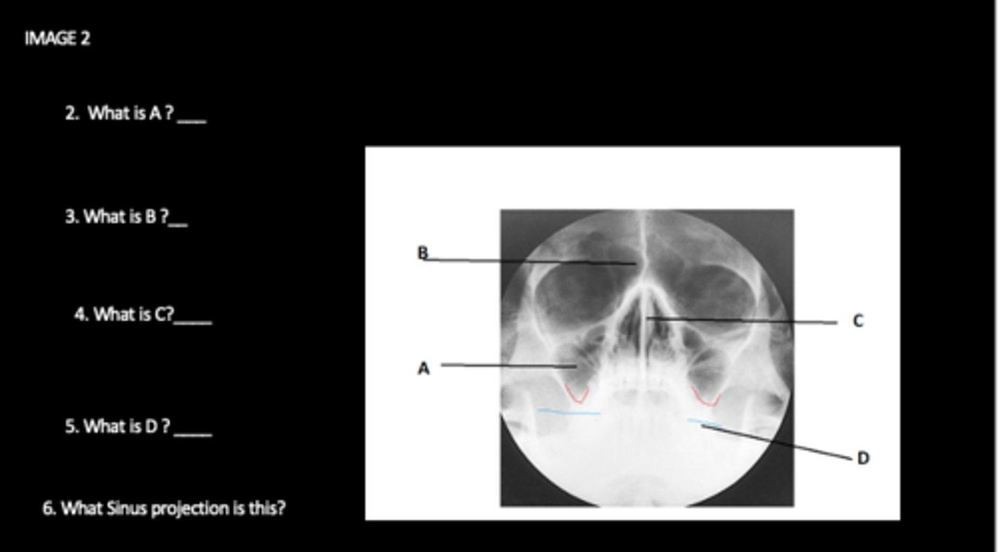
Waters projection- sinus
What projection is this?

Maxillary Sinus
A is which sinus?
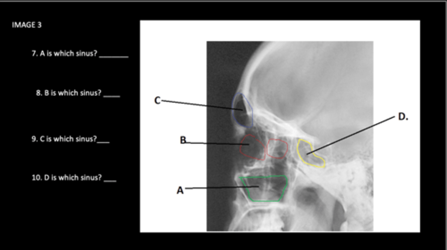
Ethmodial sinus
B is which sinus?

Frontal sinuses
C is which sinus?

Sphenoidal sinuses
D is which sinus?

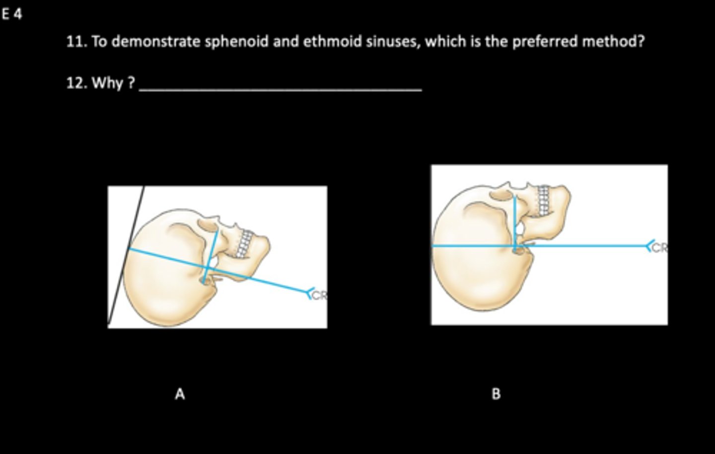
B, air/fluid levels are best demonstrated with horizontal x-ray beam
To demonstrate sphenoid and ethmoid sinuses, which is the preferred method, A or B? Why?
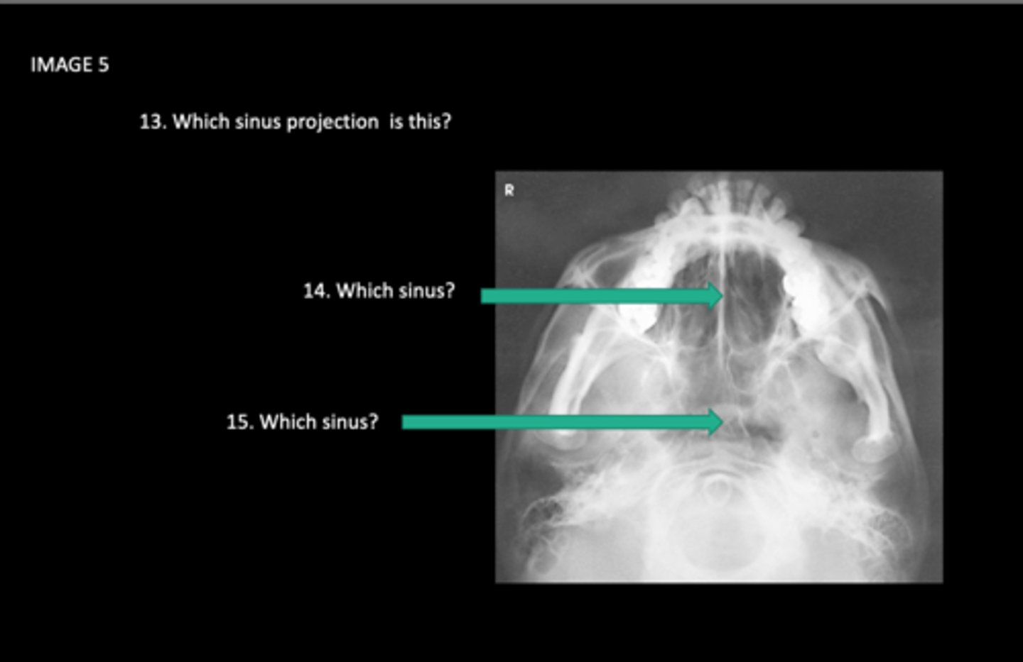
SMV sinuses
Which sinus projection is this?

ethmoid sinuses
Which sinus is 14 pointing at?

Sphenoid sinuses
Which sinus is 15 pointing at?
Frontal sinuses
What does the red line indicate?

Petrous ridges
What does the blue lines indicate?
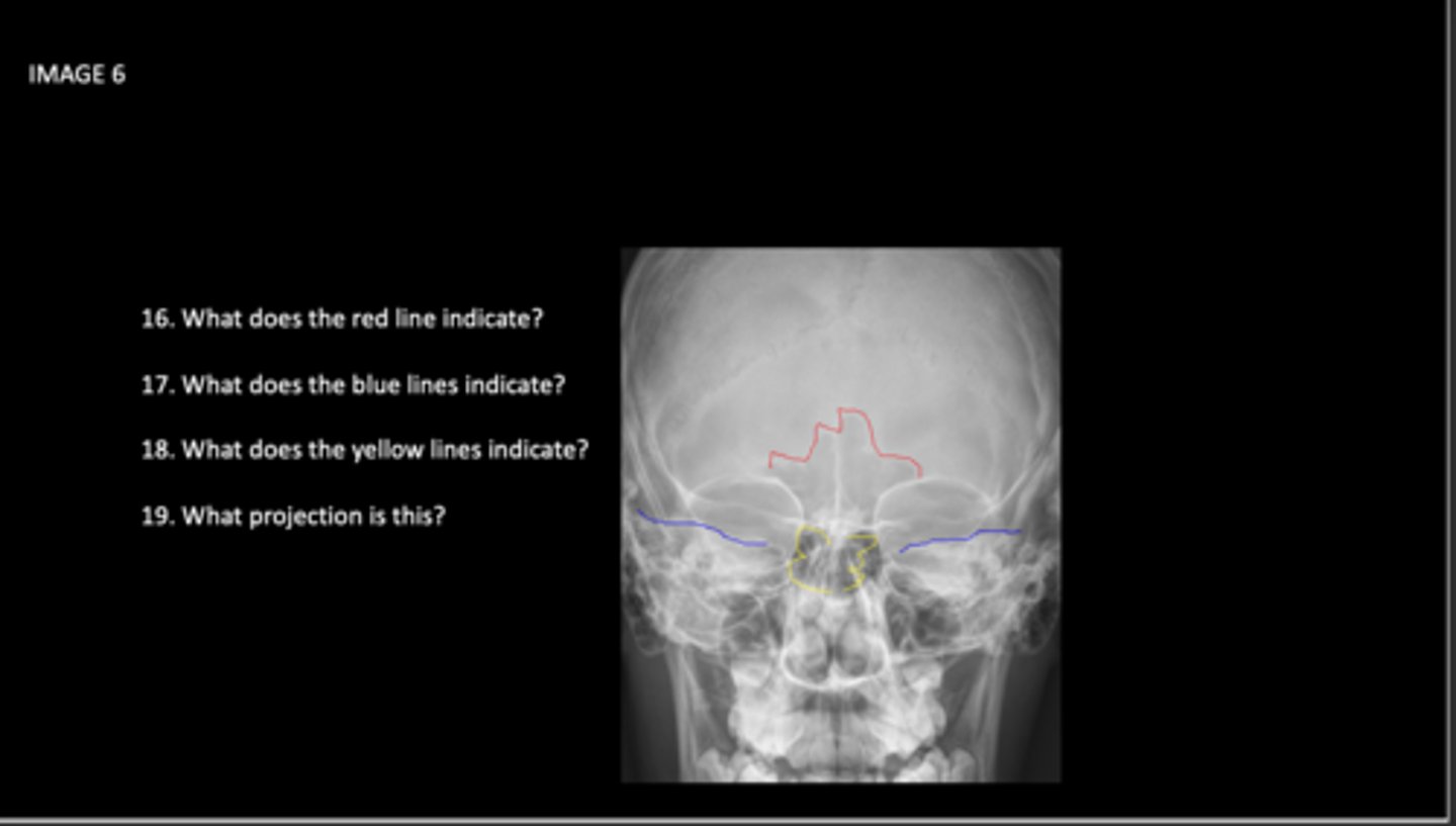
ethmoidal sinuses
What does the yellow lines indicate?

PA Axial Caldwell
What projection is this?

Open Mouth Waters Sinuses
What projection is this?
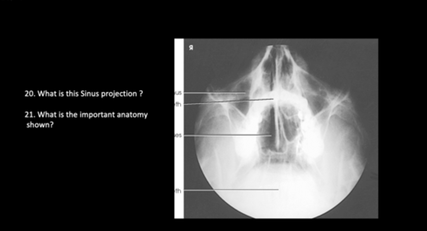
Sphenoid sinuses through the open mouth
What is the important anatomy shown?

Lateral
Which projection of the sinuses demonstrates all of the sinuses?
PA Axial Caldwell
Which projection(s) of the sinuses demonstrates the frontal and ethmoid sinuses?
MML and OML
Which topographical line is used to perform the waters for sinus?
15 degrees
How much angle is used for a Caldwell Sinus?
.5-1" posterior to the outer canthus
What is the CR location for a lateral sinus?
Acanthion
What is the CR exit for a Waters Sinus?
37 vs 55 degrees
What is the OML alignment for a waters sinus vs modified waters?
Frontal Sinuses
What is 29?

Sell turcica
What is 30?

Sphenoid sinus
What is 31?
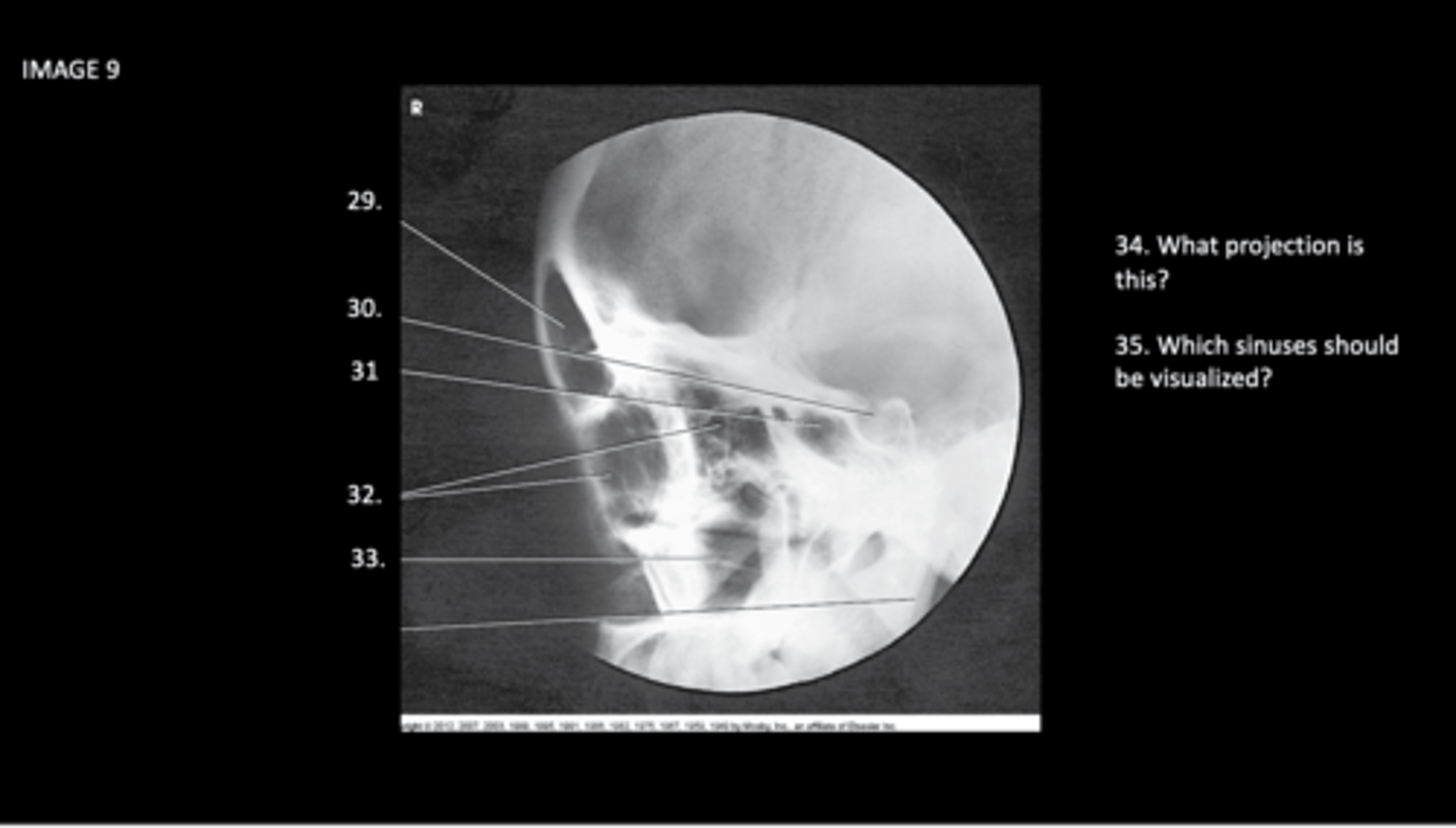
Ethmoid sinuses
What is 32?

Maxillary sinuses
What is 33?

Lateral projection & all sinuses
What projection is this, which sinuses should be visualized?

Frontal Sinus
What is 36?

Ethmoidal sinus
What is 37?
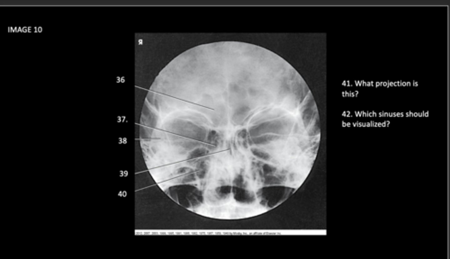
Petrous Ridge
What is 38?

Sphenoid sinus
What is 39?

Maxillary sinus
What is 40?

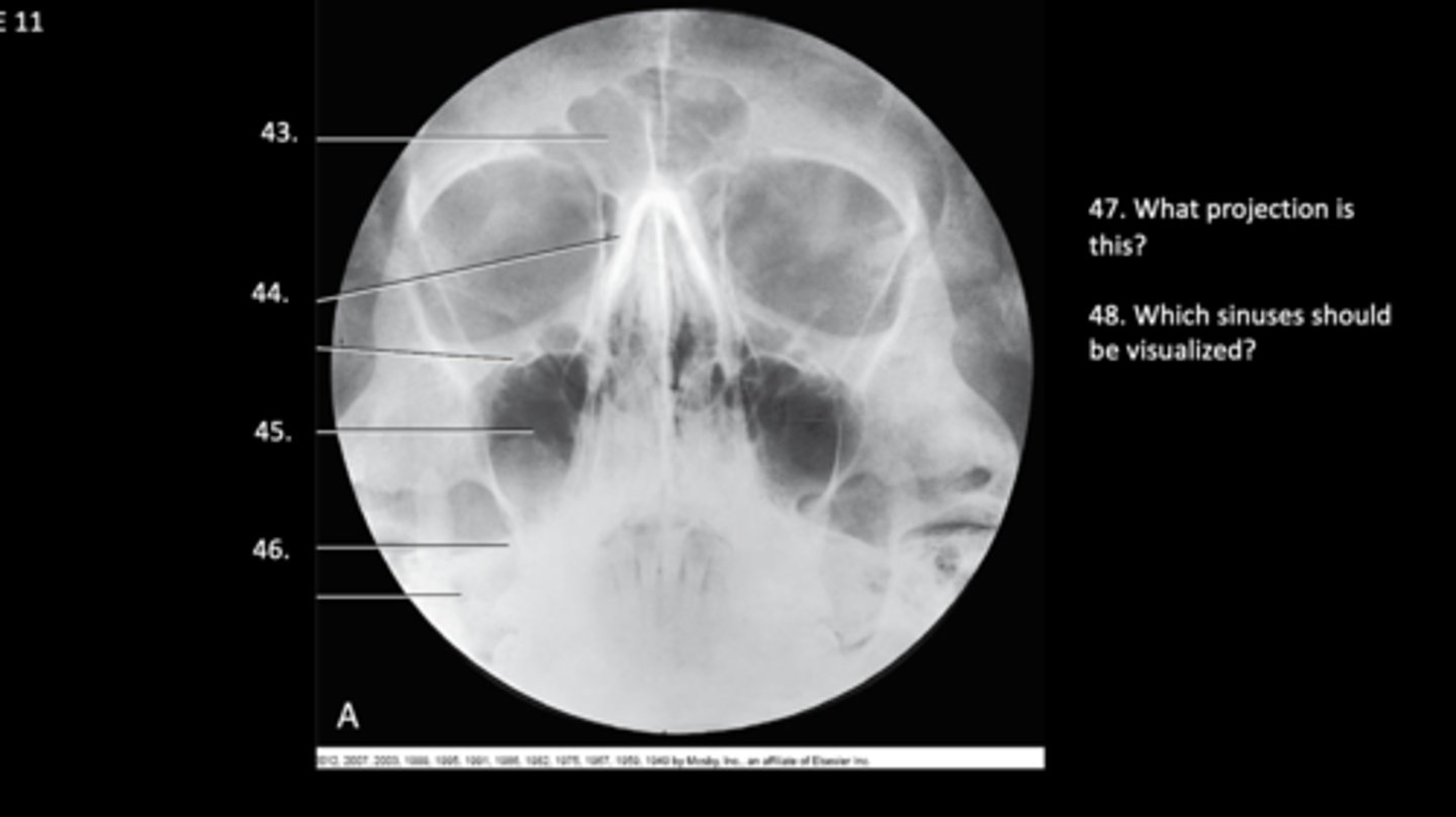
PA Axial Caldwell. frontal and ethmoid
What projection is this?
Which sinuses should be visualized?

Frontal Sinus
What is 43?

Ethmoid Sinus
What is 44?

Maxillary sinus
What is 45?

Petrous ridge
What is 46?

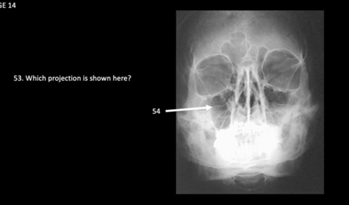
Waters method & Maxillary sinuses
What projection is this, and what sinuses should be visualized?
Waters method
What projection is this?

maxillary sinus
What is 54?
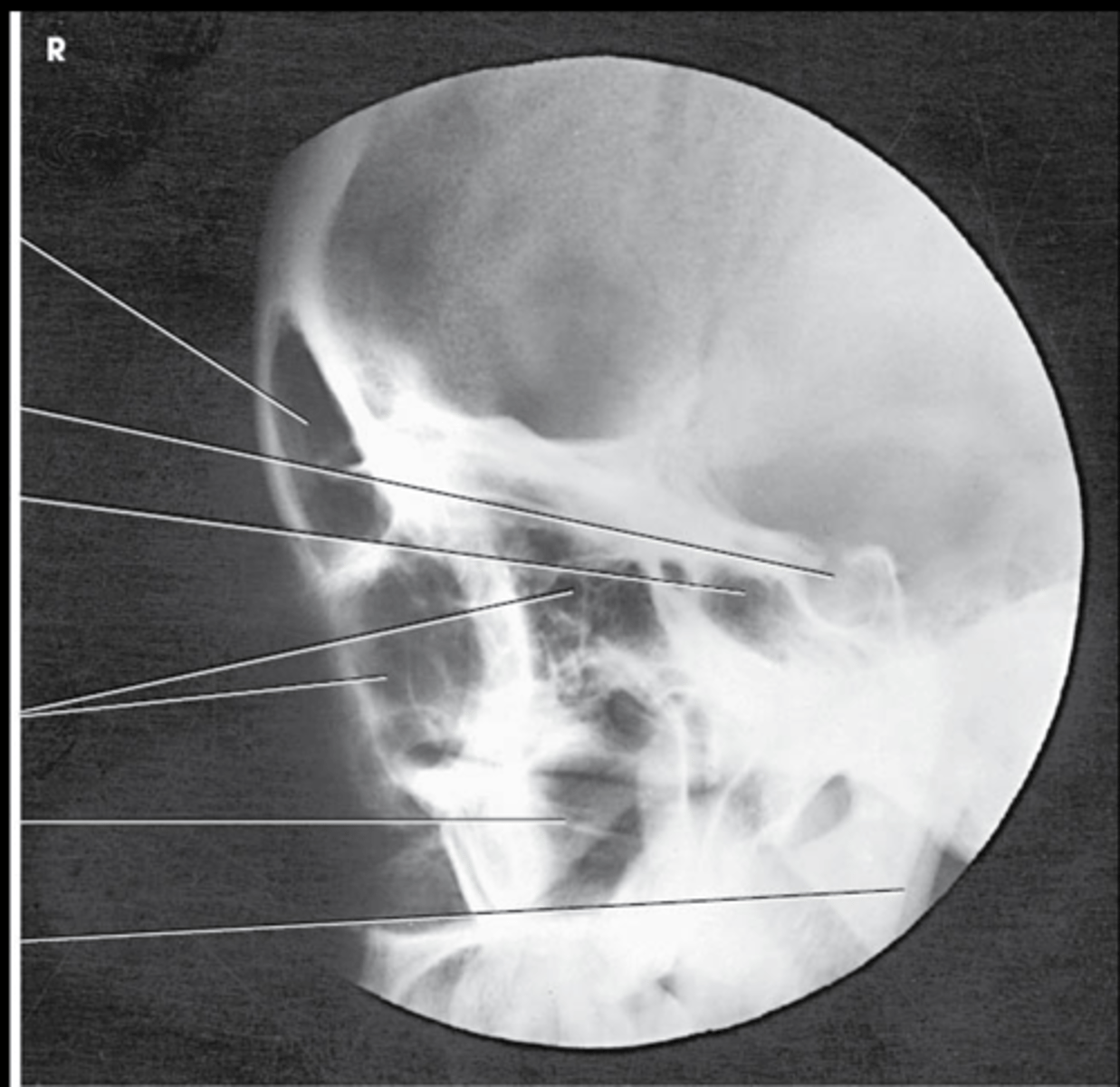
Lateral Sinuses:
- Sella turcica should not be rotated, orbital roofs and mandibular rami should be superimposed.
- all four sinus groups, but the sphenoid sinus is of primary importance
What are the eval criteria for this image?
PA Axial Caldwell:
- Distance between the lateral border of the skull to the mandibular condyles should be equal on both sides
- Petrous ridges should be symmetric on both sides lying in the lower third of the orbit.
- frontal sinuses should lie above the frontonasal suture and the anterior ethmoid sinuses should be clearly visualized
What are the eval criteria for this image?