cranial blood supply and dural venous sinuses
 Call Kai
 Learn
Learn Practice Test
 Spaced Repetition
 Match
Match Flashcards
 Knowt Play
1/50

There's no tags or description
Looks like no tags are added yet.
Name | Mastery | Learn | Test | Matching | Spaced | Call with Kai | Chat |
|---|
No analytics yet
Send a link to your students to track their progress
51 Terms
most external layer of the scalp
skin
what is in the dense connective tissue layer of the scalp
blood vessels and nerves
scalp layer superficial to the loose connective tissue
aponeurosis
what is in the aponeurosis layer of the scalp
sheetlike tendon of occipitofrontalis muscle
role of loose connective tissue in the scalp
allows for scalp movement and collects fluids
scalp layer that covers the bone
pericranium
calvaria
dome like portion of the skull
layers superficial to the calvaria
skin, dense connective tissue, aponeurosis, loose connective tissue, pericranium
layers deep to the calvaria
dura, arachnoid, and pia mater
periosteal layer
superficial layer of dura mater that is flush with the calvaria
meningeal layer
deep layer of dura mater that is against brain and spinal tissue
tough fibrous layer under the calvaria
dura mater
layer that provides support for blood vessel network and allows for CSF flow
arachnoid mater
layer that directly adheres to brain or spinal cord
pia mater
shock absorbing fluid that also provides waste filtration and nutrients to the brain
cerebral spinal fluid
cranial epidural space
pathological space in brain between the dura layer and bone
spinal epidural space
fat filled space between dura layers in the spinal cord
subdural space
pathological space in between the dura and arachnoid mater
sub arachnoid space
where the cerebral spinal fluid circulates in the arachnoid mater
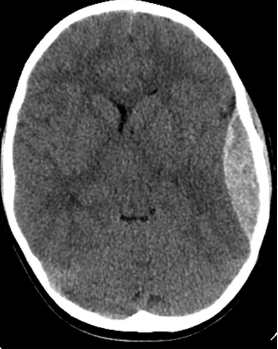
accumulation of blood in cranial epidural space
epidural hemorrhage
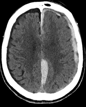
accumulation of blood between dura and arachnoid mater traveling to the dural sinuses
subdural hemorrhage
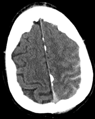
artery or vein rupture that causes accumulation of blood in the arachnoid space where just CSF should be
subarachnoid hemorrhage
arteries that carry blood to your brain branching off of the common carotid arteries
internal carotid arteries
arteries that carry blood to the brain branching off the subclavian artery through the foramen transversarium
vertebral arteries
anastomoses between vertebral and internal arteries that provide multiple pathways to the brain
cerebral arterial circle
artery where the internal carotid arteries merge together
basilar artery
terminal branches in the cerebral arterial circle that supply blood to brain tissue
anterior, middle, and posterior cerebral arteries
artery that connects the two anterior cerebral arteries together
anterior communicating artery
artery that connects the middle and posterior cerebral artery together
posterior communicating artery
supplies blood to midline frontal and parietal lobes
anterior cerebral artery
supplies blood to lateral temporal, parietal, and portion of frontal lobe
middle cerebral artery
supplies blood to occipital and inferior temporal lobes
posterior cerebral
artery that supplies blood to the cranium but not the brain branching off the common carotid artery
external carotid artery
branch off the external carotid artery that also branches off into the middle meningeal artery
middle maxillary artery
branch off of the internal maxillary artery that supplies blood to the dura
middle meningeal artery
draws blood out of the subarachnoid space and dumps it into the veinus dural sinus
bridging vein
big sickle shaped layer in the middle of the dural layer
falx cerebri
part of the dural layer that comes down and separates the two halves of the cerebellum
falx cerebelli
part of the dural layer that separates the cerebrum and cerebellum
tentorium cerebelli
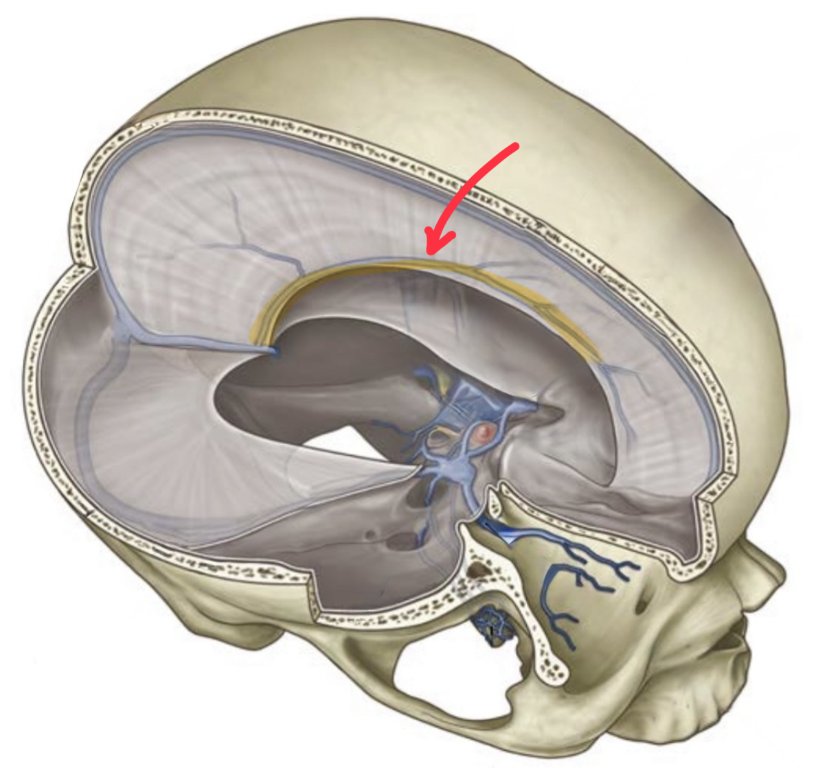
inferior sagittal sinus
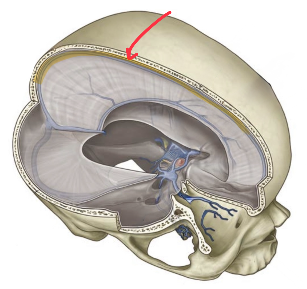
superior sagittal sinus
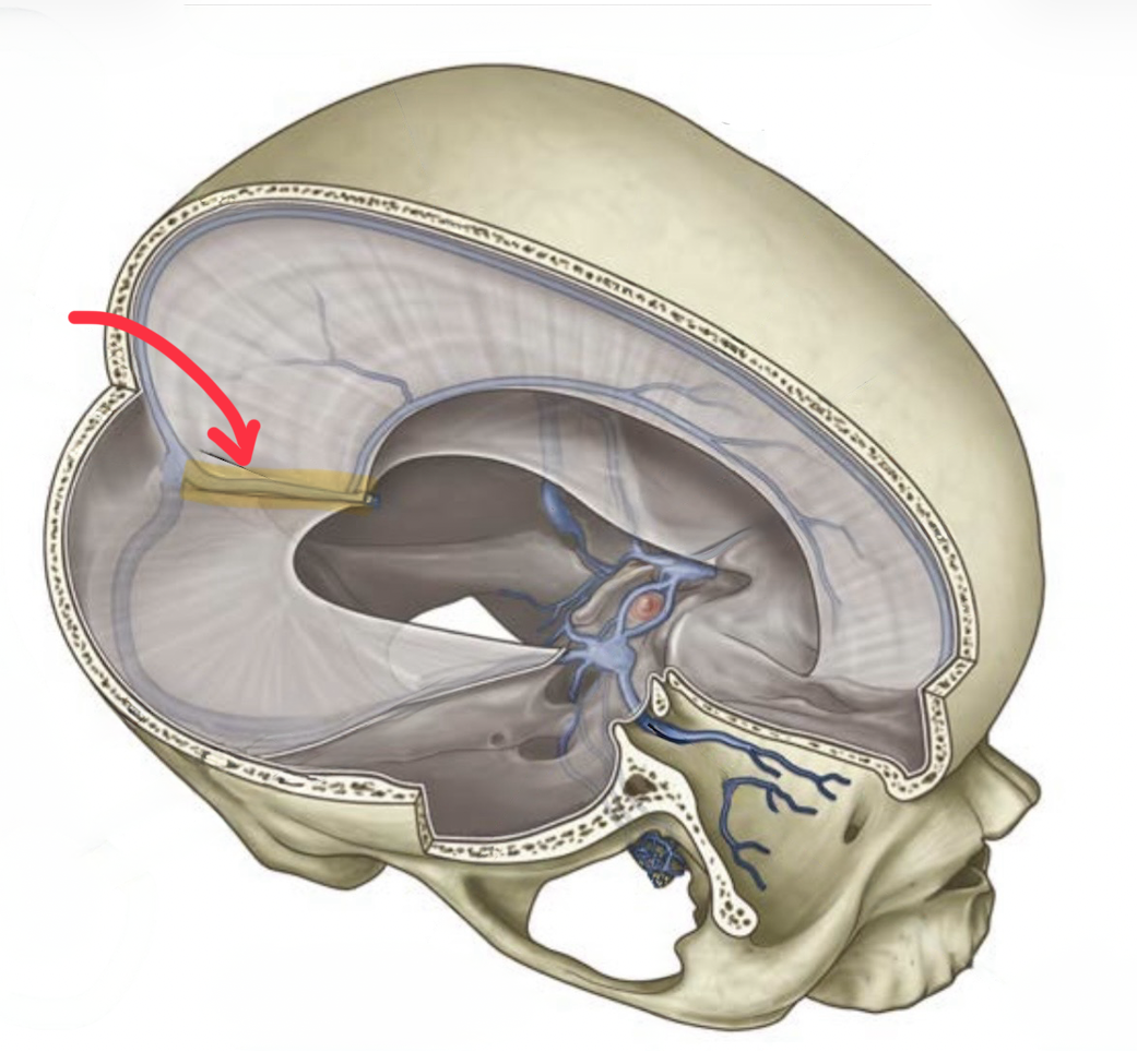
straight sinus
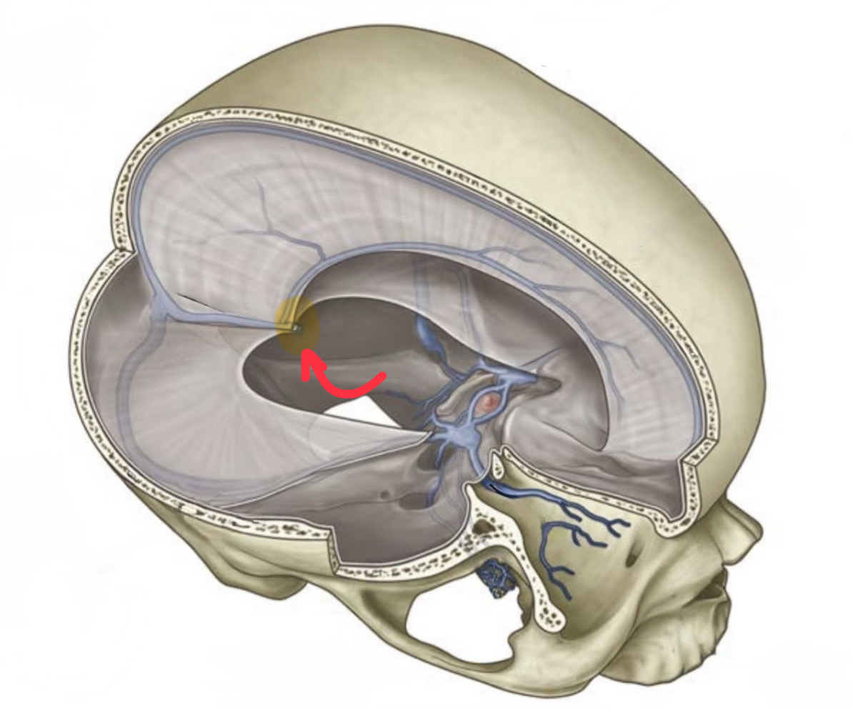
great cerebral vein
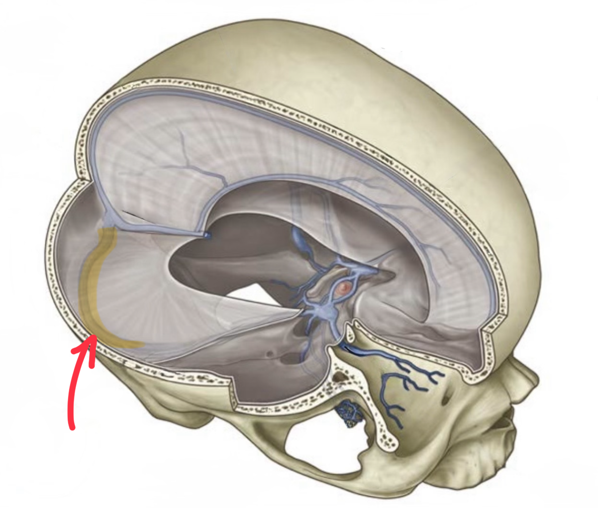
right transverse sinus
sigmoid sinus
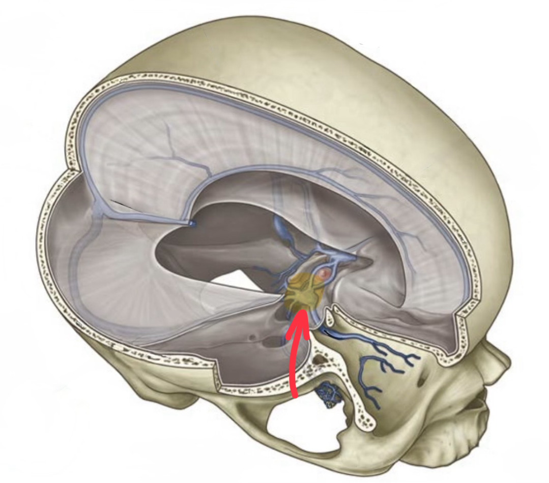
cavernous sinus
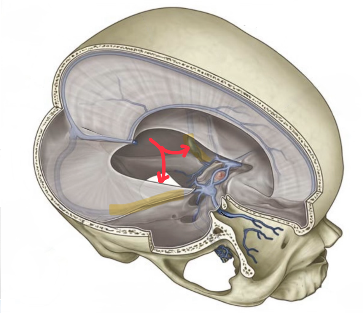
sigmoid sinus
where do the external jugular veins drain
into the internal jugular vein
where do the internal jugular veins drain
into the brachiocephalic veins
where do the inferior petrosal sinus and sigmoid sinus drain
into the external jugular vein
where do our brachiocephalic veins drain
into the superior vena cava