3. Ocular Imaging in Glaucoma - Management of Glaucoma Summer 2026
1/144
There's no tags or description
Looks like no tags are added yet.
Name | Mastery | Learn | Test | Matching | Spaced | Call with Kai | Chat |
|---|
No analytics yet
Send a link to your students to track their progress
145 Terms

The Glaucoma Continuum (Pic)
The Glaucoma Continuum (Pic)

True or False:
Structural damage occurs slowly, gradually, and prior to the loss of visual function
true

A patient will need to. lose ____% of RNFL before VF loss will occur
30
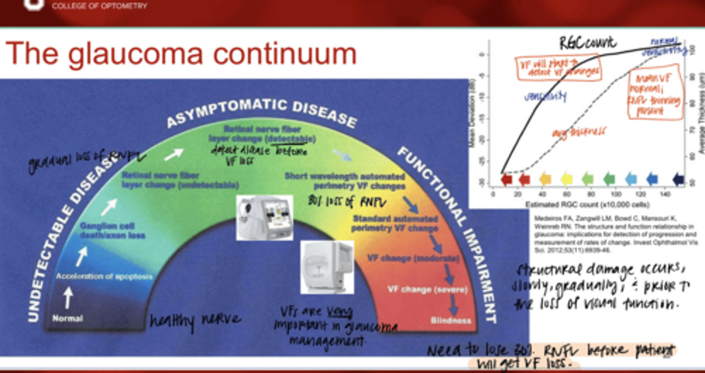
Are OCT & VF important in glaucoma management?
Yes

It is sometimes difficult to detect individual ganglion cell loss within the optic nerve head? Why?
Yes -- since all the ganglion cell axons accumulate within the optic nerve
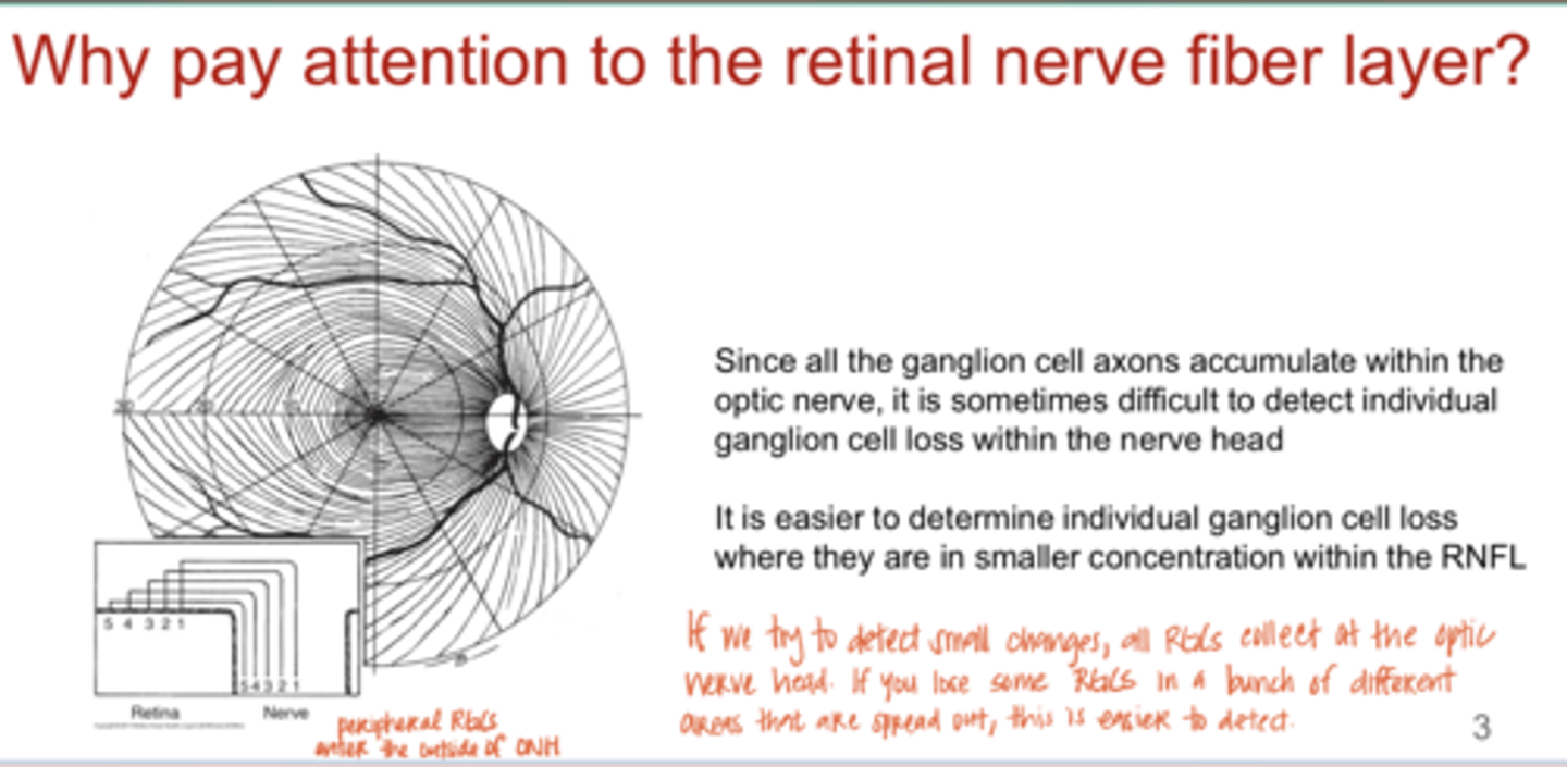
When is it easier to determine individual ganglion cell loss?
where they are in smaller conc within the RNFL

Fundus Photography -- Red Free Images (Pic)
Fundus Photography -- Red Free Images (Pic)
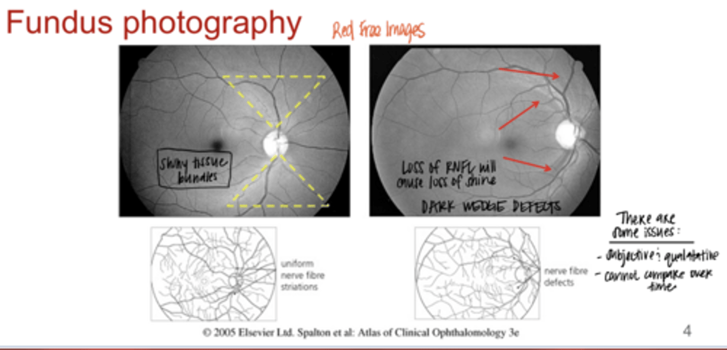
What can be seen in these red free images of the ONH?
wedge defects -- dark areas that indicate loss of RNFL

What are the issues with Fundus Photography when managing glaucoma?
-subjective and qualitative
-cannot compare over time

Optical coherence tomography (OCT) is based on the principle of what?
low-coherence tomography

OCT is similar to what?
multiple ultrasound A-scans

What does OCT use?
near infrared laser beam around 840nm
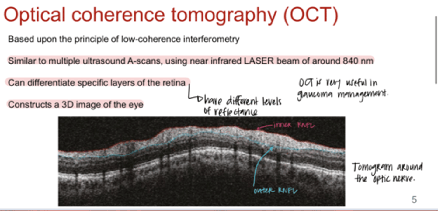
Can OCT differentiate specific layers of the retina?
yes

What does OCT construct?
3D image of the eye

The image detail of OCT will be created from what?
"reflectivity" of the laser from the tissues of the eye

What does RED color mean in an OCT of the macula?
high reflectivity

What does YELLOW color mean in an OCT of the macula?
Intermediate reflectivity

What does GREEN color mean in an OCT of the macula?
low reflectivity

What does BLACK color mean in an OCT of the macula?
No reflectivity
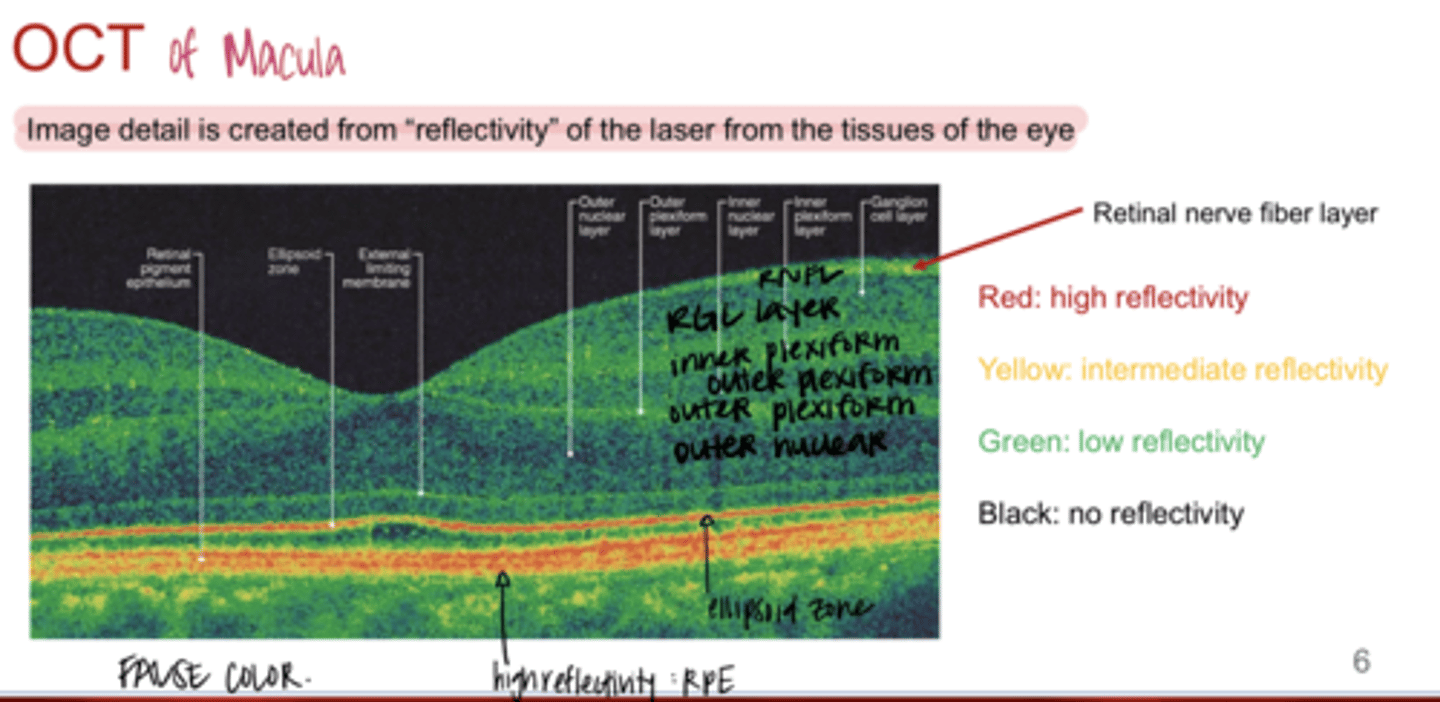
What layers of the retina have HIGH reflectivity (Red)?
-RPE
-ellipsoid zone
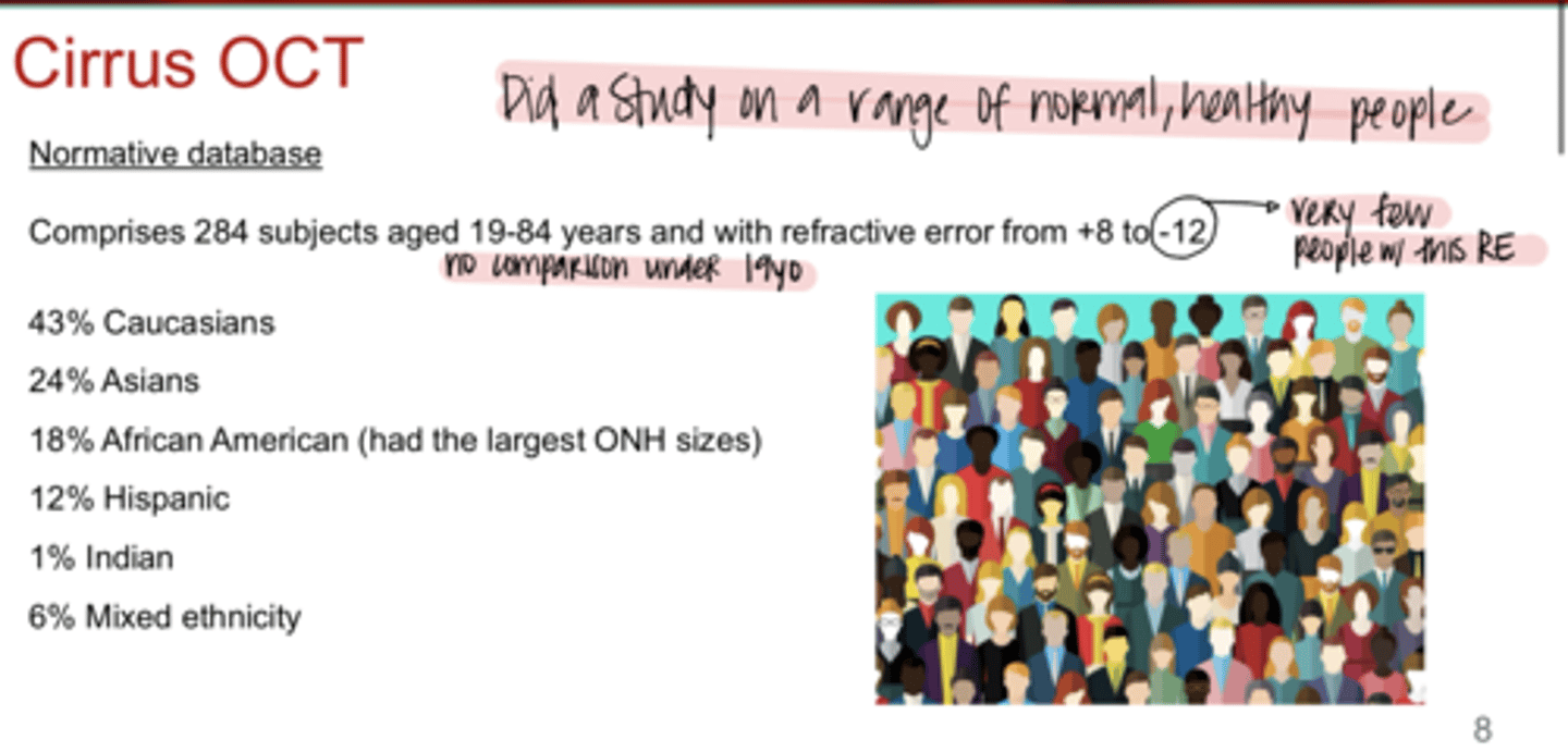
How did Cirrus OCT gather a normative database for their measurements?
284 subjects aged 18-84 and with refractive error +8 to -12

WHITE color on Cirrus OCT meaning
5% falls within white band, considered outside normal limits
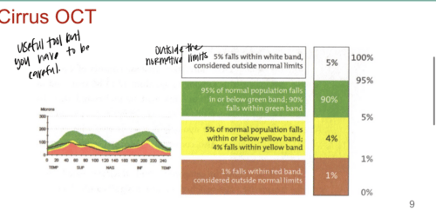
GREEN color on Cirrus OCT meaning
95% of normal population falls in or below the green band; 90% fall within the green band

YELLOW color on Cirrus OCT meaning
5% of normal population falls within or below the yellow band; 4% falls within the yellow

RED color on Cirrus OCT meaning
1% falls within the red band; considered outside normal limits
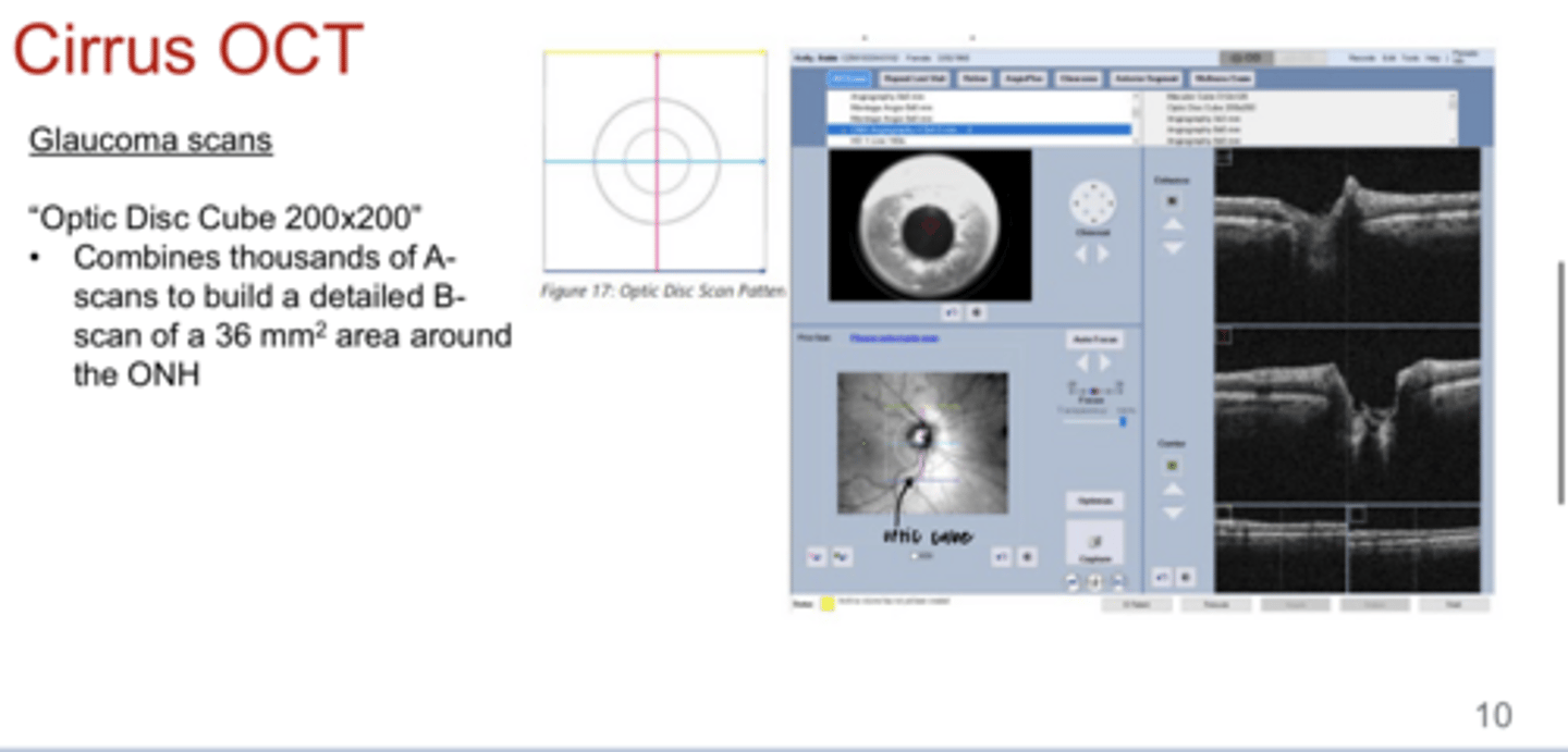
What is the purpose of the Optic Disc Cube 200x200 scan?
Combines thousands of A-scans to build a detailed B-scan of a 36mm^2 area around the ONH

What does the Optic Disc Cube 200x200 scan provide?
-measurement of RNFL thickness
-analysis of ONH characteristics
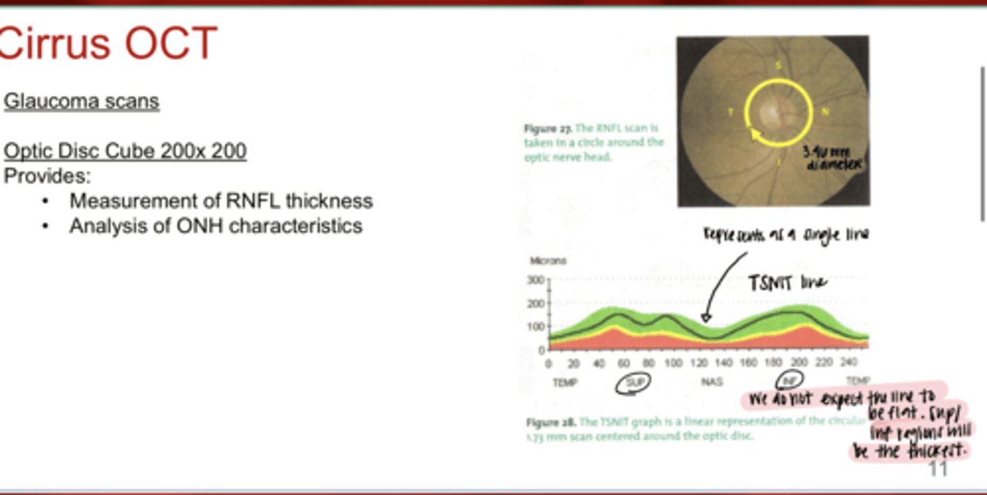
The Optic Disc Cube scan will be laid flat and provide the _______ line
TSNIT

On TSNIT graph, what areas of the retina will be the thickest?
Superior/inferior
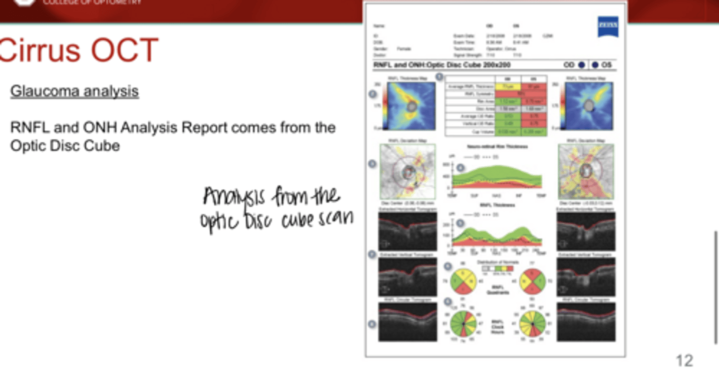
RNFL and ONH Analysis Report will come from the _______ scan
Optic Disc Cube

What is the function of a Macular Cube 512x128 scan?
-Combines thousands of A-scans to build a detailed B-scan of a 36mm^2 area around the fovea
-Higher resolution horizontally
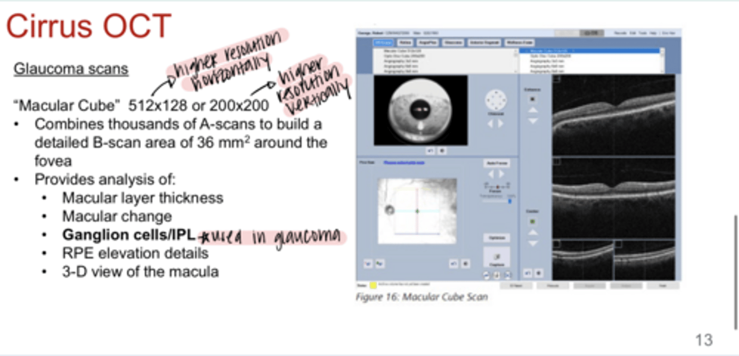
What is the function of a Macular Cube 200x200 scan?
-Combines thousands of A-scans to build a detailed B-scan of a 36mm^2 area around the fovea
-Higher resolution vertically

Macular Cube Scans provide analysis for what?
-macular layer thickness
-macular change
-ganglion cells/IPL
-RPE evaluations details
-3D view of the macula
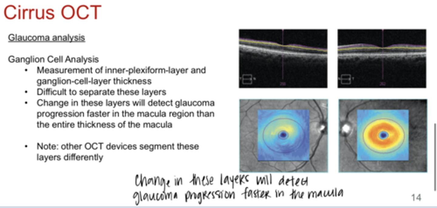
What is the purpose of the ganglion cell analysis on Cirrus OCT?
-measurement of the inner plexiform layer and the retinal ganglion cell layer thickness
-difficult to separate these layers
-change in these layers will detect glaucoma progression faster in the macula region that the entire thickness of the macula
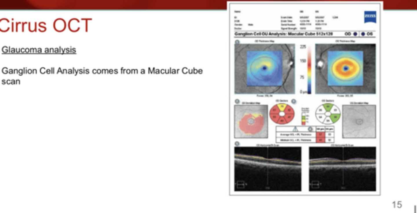
Ganglion Cell Analysis comes from a ______ scan
Macular Cube

The OCT measures what?
The entire thickness of the RNFL

What comprises the RNFL, which is measured with OCT?
-glial cells
-blood vessels
-astrocytes
-RGC axons

The RNFL is segmented between the _____ and ____ lines
red; purple
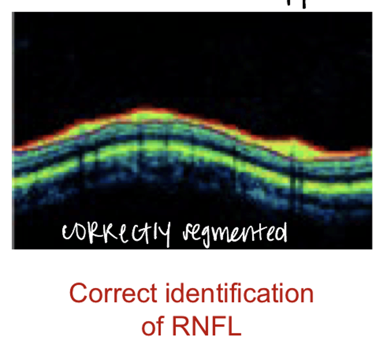
Correct Identification of the RNFL (Pic)
Correct Identification of the RNFL (Pic)
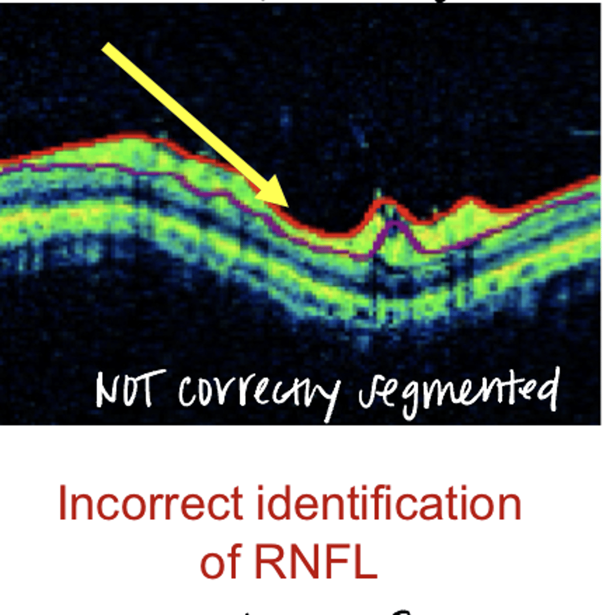
Incorrect Identification of the RNFL (Pic)
Incorrect Identification of the RNFL (Pic)
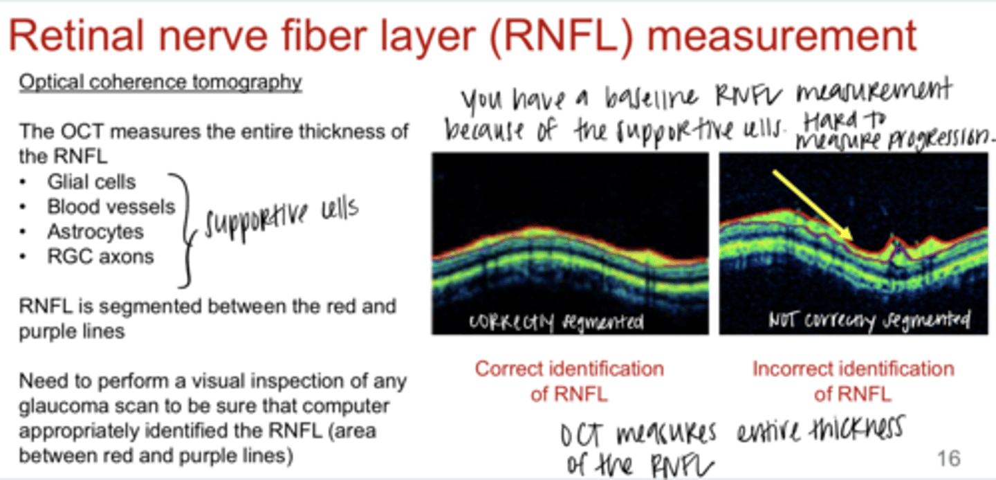
Is it hard to measure progression of glaucoma with OCT in severe stages d/t the presence of supportive cells in the RNFL?
Yes -- there will always be a baseline thickness d/t the presence of these supportive cells in the RNFL

Fundoscopic examination of the ONH will have both _____ and _____ variability
-inter-examiner
-intra-examiner
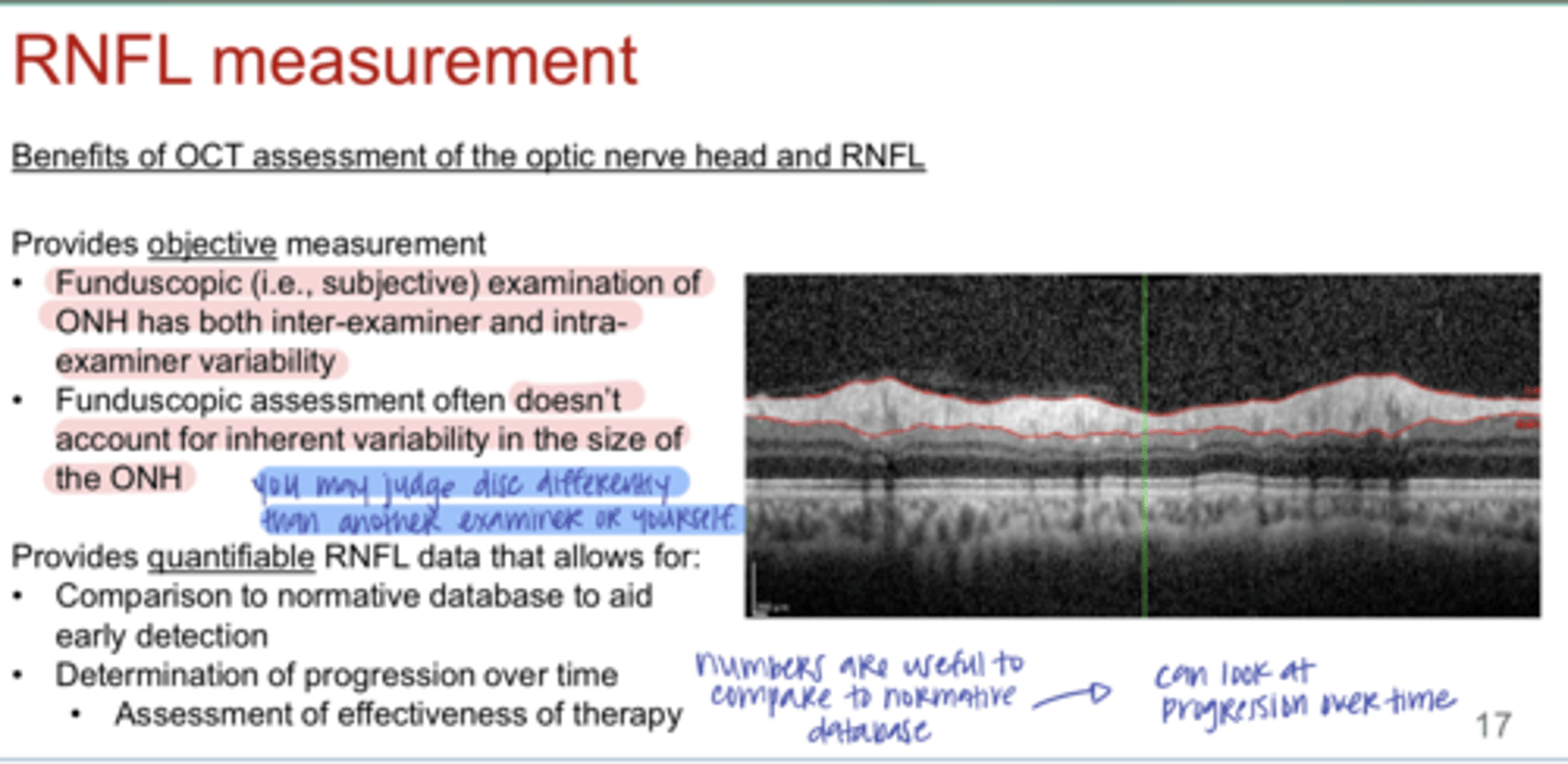
Fundoscopic examination of the ONH often (does/does not) account for inherent variability in the size of the ONH
does not

What does OCT provide quantifiably?
-comparison to normative database to aid in early detection of disease
-determination of progression over time (assessment of effectiveness of therapy)
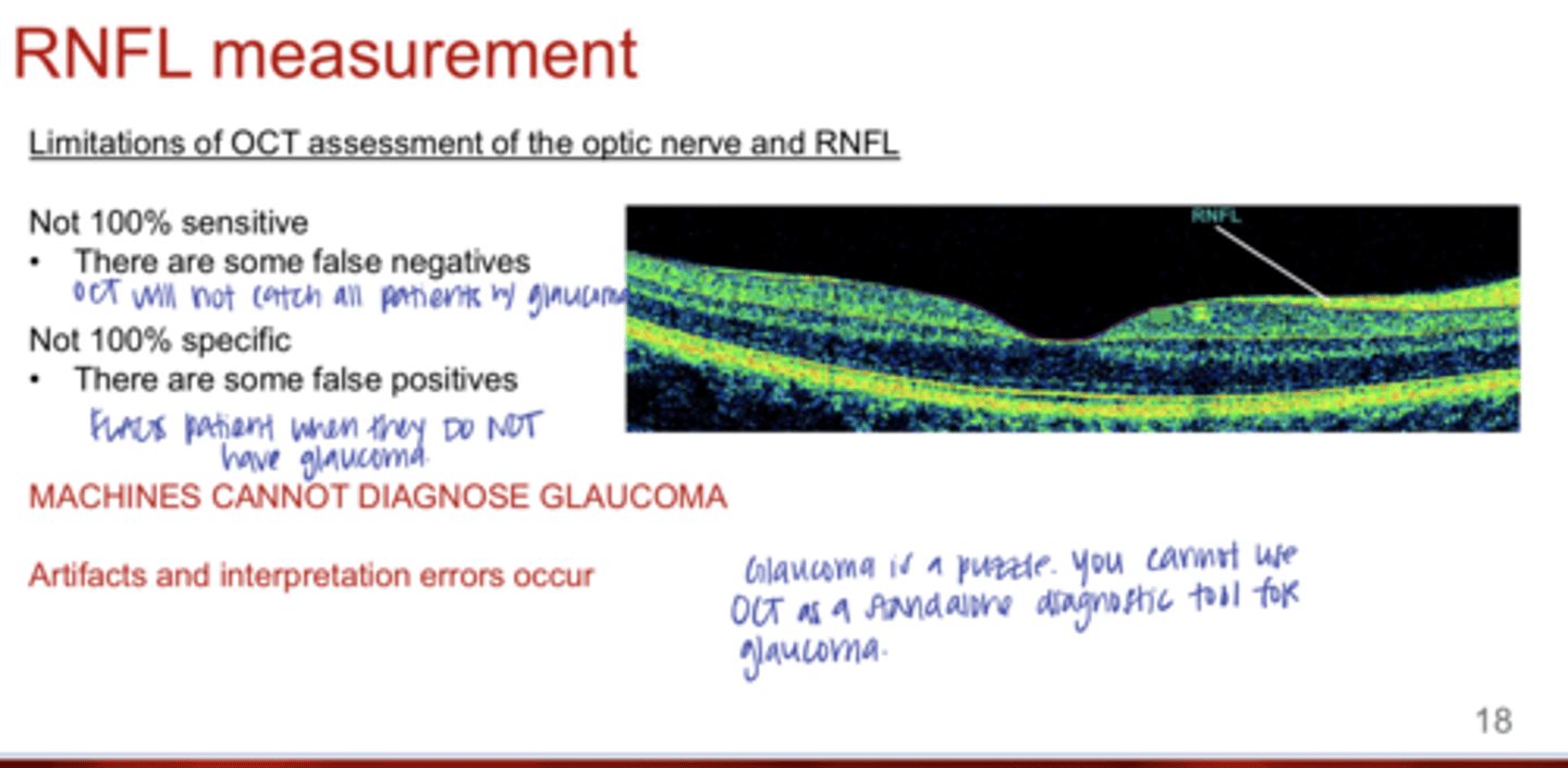
What are the limitations of OCT assessment of the ONH and RNFL?
-Not 100% sensitive (some false negatives)
-Not 100% specific (some false positives)

Can machines, such as OCT, diagnose glaucoma?
No -- machines cannot diagnose glaucoma

True or False:
Artifacts and interpretation errors can occur on OCT measurements
true
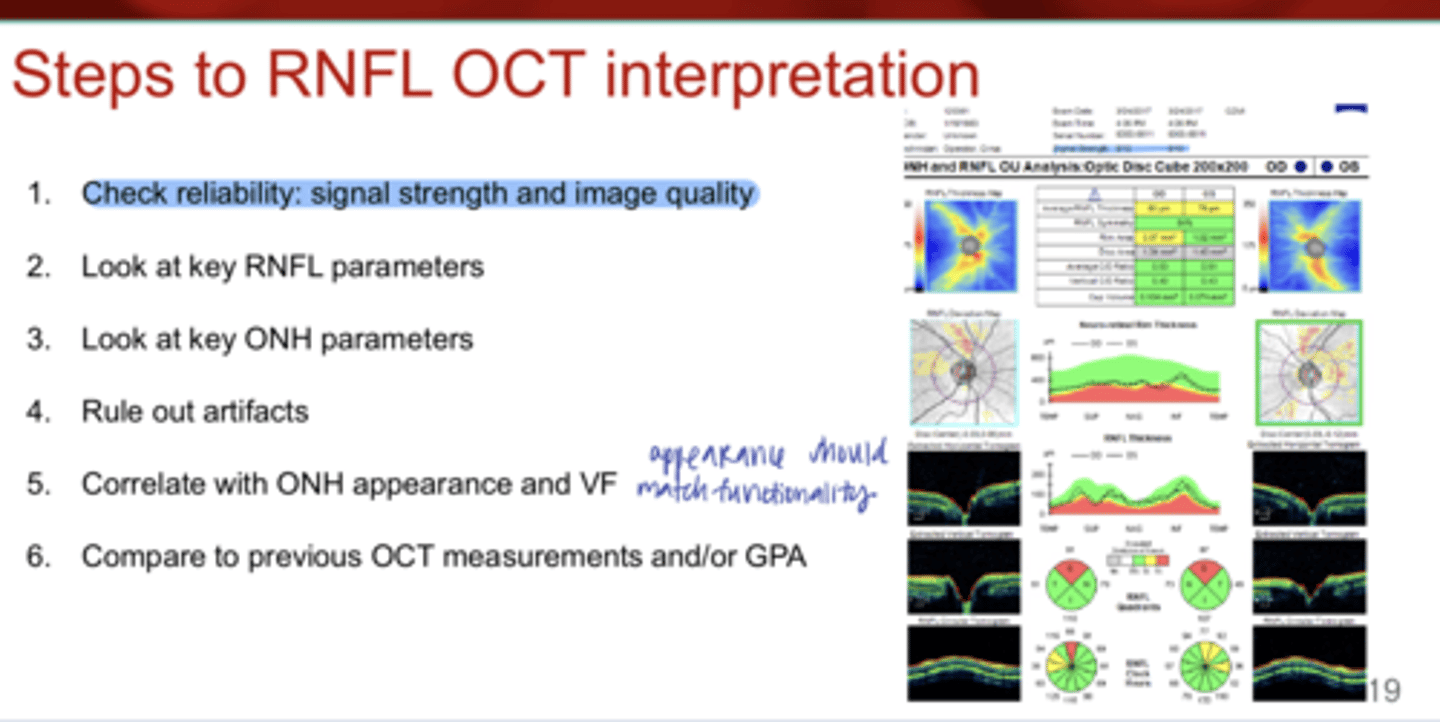
What are the steps to RNFL OCT Interpretation?
1) Check reliability; signal strength and image quality
2) Look at key RNFL parameters
3) Look at key ONH parameters
4) Rule out artifacts
5) Correlate with ONH appearance and VF
6) Compare to previous OCT measurements and/or GPA

Signal strength of OCT measurement should be ____ or above
6
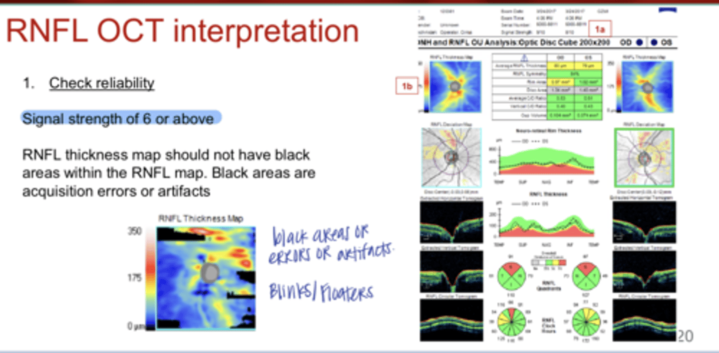
What could black areas in the OCT RNFL thickness map indicate?
acquisition errors or artifacts (blinks/floaters)
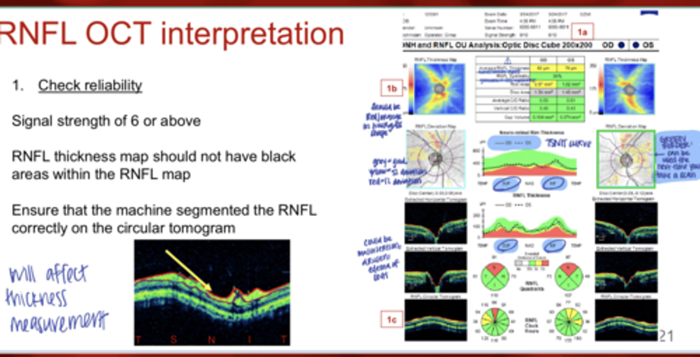
What should you ensure when interpreting OCT RNFL measurement?
Ensure that the machine segmented the RNFL correctly on the circular tomogram
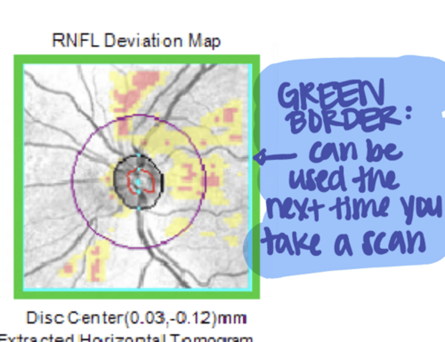
What does the green border around the RNFL Deviation Map indicate on "ONH and RNFL OU Analysis"?
This scan can be used next time for comparison over time
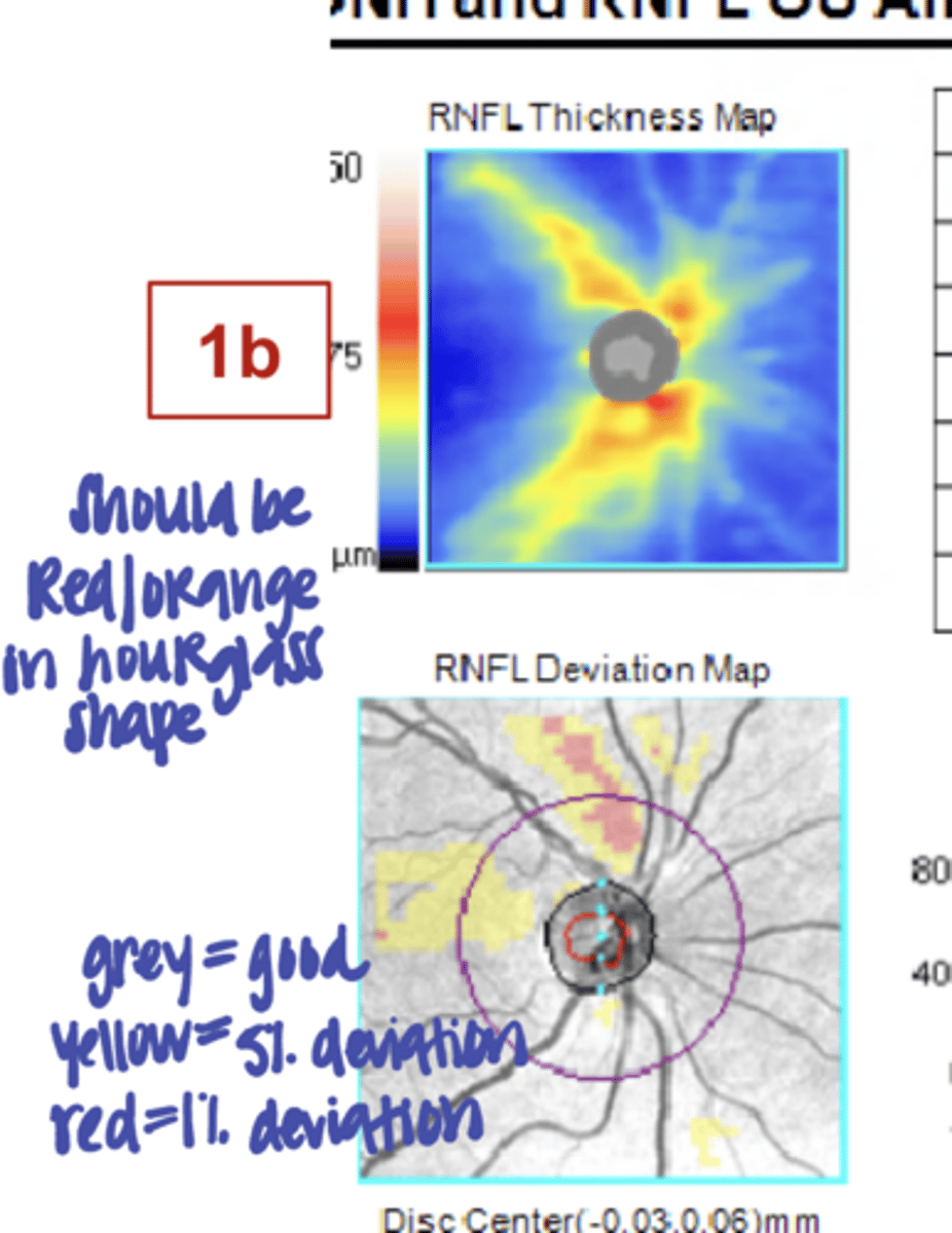
On the RNFL thickness map, the red/orange color should be in a _____ shape
hourglass

Average RNFL thickness measures across the entire ______
TSNIT
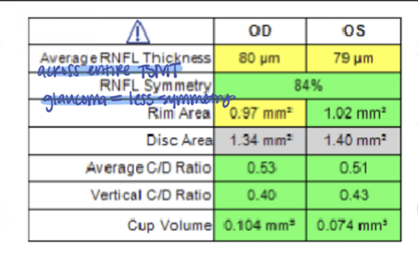
In glaucoma, there will be a (higher/lower) symmetry value
lower
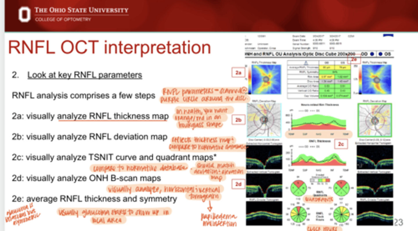
REVIEW: What is important about the RNFL Thickness Map?
In health, you want the orange/red colors in an hourglass shape around the ONH

The RNFL Deviation map reflects what? What is it compared to?
The RNFL Thickness Map & is compared to normative data

TSNIT curve & Quadrant maps should match what?
the RNFL Deviation and Thickness Map

Are the TSNIT curve and Quadrant Maps compared to normative data?
Yes

What ONH B-scan maps need to be analyzed on a ONH and RNFL OCT Analysis?
Horizontal & vertical tomograms

Usually glaucoma starts to show up in a ____ area
local
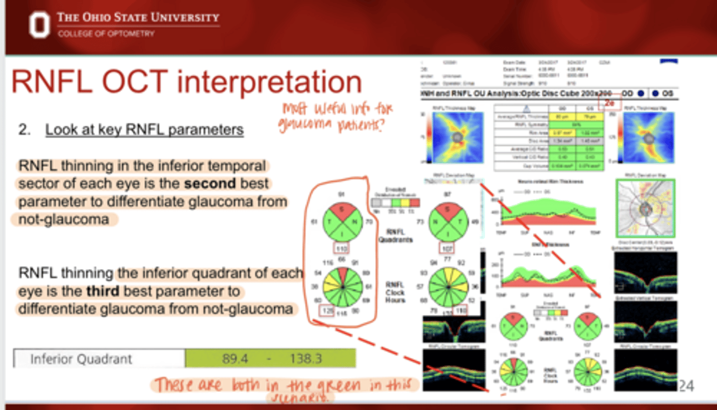
What is the SECOND best parameter to differentiate glaucoma from not-glaucoma?
inferior temporal (IT) sector RNFL thinning in each eye

What is the THIRD best parameter to differentiate glaucoma from not-glaucoma?
inferior quadrant of each eye
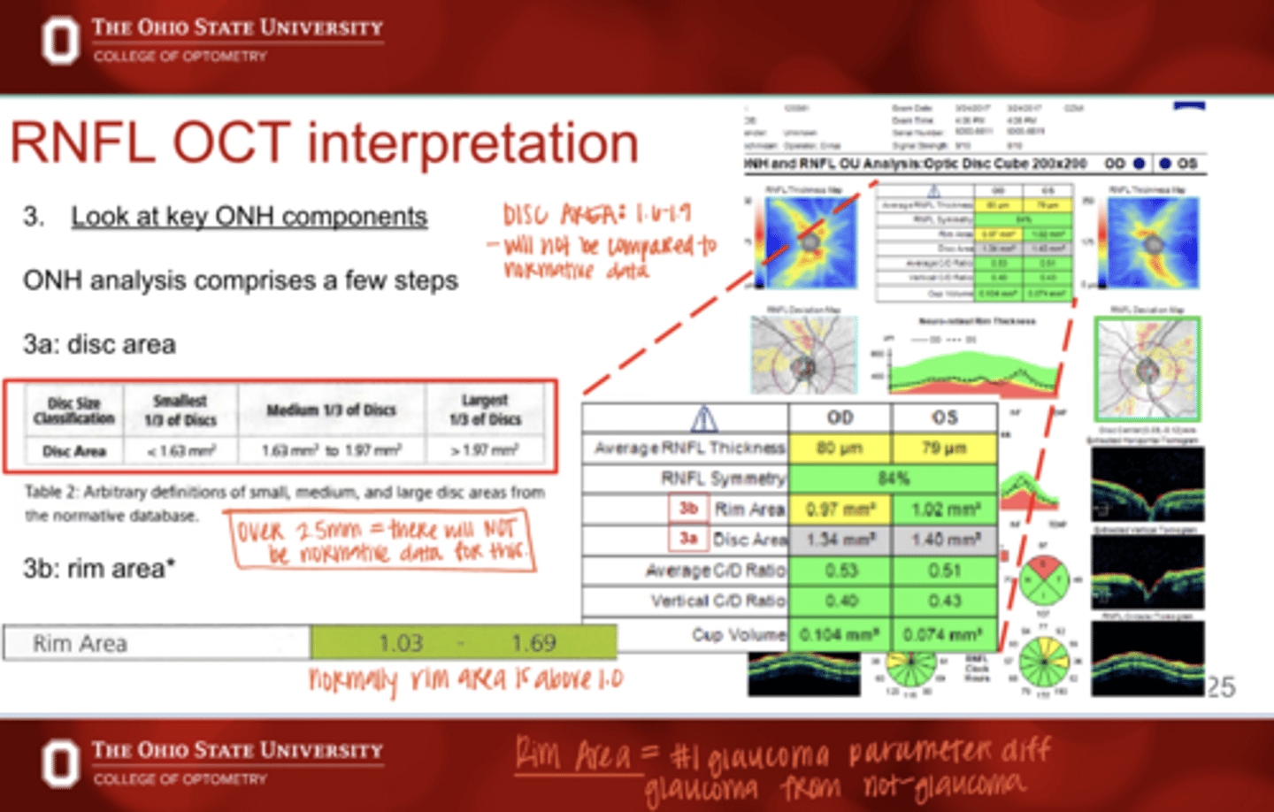
What is the measurement for a SMALL OPTIC NERVE DISC AREA?
<1.6mm^2

What is the AVG measurement for a OPTIC NERVE DISC AREA?
1.6-1.9mm^2

What is the measurement for a LARGE OPTIC NERVE DISC AREA?
>1.9mm^2

Is disc area on OCT compared to normative data?
No -- have to memorize these values

Normally rim area is above _______
1.0mm^2

What is the #1 glaucoma parameter that differentiates glaucoma from not-glaucoma

True or False:
OCT errors can manifest false positive RNFL thinning
true
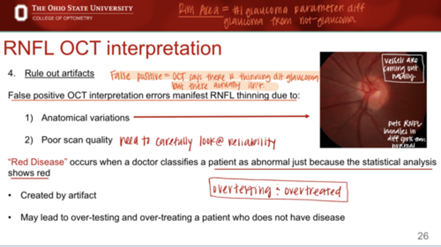
What OCT errors can manifest false positive RNFL thinning?
-anatomical variations
-poor scan quality

What is "red disease"?
occurs when a doctor classified a patient as abnormal just because the statistical analysis shows red, the red is actually d/t an artifact

What can "red disease" lead to?
-over testing
-over treating

What are contributors of red disease (OCT artifacts)?
-poor image acquisition
-PPA
-Scleral crescent
-choroidal crescent
-malinserted discs
-tilted discs
-split bundle deficits
-disc drusen
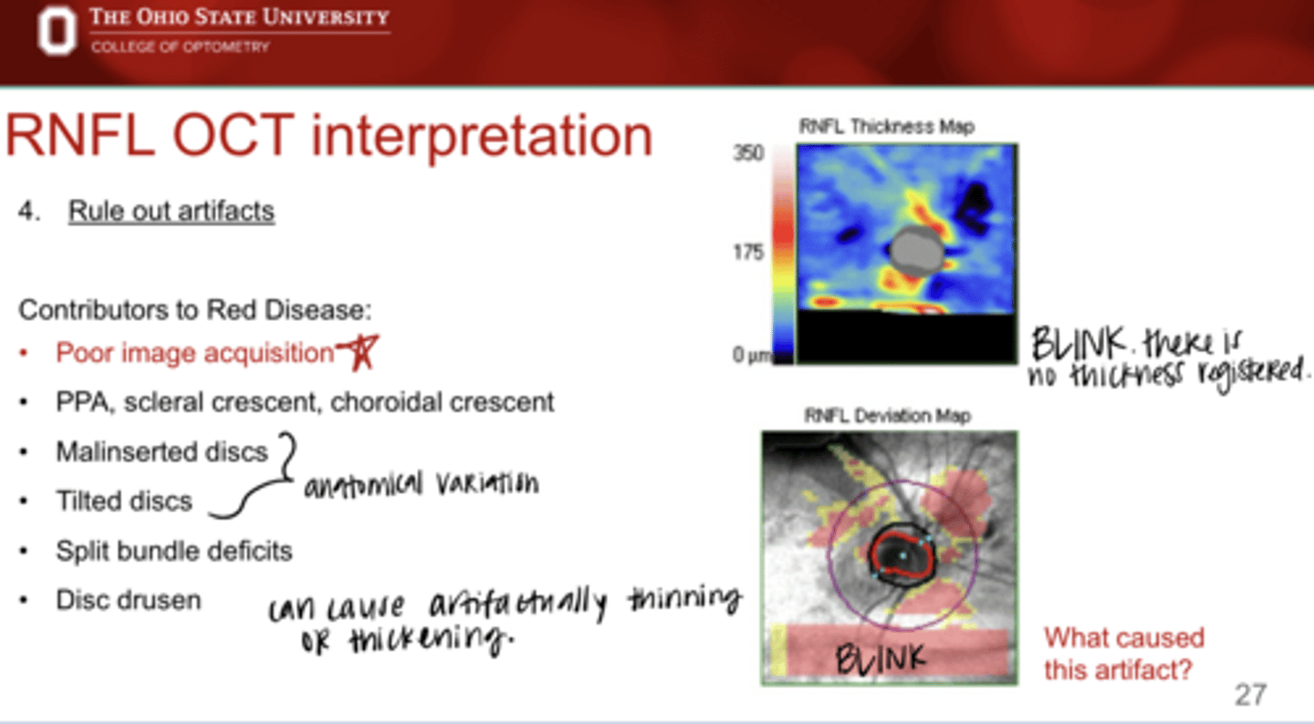
Artifact on OCT (Pic)
Artifact on OCT (Pic)
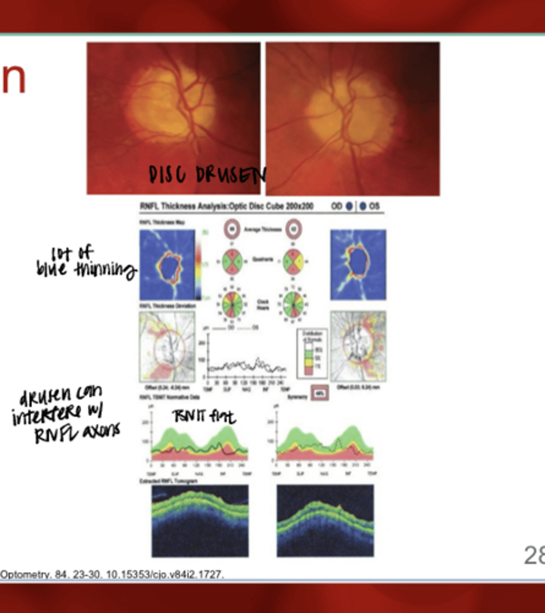
Disc Drusen -- Fundus & on OCT (Pic)
Disc Drusen -- Fundus & on OCT (Pic)

False negative OCT interpretation errors manifest what?
normal OCT thickness in the presence of pathology
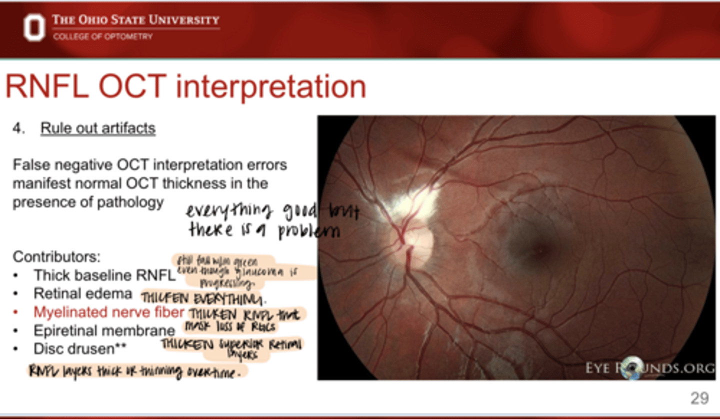
What are the contributors to false negative OCT interpretation errors?
-thick baseline RNFL
-retinal edema
-myelinated nerve fiber
-epiretinal membrane
-disc drusen
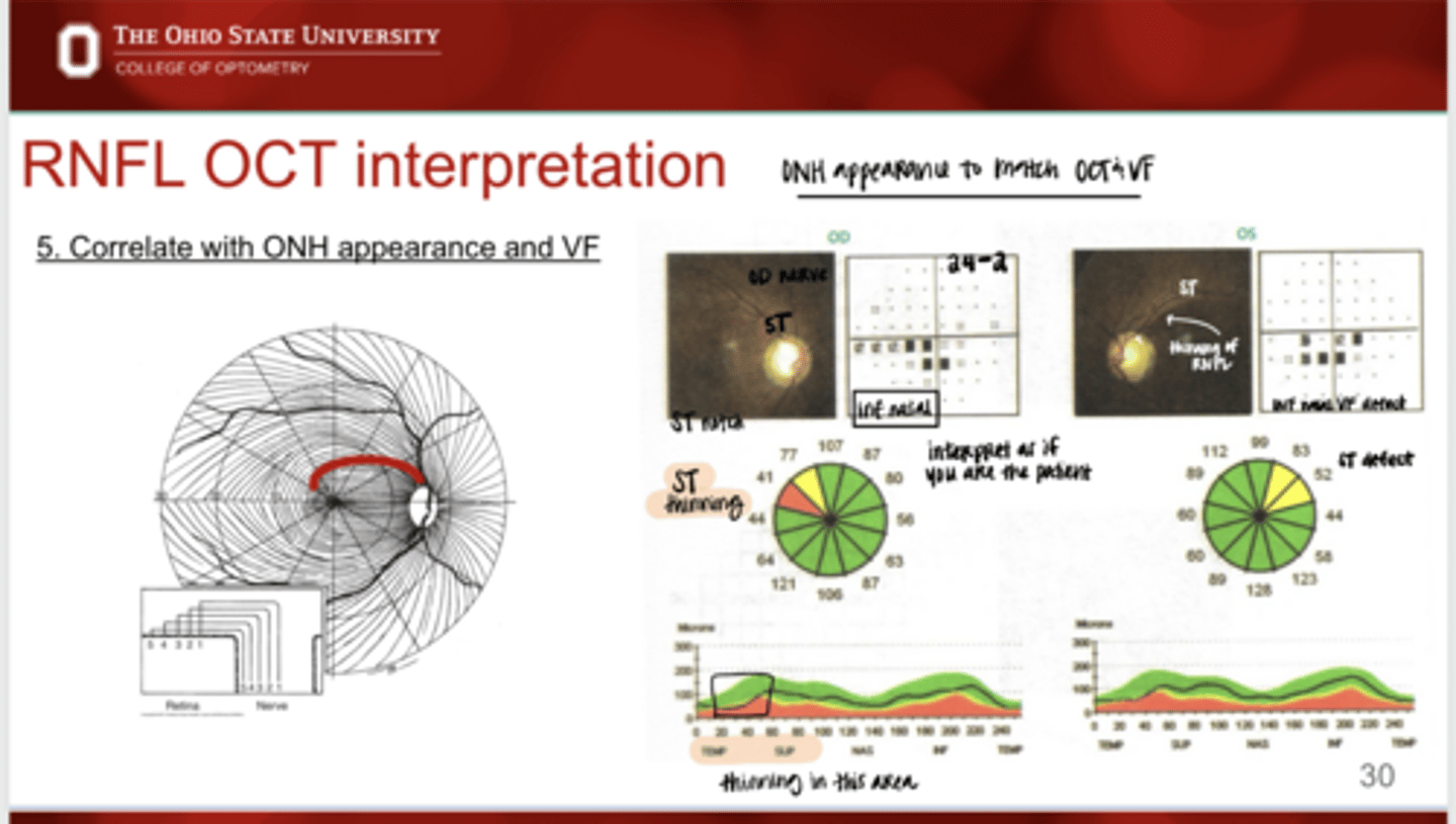
Should you correlate the ONH appearance to the VF?
Yes
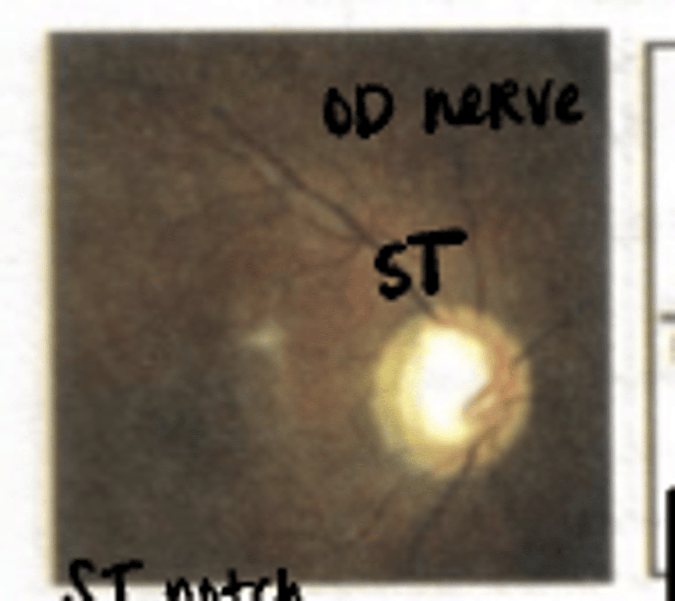
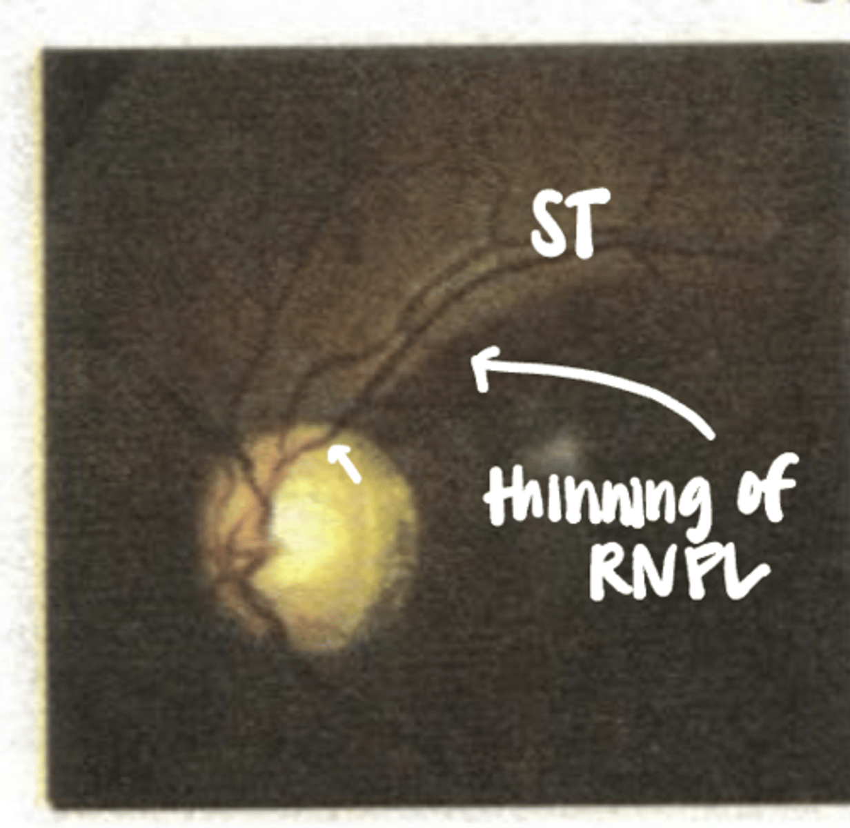
Where is the defect in this ONH?
superior temporal notching
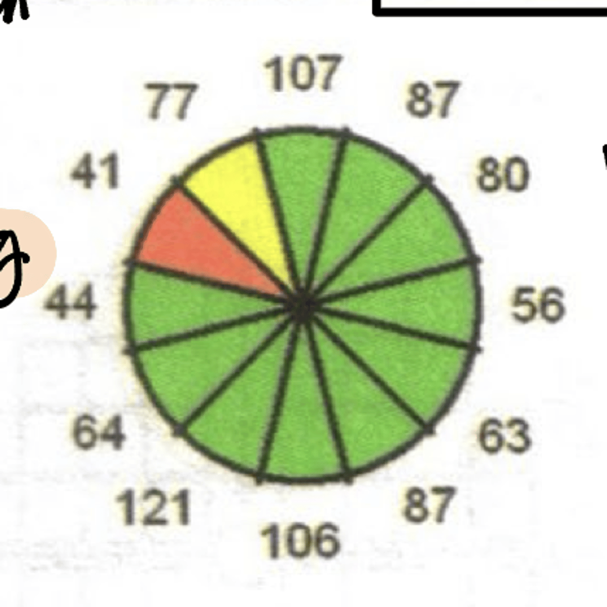
Where is the defect in this OCT?
superior temporal thinning
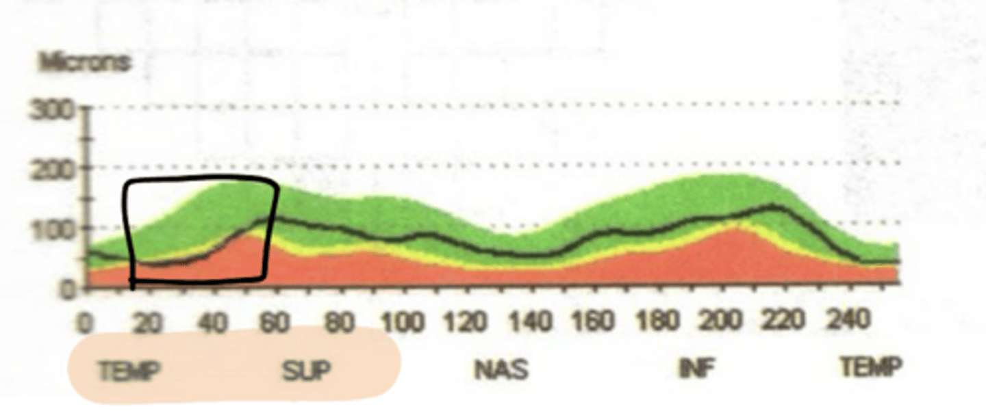
Where is the thinning in this TSNIT curve?
Sup temp thinning
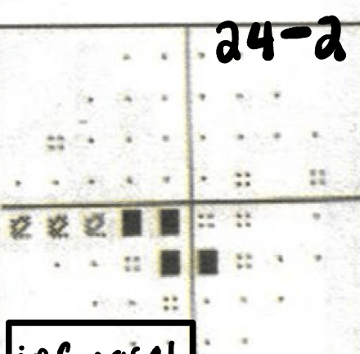
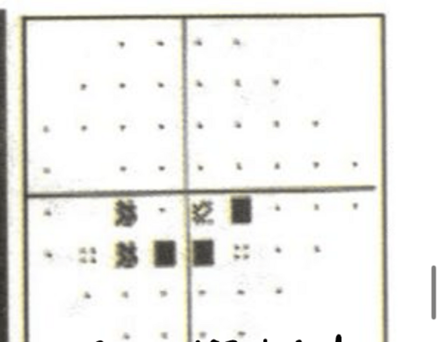
Where is the VF defect?
inferior nasal defect
Where is the defect in this ONH?
superior temporal thinning of RNFL
Where is the defect in this OCT?
superior temporal thinning
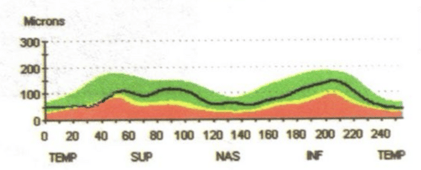
Where is the thinning in this TSNIT curve?
sup temp thinning
Where is the VF defect?
inferior nasal defect
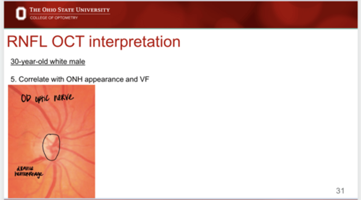
Example: Correlate this nerve ONH appearance with a VF (See Pic)
-vertical elongation
-drance hemorrhage inferior temporal
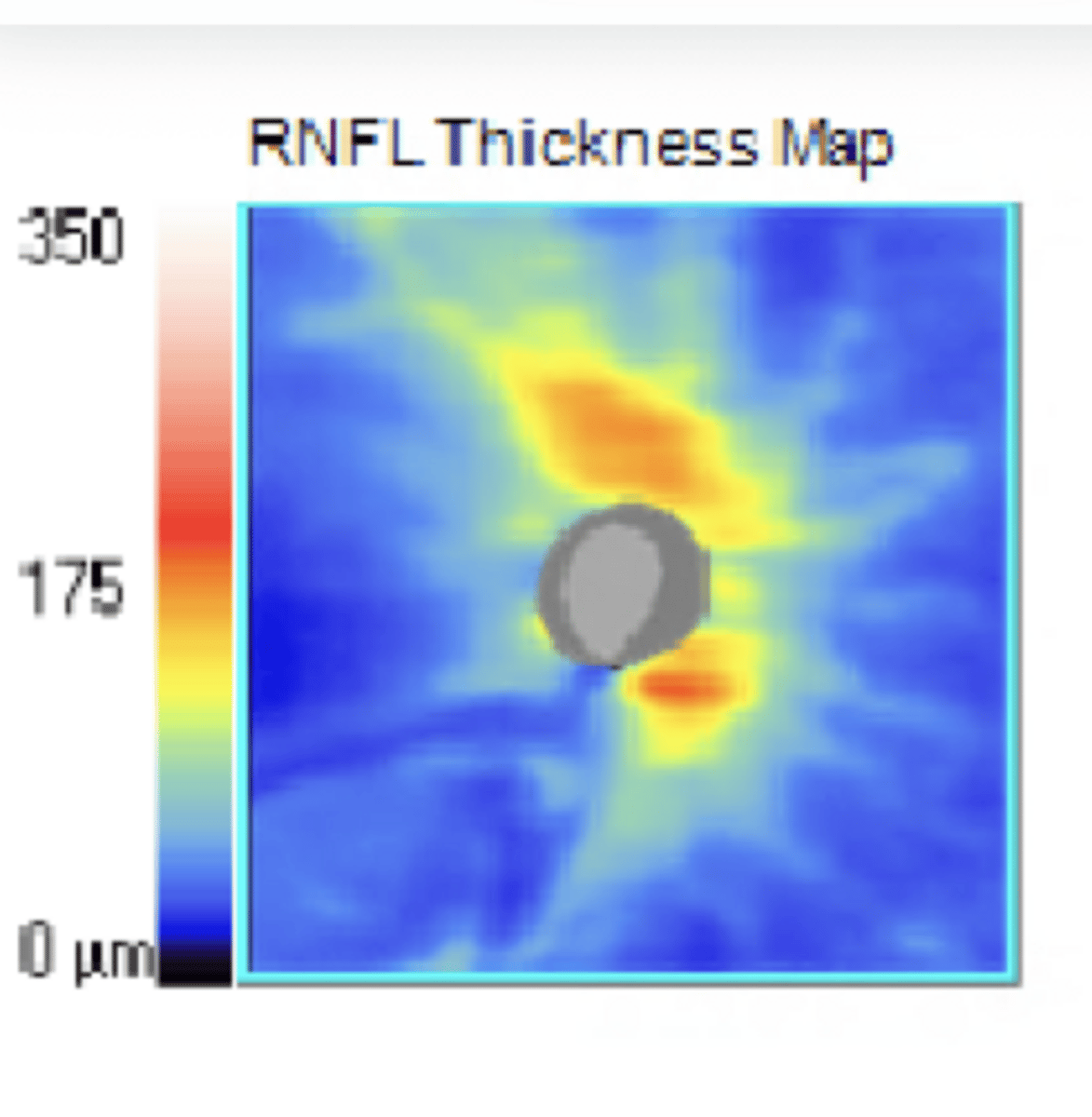
RNFL Thickness Appearance for IT Drance Hemorrhage
**IT thinning shown
RNFL Thickness Appearance for IT Drance Hemorrhage
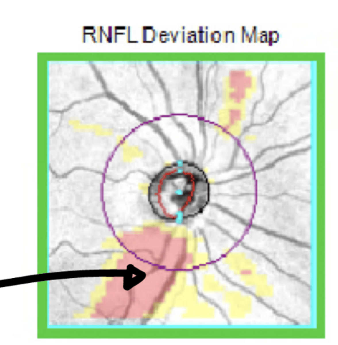
RNFL Deviation Map for IT Drance Hemorrhage
**thin compared to normative data
RNFL Deviation Map for IT Drance Hemorrhage
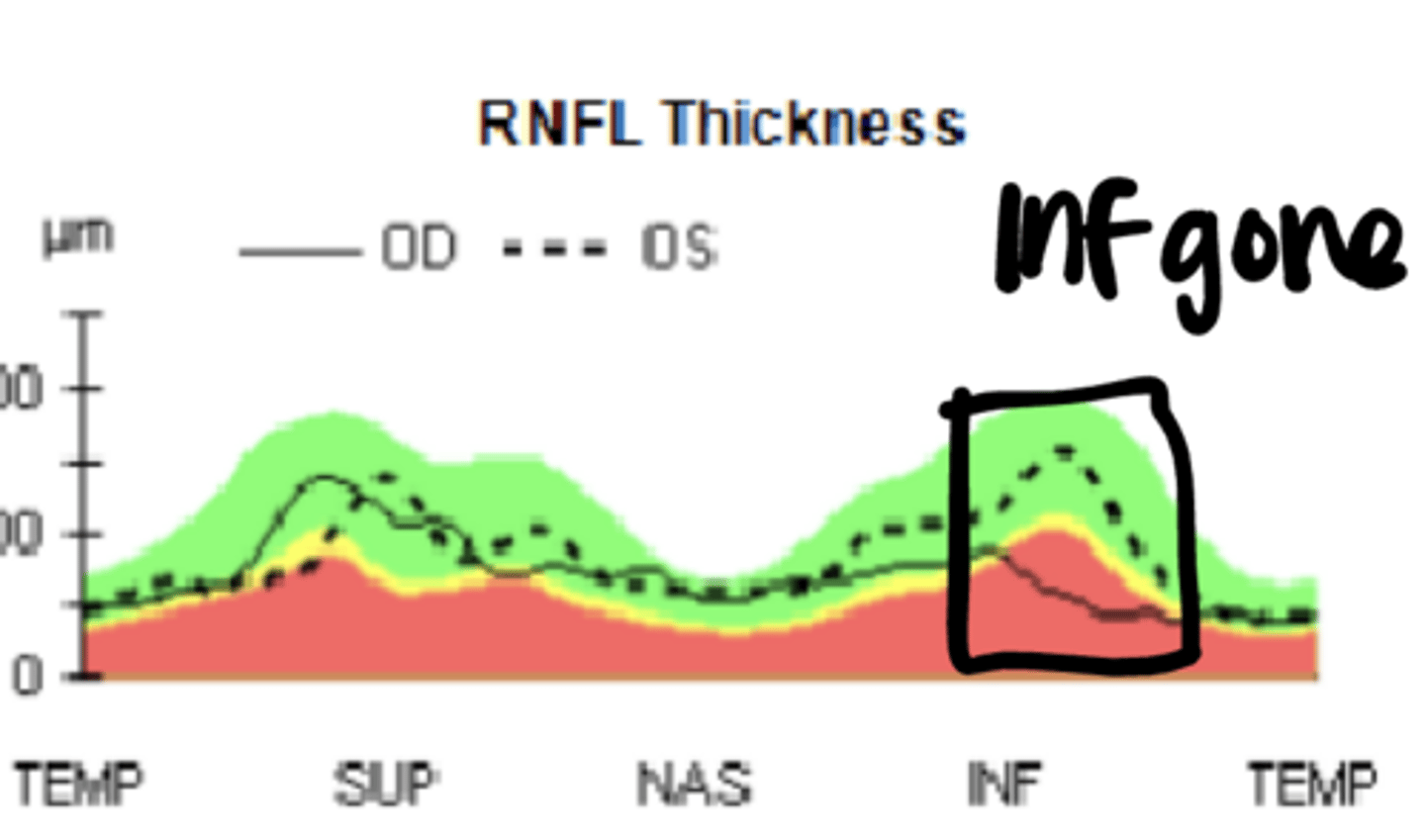
TSNIT Curve for IT Drance Hemorrhage
Inferior temporal thinning
Where would you expect a VF defect to be present for an Inferior Temporal Drance Hemmorhage?
superior nasal
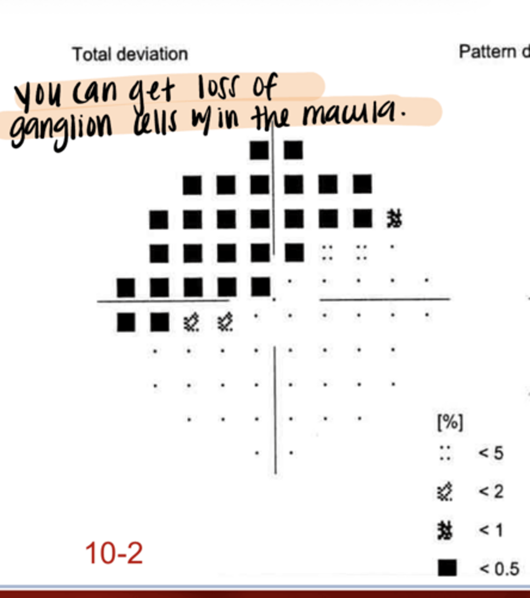
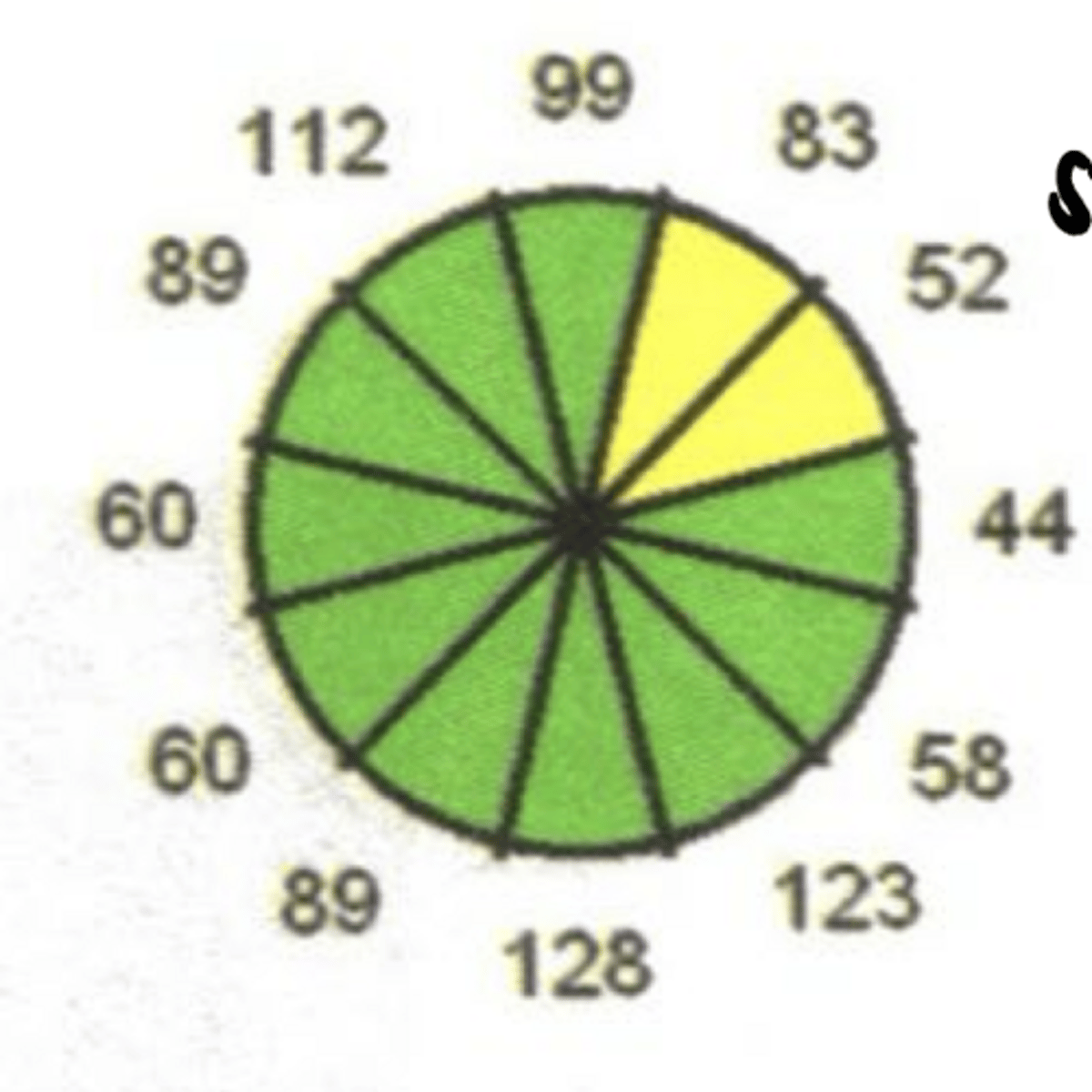
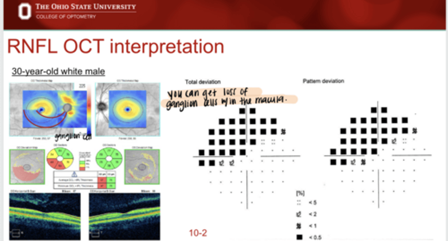
Total Deviation VF Plot for IT Drance Hemorrhage
See pic
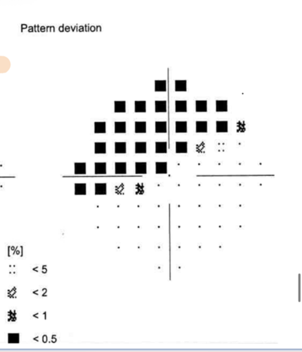
Pattern Deviation VF Plot for IT Drance Hemorrhage
See pic

Do you need to pay attention to the reliability of OCT and VF measurements?
Yes -- need to figure out if something really changed or if the quality of the scan changed

For an avg RNFL thickness, what is the "fudge factor" used to detect significant changes in repeat OCT testing?
5 microns

For a quadrant RNFL thickness, what is the "fudge factor" used to detect significant changes in repeat OCT testing?
10 microns
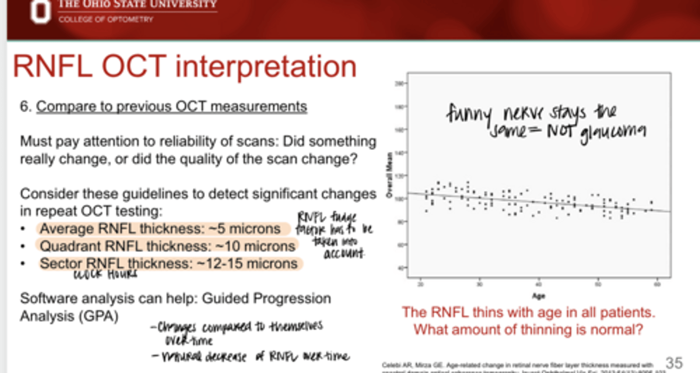
For a sector RNFL thickness, what is the "fudge factor" used to detect significant changes in repeat OCT testing?
12-15 microns

What can help you determine if something really changed or if the quality of the scan changed?
Guided Progression Analysis (GPA) -- changes over time compared to their own past scans
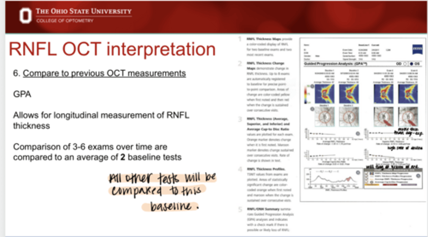
What does Guided Progression Analysis (GPA) allow for?
-allows for longitudinal measurements of RNFL thickness over time
-comparison of 3-6 exams over time are compared to an average of 2 baseline tests