Palmer- PNS- FINAL- TQs- Pt. 1 & Pt. 2 COMBINED
 Call Kai
 Learn
Learn Practice Test
 Spaced Repetition
 Match
Match Flashcards
 Knowt Play
1/131

Earn XP
Description and Tags
Deck courtesy of Brooklyn Reading!
Name | Mastery | Learn | Test | Matching | Spaced | Call with Kai | Chat |
|---|
No analytics yet
Send a link to your students to track their progress
132 Terms
The eye is derived from what?
ectoderm
What gives rise to the retina and pigmented structures of the eye? FIB TQ
neuroectoderm of forebrain
Neuroectoderm of the forebrain gives rise to what structures of the eye? TQ
Retina and pigmented structures
What gives rise to the lens and cornea of the eye? TQ
Surface ectoderm
Surface ectoderm gives rise to which structures of the eye? TQ
Lens and cornea
What gives rise to the sclera of the eye? TQ
neural crest mesenchyme
Neural crest mesenchyme gives rise to which structure of the eye? TQ
sclera (fibrous coats)
Know the eye image! multiple TQs
!
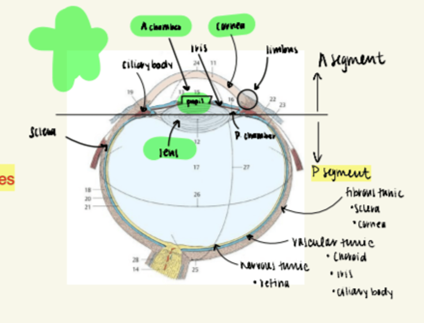
What comprises the fibrous tunic (outermost tunic of eye)? Quiz Question
Sclera and cornea
what comprises the vascular tunic?
choroid, ciliary body, iris
what comprises the nervous tunic?
retina
What is the nerve supply to the eye?
nasociliary N of opthalmic division of trigeminal
What is the first structure that light passes through and is most important for focusing light on the retina?
**known as the primary refractory structure**
Cornea
Which structure maintains the shape of the eye if pressure increases?
sclera
Where do extraocular muscels attach to ?
sclera
the sclera is continuous posteriorly with the ________ _____.
fibrous sheath
What is the primary refractory structure of the eye? TQ
Cornea
What are the 5 layers of the cornea?
(not a likely TQ)
1. corneal epithelium (continuous with conjunctiva)
2. ant. limiting lamina
3. substantia propria (largest!)
4. posterior limiting lamina
5. endothelium (lines iridocorneal layer)
Name the order of how a tears travel, starting with the lacrimal apparatus.
lacrimal apparatus--> excretory ducts-->eye--> lacrimal ducts--> lacrimal sac--> nasolacrimal duct--> inferior nasal meatus
An irregularity in the shape of the cornea known as what? FIB TQ...watch spelling
astigmatism
The transition between the sclera and cornea is known as what?
the limbus
What are the 2 muscles of the iris?
sphincter pupillae and dilator pupillae
Which structure connects the optic nerve and posterior aspect of the lens?
hyaloid ligament
What structure separates the anterior segment into the anterior and posterior chamber? TQ
iris
What is the area of clearest vision?
macula lutea
an increase in pressure/fluid of the anterior segment can lead to what clinical presentation?
glaucoma
the macula lutea contains the _____ ______, which is where we focus all light.
fovea centralis
(cones are only photoreceptors here)
Where do we have a "blind spot", aka, where in the eye is absent of photoreceptors?
optic disc
Which muscle of the iris has circumferential fibers?
sphincter pupillae
Which mm of the iris has radially oriented fibers?
Dilator pupilae
Which muscle of the iris has parasympathetic innervation?
Which muscle of the iris has sympathetic innervation?
**TQs!
-sphincter pupillae = parasympathetic
- dilator pupilae= sympathetic
IMAGE TQ:
Which structure/area of the eye is filled with aqueous humor?
Anterior segment
(be able to identify this on image)
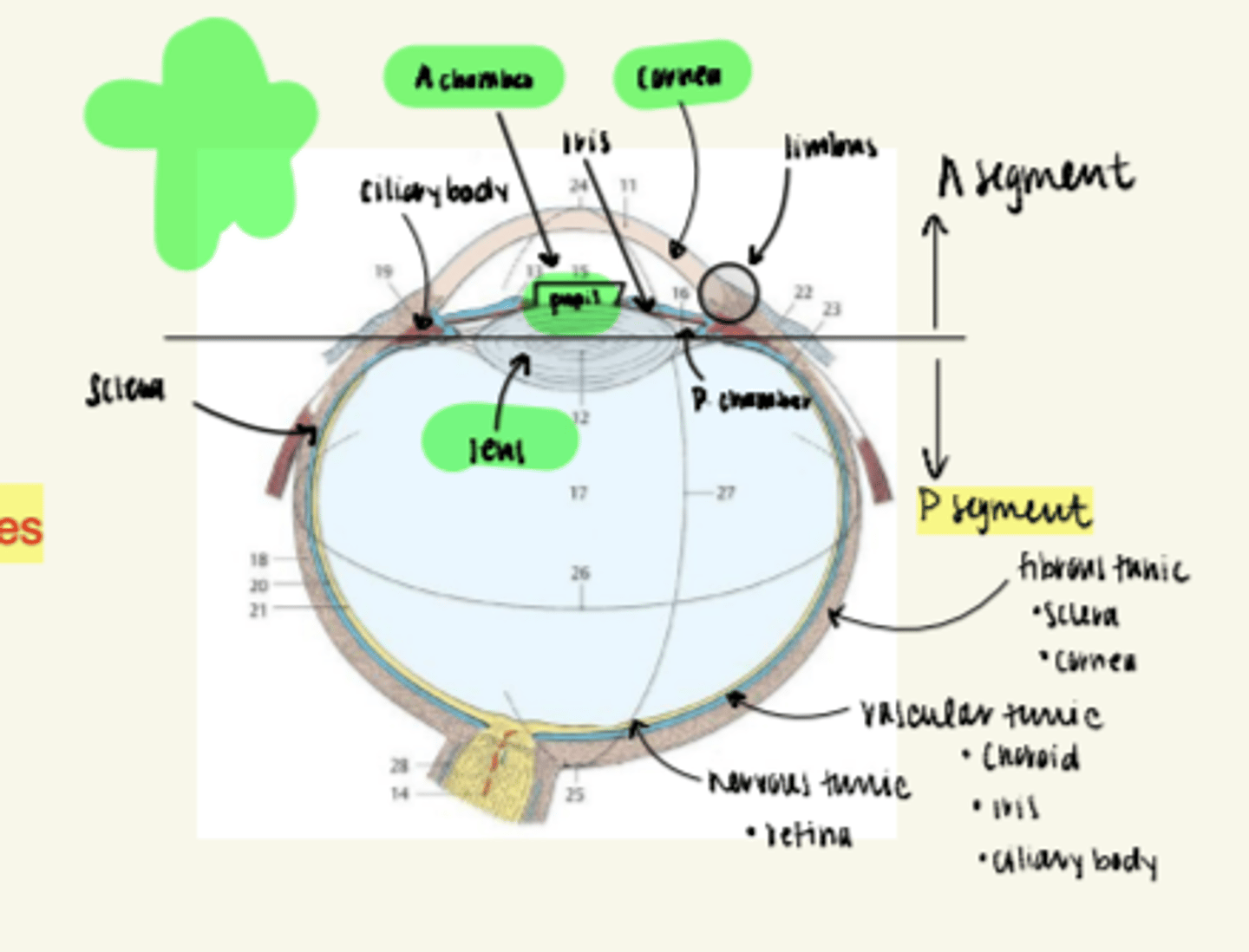
aqueous humor is a clear fluid secreted by the ______ ___________. FIB TQ
ciliary processes
Which layer of the retina do we find synapses of rod and cone cells?
outer plexiform layer
(#30 in image)
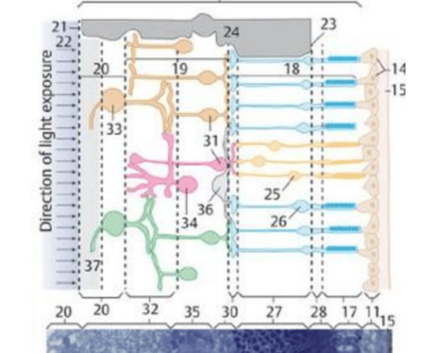
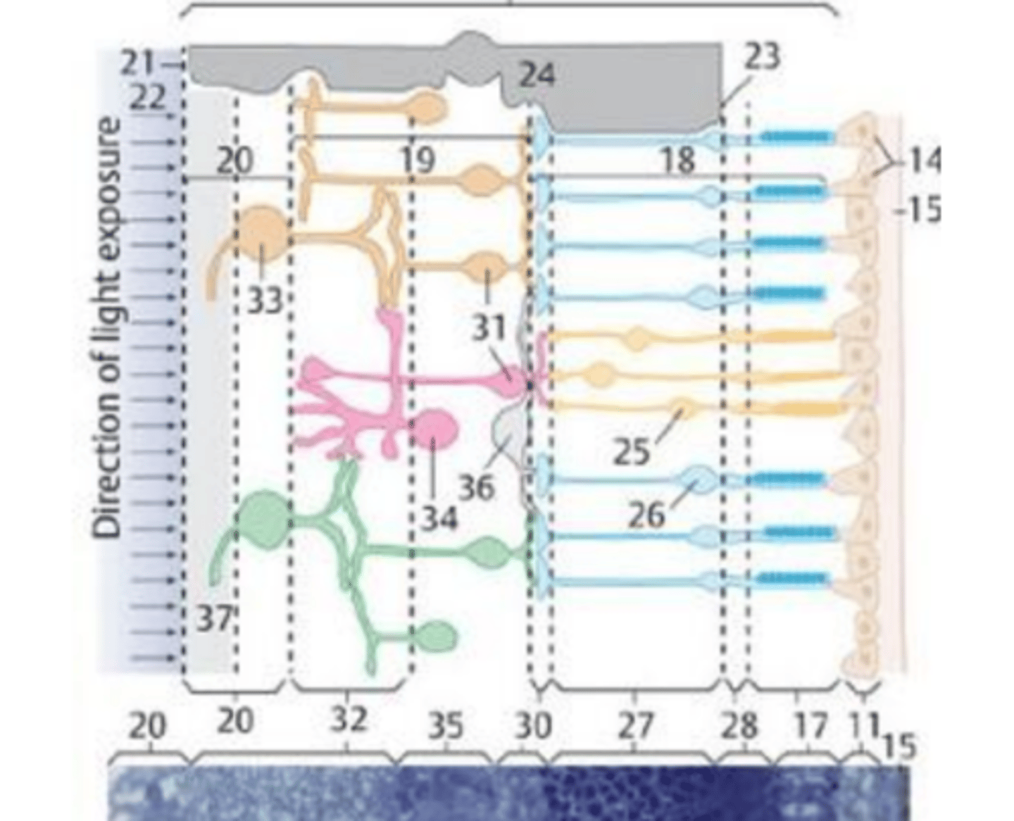
which layer of the retina do we find bipolar cells synapsing on ganglia?
Inner plexiform layer
(#32 in image)
What is the order in which aqueous humor moves? TQ and Quiz Question
secreted by ciliary processes-->goes into posterior chamber--> through pupil--> into anterior chamber--> leaves via sclera venous sinus (canal of schlemm)
This structure "suspends" the lens and produces aqueous fluid that fills the anterior segment of the eye
ciliary body
Loss of resiliency of the lens is known as what condition? TQ
(lens is harder, making it more difficult for ciliary muscle to increase convexity and see things that are near to us)
presbyopia
Which condition is when the lens of the eye loses transparency?
Catartacts
What fills the posterior segment?
vitreous humor/body
In the lacrimal apparatus, which structure secretes tears via excretory ducts?
Lacrimal Glands
Which structure of the lacrimal apparatus drains tears into the lacrimal sac? TQ
Lacrimal ducts
The lacrimal ducts drain tears into what structure? TQ
Lacrimal sac
Which structure drains tears into the inferior nasal meatus? TQ
nasolacrimal ducts
What is the most superior part of the nasolacrimal duct?
lacrimal sac
What are the modifies sebaceous (oil) glands located in each eyelid? TQ
**they keep tears from evaporating
Tarsal glands
What type of gland are tarsal glands (located in each eyelid)? TQ
modified sebaceous (oil) glands
What is the only artery to supply the retina? (one in each eye)
Central Retinal Artery
Image TQ!
Name the structure that is located between the tragus and antitragus of the external ear.
intertragic notch
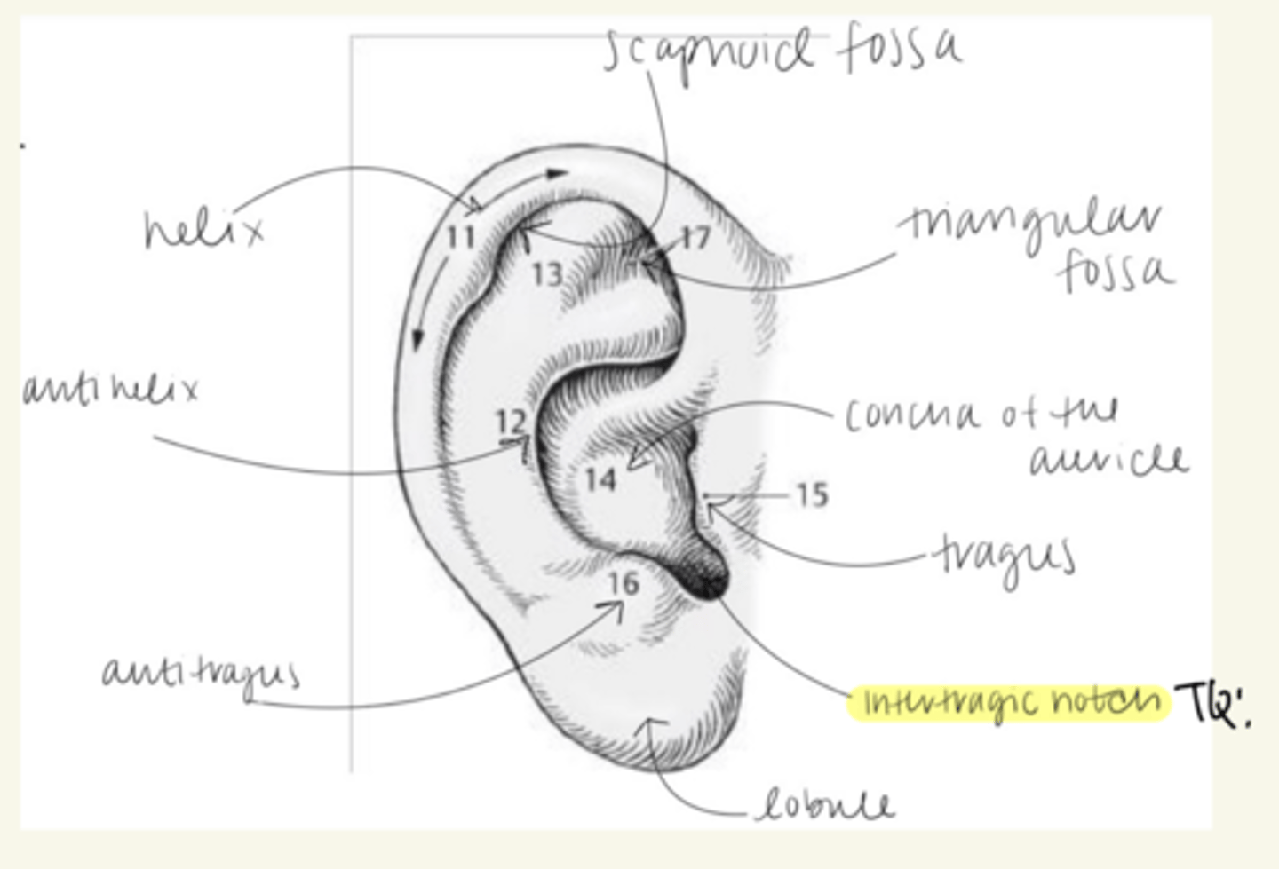
Be able to distinguish between extrinsic and intrinsic ear muscles. Included with this card!!
Extrinsic mm: (have directional terms)
- Anterior auricular mm
- Superior auricular mm
- posterior auricular mm
Intrinsic auricular muscles:
- helices major/minor
- tragicus and antitragus
-transverse muscle of the auricle
-oblique mm of auricle
What are the glands of the ear that secrete ear wax (cerumen)?
What type of glands are these?
-Ceruminous glands
- Modified sudoriferous (sweat glands)
What are the three sensory nerves that supply the external ear? TQ
1. auriculotemporal N (V3)
2. Auricular n (CN 10)
3. Great Auricular N (cervical plexus)
What nerves provide motor innervation to the external ear? TQ
Temporal and posterior auricular nerves (CN7)
What supplies sympathetic innervation to the external ear? TQ
branches from superior cervical ganglion
What is the connection between the middle ear and the mastoid air cells called? IMAGE TQ
(potential route for infection)
Aditus of mastoid antrum
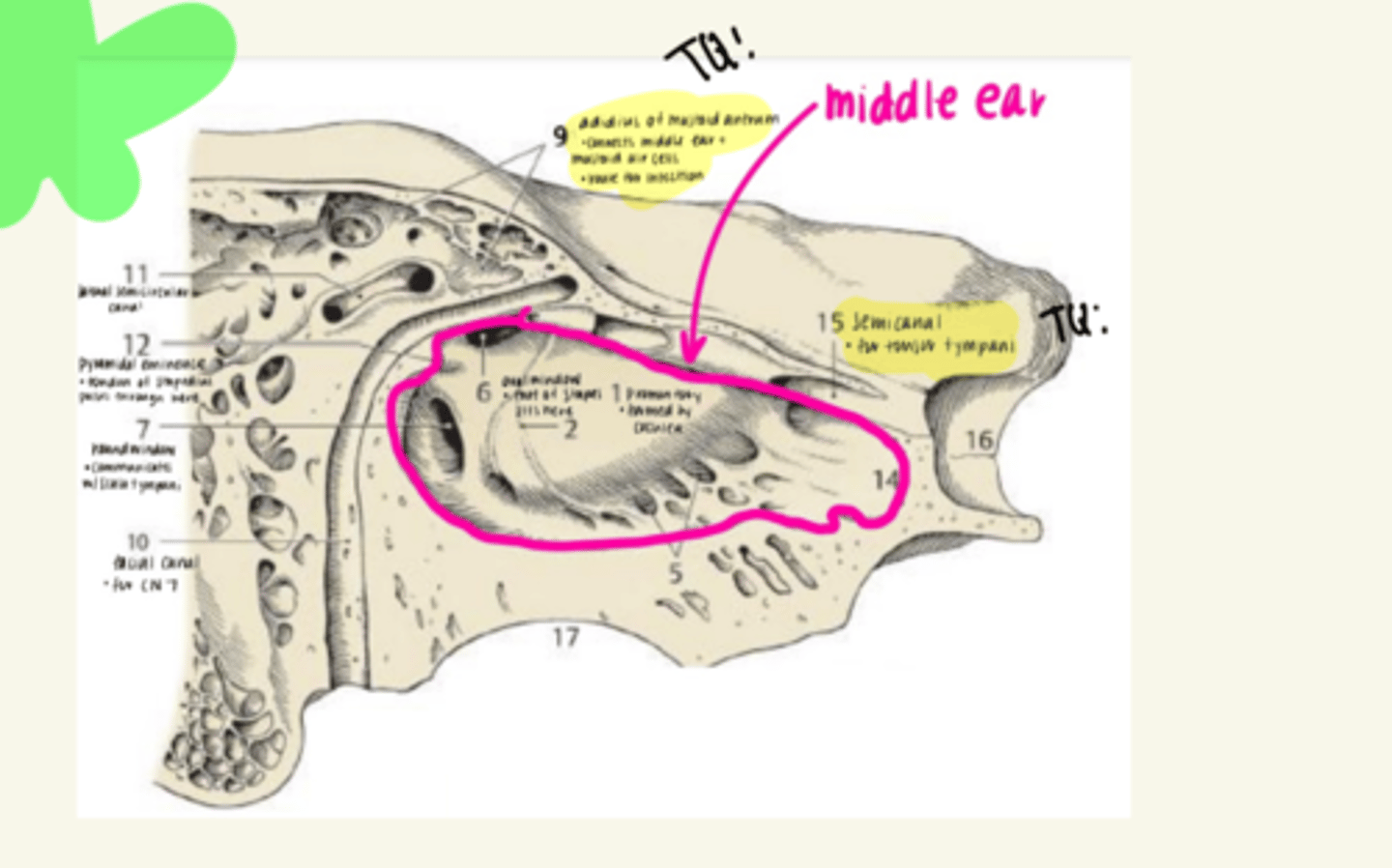
T/F: there is an opening in the middle ear that the chorda tympani can pass through and synapse. TQ!
FALSE: There is an opening, however, it DOES NOT synapse here!
Know this image!
!
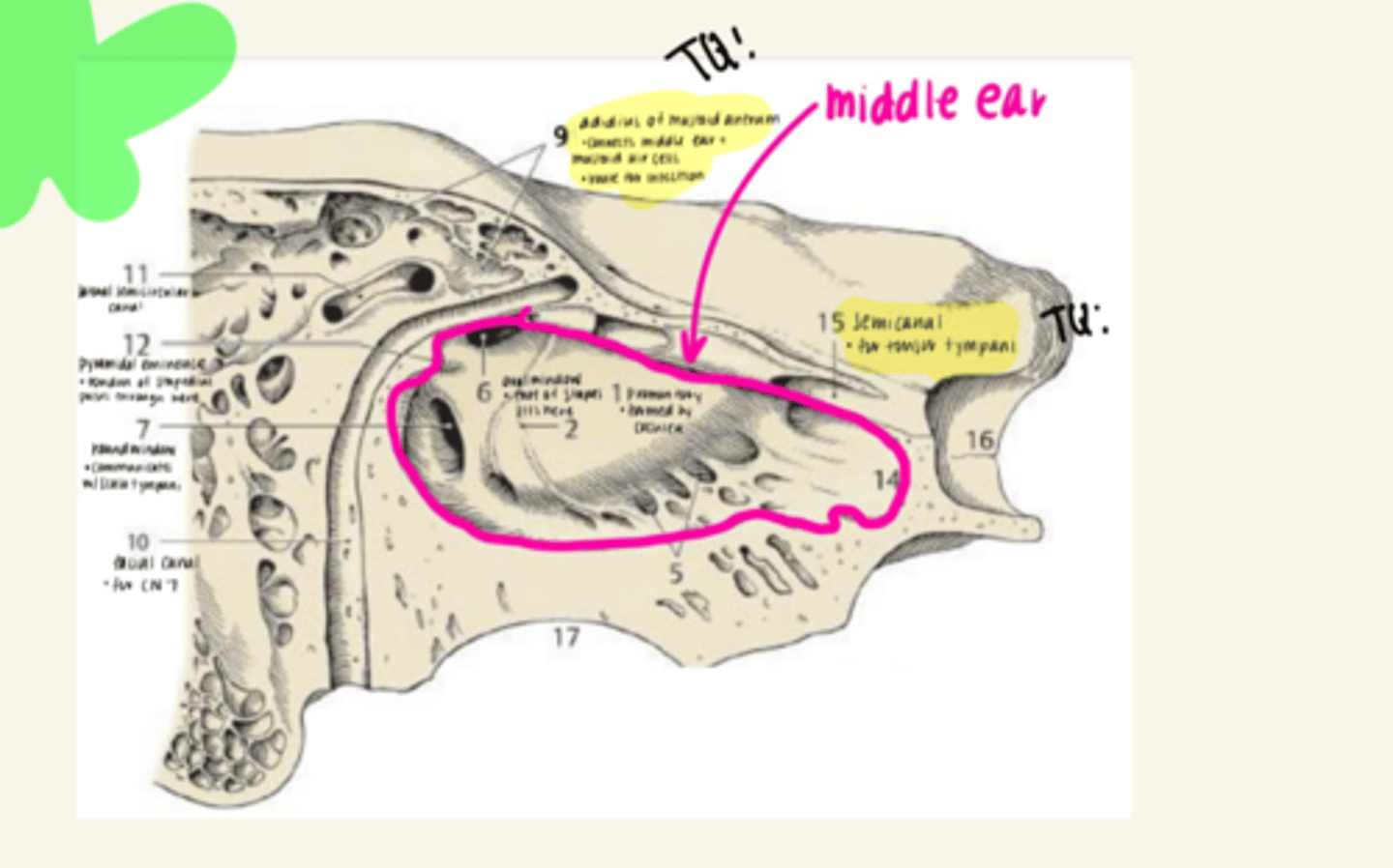
Image TQ
Where is the canal for the tensor tympani on the image?
..
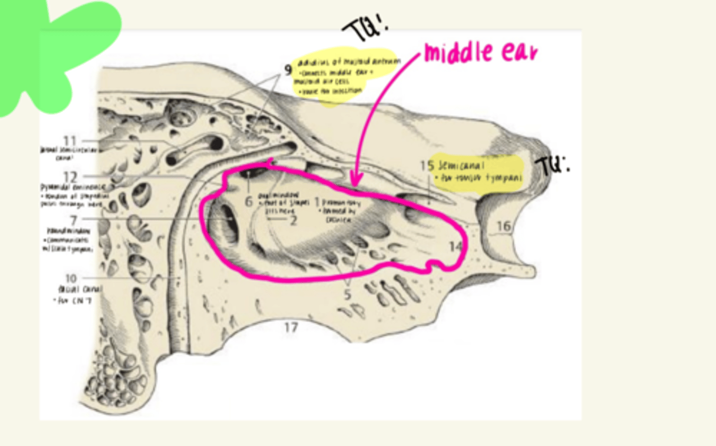
Image TQ:
The cochlea makes this impression...
promontory
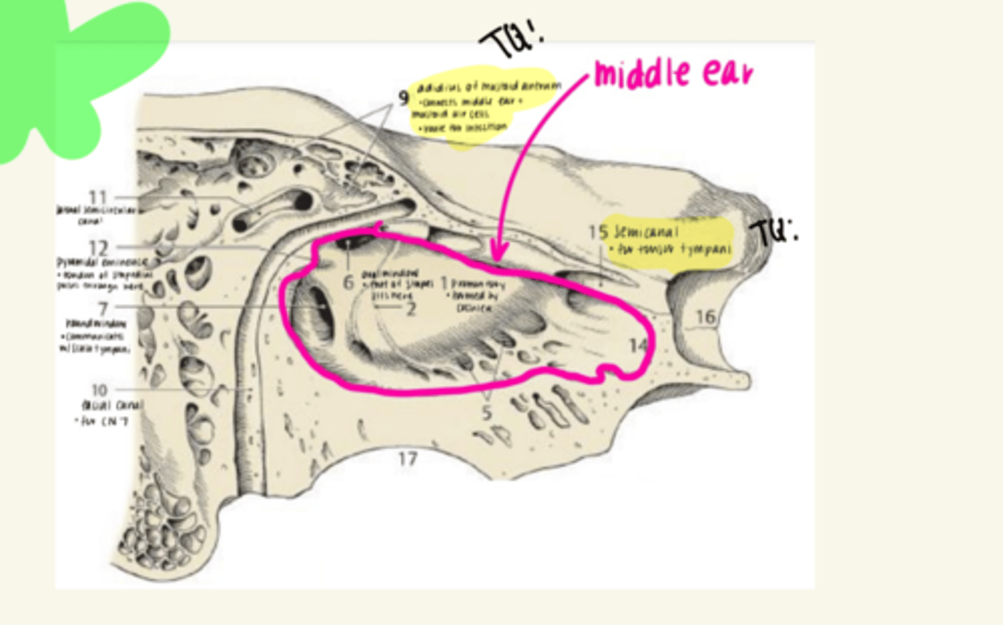
Image TQ:
the foot of the stapes is found here:
oval window
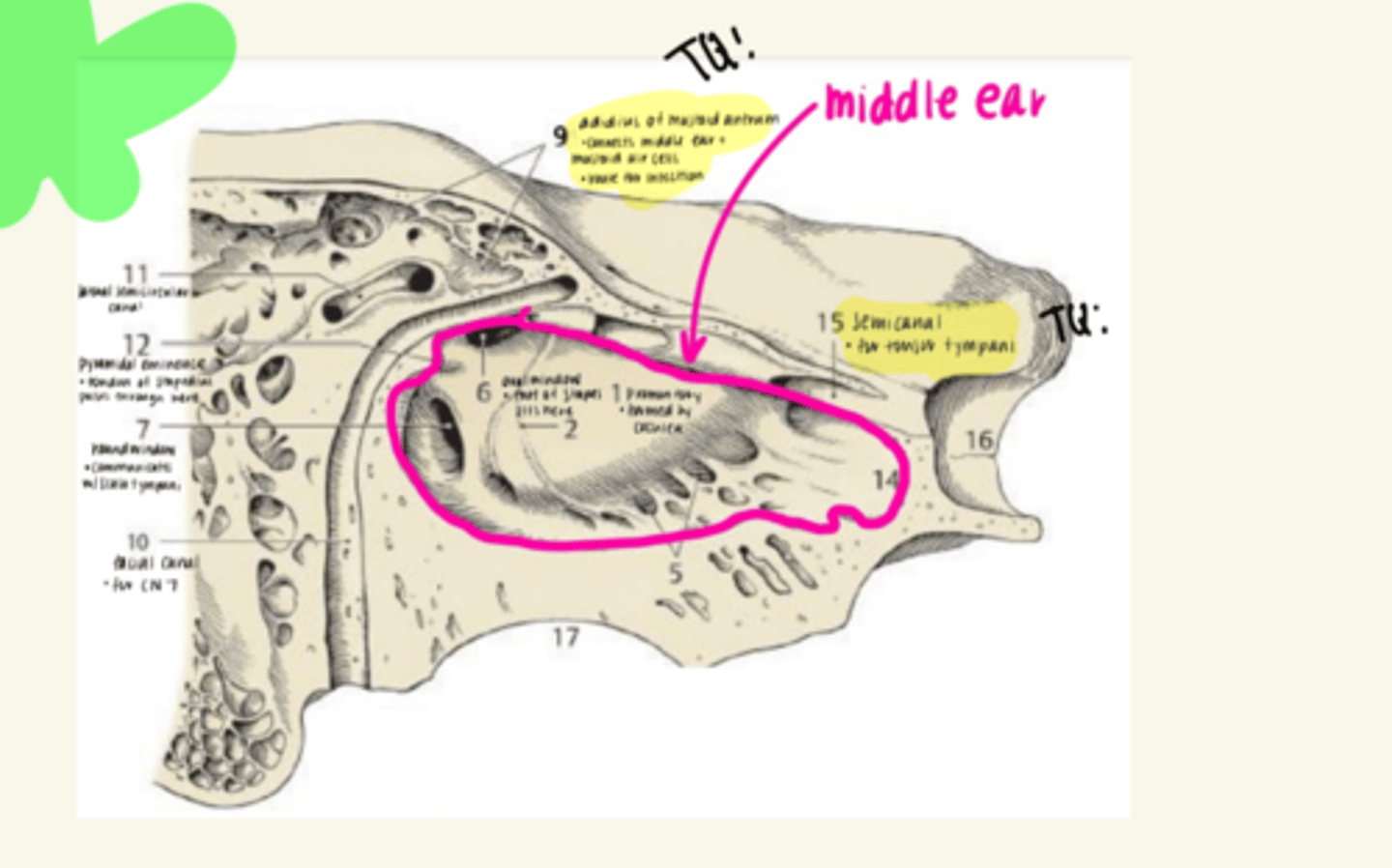
Image TQ:
This structure allows communication with scala tympani but NOT BONE
Round Window
What are the three parts of the bony labyrinth ? TQ
1. Cochlea
2. Vestibule
3. Semicircular canals
(semicircular ducts are part of the membranous labyrinth ***)
Which of the following is part of the membranous labyrinth?
A. Cochlea
B. Vestibule
C. Semicircular ducts
D. Semicircular canals
TQs
C. Semicircular ducts
* all other parts are a part of the BONY labyrinth*
the bony labyrinth is filled with _____________.
the membranous labyrinth is filled with ____________.
perilymph; endolymph
Which structure connects the utricle and saccule?
Utrisaccular duct
Which structure connects and allows communication between saccule with cochlear duct? FIB TQ
ductus reunions
Where do scala vestibuli and scala tympani communicate with each other? FIB TQ
helicotrema
Image TQ:
Which structure is located above the cochlear duct and is filled with perilymph?
Scala vestibuli
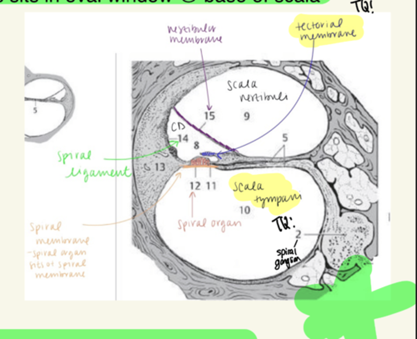
The scala vestibuli communicates with the middle ear via the ______ _________? TQ
What is also found in this spot?
-oval window
-foot of stapes sits in oval window
IMAGE TQ:
What structure is located below the cochlear duct and is filled with perilymph?
Scala tympani
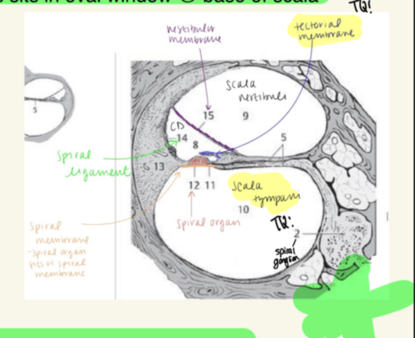
the scala tympani communicates with the middle ear via the ________ ________. TQ
round window
Hair cells of the spiral organ are covered by the _________ __________.
tectorial membrane
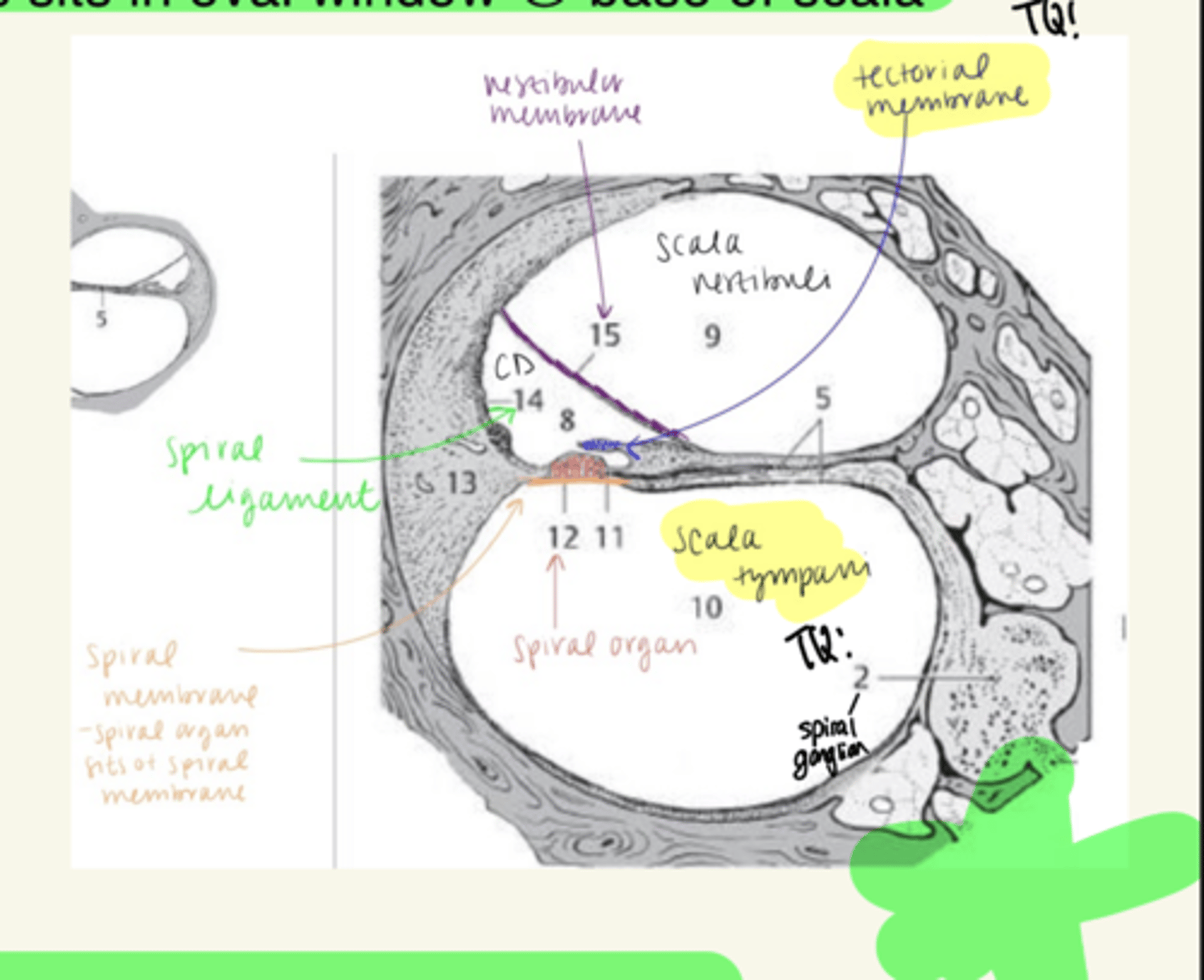
the spiral organ is located where?
What is it attached to?
TQ
-located on floor of cochlear duct
- attached to basilar/spiral membrane
T/F: The spiral organ is attached to the tectorial membrane
FALSE: attaches to the basilar/spiral membrane
Receptors/sense organs in the utricle and saccule (sense organs) are called __________
maculae
The utricles sense ______________ and ____________ accelerations in any plane.
centrifugal; linear
the saccule senses ___________ acceleration in what two planes?
linear; vertical and A-P planes
What are the sense organs in the semicircular ducts called? TQ
crista
(located in ampulla of each duct)
which nerve of the superior branch carries information from the saccule? FIB TQ
Voit's Nerve
which nerve from the inferior branch carries information from the saccule?
saccule nerve
What is the blood supply to the labyrinth?
- internal auditory artery from basilar artery
- stylomastoid artery from posterior auricular artery
Referring to the embryology of the ear, the thickening of ectoderm is called the ________
placode
Auditory vesicles give rise to what part of the ear (embryologically)?
membranous inner ear
Post-ganglionic sympathetic release which neurotransmitter? (Quiz Question)
Norepinephrine (NE)
Central processes (axons) carries by spinal nerves enter the cord via what? Past TQ
posterior rootlets (dorsal-->afferent)
Which nucleus of the oculomotor nerve contains preganglionic parasympathetic cell bodies? TQ
accessory oculomotor nucleus (Edinger wesphal's)
Preganglionic parasympathetic fibers from the accessory oculomotor nucleus follow the oculomotor nerve into orbit and synapse where? FIB TQ
ciliary ganglion
the ciliary ganglion contains the cell bodies of which fibers? TQ
post-ganglionic parasympathetics
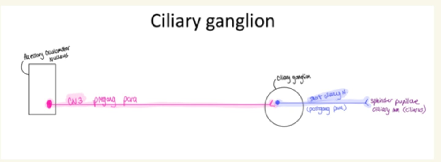
Which muscle is responsible for constricting the pupil (from bright light or rest/digest)? TQ
sphincter pupilae
the greater petrosal nerve carries which fiber type?
preganglionic parasympathetics
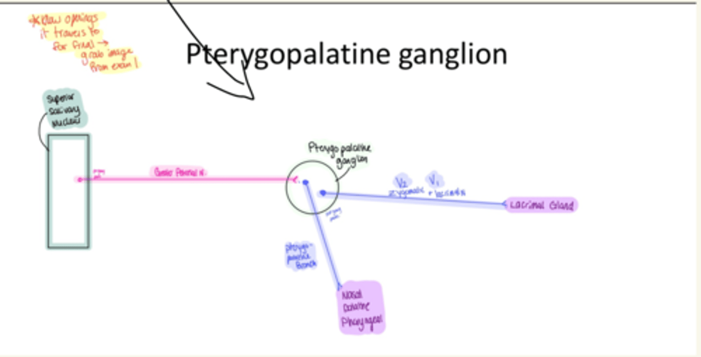
Which nerve travels in the pterygoid canal to bring pre-ganglionic parasympathetics to the pterygopalatine ganglion? TQ
Greater Petrosal N (of CN7)
Fibers of the chorda tympani combine with the lingual nerve (V3) to communicate taste fibers. These fibers then leave the lingual nerve to bring post-ganglionic parasympathetic fibers to which ganglion? TQ
submandibular ganglion
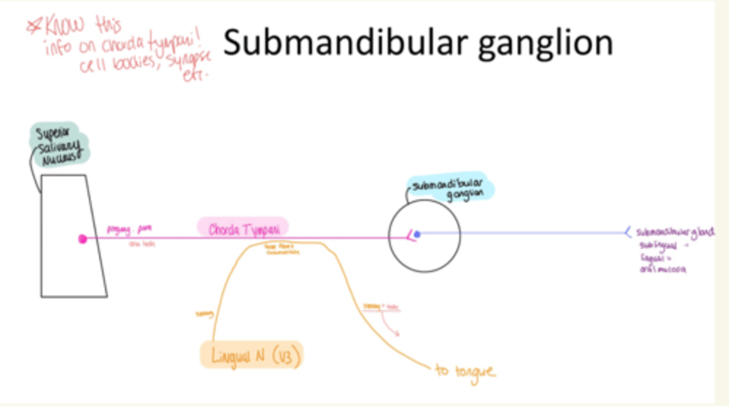
Which nucleus of the facial nerve (CN7) contains the cell bodies of preganglionic parasympathetic fibers?
superior salivary nucleus
Which nucleus of the glossopharyngeal nerve (CN 9) contains the cell bodies of preganglionic parasympathetic fibers? TQ
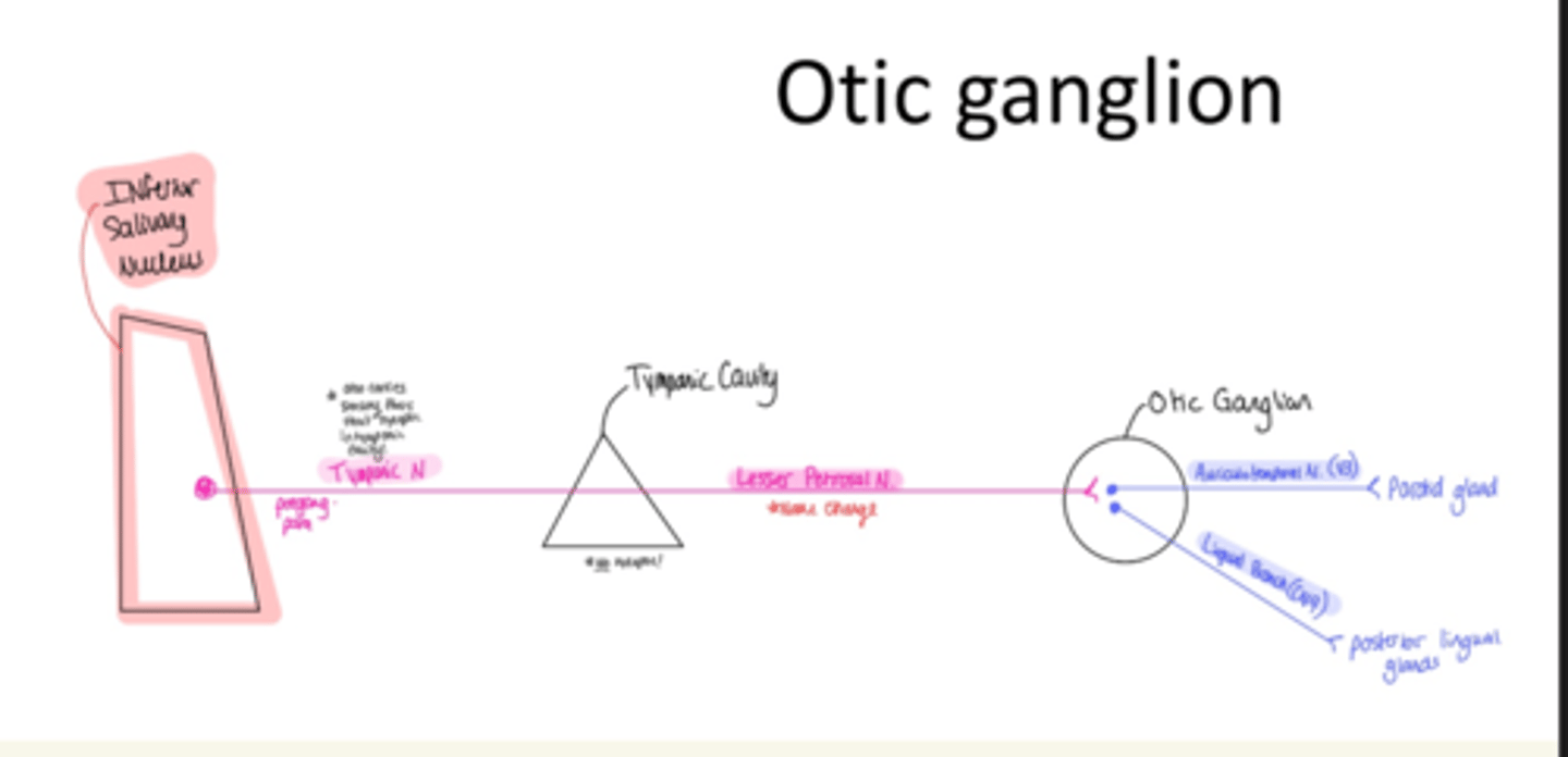
inferior salivary nucleus
The lesser petrosal nerve only carries what fiber type? TQ
preganglionic parasympathetic fibers
(same as greater petrosal N)
The lesser petrosal nerve of CN9 carries __________ _____________ fibers to which ganglion? TQ
preganglionic parasympathetic fibers; otic ganglion
The otic ganglion contain the cell bodies of ______________ ___________ fibers. FIB TQ
postganglionic parasympathetic
which nucleus of the vagus nerve has the cell bodies of preganglionic parasympathetics? TQ
posterior nucleus of vagus nerve
(aka dorsal nucleus of vagus nerve)
The vagus nerve supplies preganglionic parasympathetic fibers to the heart with the superior and inferior cardiac nerves.
the superior cardiac nerve is a branch directly from the ___________ nerve.
the inferior cardiac nerve is a branch from the _____________ _____________ nerve. TQ
Vagus; recurrent meningeal
increasing parasympathetic activity to the heart has what effect on myocardium?
what effect does it have on coronary vessels? 100% TQ
-inhibits myocardium (decrease HR, SV and CO)
-constricts coronary vessels (decrease blood to heart mm, dec pressure in myocardium)
*every other artery in body will dilate with increased parasympathetic activity*