MMG 2010 Exam 1
 Call Kai
 Learn
Learn Practice Test
 Spaced Repetition
 Match
Match Flashcards
 Knowt Play
1/54

There's no tags or description
Looks like no tags are added yet.
Name | Mastery | Learn | Test | Matching | Spaced | Call with Kai | Chat |
|---|
No analytics yet
Send a link to your students to track their progress
55 Terms
Describe how microorganisms are classified and distinguished as unique species
Microorganisms are classified with a taxonomic binomial/nomenclature system.
Taxa: Domain -> kingdom -> phylum -> class -> order -> family -> genus -> species
Compare historical and current systems of taxonomy used to classify microorganisms
Used to be classified by anatomical or behavioral differences, or traits. Groups could often be groups wrong (especially with convergent traits etc.)
Modern taxonomy is now based off of genetic data: 16S rRNA sequencing for bacteria and archaea and 18S rRNA sequencing for microbial eukaryotes.
List the various types of microorganisms and describe their defining characteristics
Prokaryotes: only unicellular, lack a nucleus, 2 domains, divide by binary fission, no organelles. Bacteria have peptidoglycan cell wall, archaea have pseudomurein cell wall
-Bacteria and Archaea
Eukaryotes: unicellular or multicellular, distinct nucleus, divide by mitosis, polysaccharide cell wall
-Animals, Plants, fungi, protists
Viruses: Not a cell - an agent that invades a host cell
Identify and define the characteristics of electromagnetic radiation (EMR) used in microscopy
EMR includes the light spectra, X rays, gamma ray, radio waves, and more. This can be utilized with certain fluorescent dyes to get them to emit photons at a higher energy wavelength that we can then see.
Describe historical developments and individual contributions that led to the invention and development of the microscope
Van Leeuwenhoek - simple microscope where light passed through one lens
Galileo - compound microscope where light passes through 2 lenses
Describe the distinguishing features and typical uses for various types of light microscopes, electron microscopes, and scanning probe microscopes
Brightfield microscopy is very standard, image on bright background
Darkfield increases contrast without staining, bright image on dark background. Useful for live specimens
Phase contrast - Makes high contrast images, high resolution useful for observing structures like endospores and organelles.
Fluorescence - can be used for various stains, living/dead, gram, autofluorescence, etc.
Electron Microscopy uses electron beams focused with magnets to produce an image rather than light. It is very detailed and can be useful to observe the 3D structure of a surface or small thin sections.
Scanning probe microscopes - Uses a probe passes horizontally at a constant distance above specimen, can map the structure of surfaces at an atomic level. Works best with conducting materials.
Contrast vs resolution vs magnification
magnification - how zoomed in
Contrast - how distinguished structures are from each other
Resolution - how focused/blurry
Differentiate between simple and differential stains
Simple stain - a single dye is used to emphasize a particular structure in specimen
Differential staining - 2 or more dyes distinguish organisms based on interactions with different stains.
Explain the procedures and name clinical applications for Gram stain
Gram Stain - Uses crystal violet, grams iodine, decolorizer, and safranin on heat or chemical fixed cells. (+ purple and - pink) outcomes used to distinguish cells by cell-wall type. A positive stain demonstrates a thick PG layer with no outer membrane. A gram negative tells us its a thin PG layer with an outer membrane.
Endospore stain
Endospore stain - heat stains with malachite green, washed and counterstained with safranin. Used to distinguish organisms with endospores (green) from those without.
Acid-fast stain
Acid-Fast stain - Stained with basic fuchsin, rinsed with alcohol, counterstain methylene blue. Used to distinguish acid-fast bacteria from non acid fast bacteria. Acid fast bacteria have mycolic acid in their cell wall which makes them resistant to decolorization and will appear pink, while the decolorized ones will take the counterstain blue.
Negative Capsule stain
Negative capsule stain - Background stained with india ink, leaving clear area of the cell and capsule. Used to distinguish cells with capsules from those without, capsules will appear clear or as halos if present.
Flagella stain
Flagella stain - Coated with a tannic acid then stained with fuchsin or pararosaline. Used to view and study flagella, makes them visibly present.
Explain the key points of endosymbiotic theory and cite the evidence that supports this concept
Mitochondria and chloroplasts arose as a result of prokaryotic cells engulfing them. Evidence that supports this is the structural similarities in ribosomes, mitochondrial and chloroplast DNA. Also binary fission of these organelles is similar to binary fission in bacteria. DNA sequencing supports this as well.
Explain the contributions of Semmelweis, Snow, Pasteur, Lister, and Koch to the development of germ theory
Germ theory states that diseases may result from microbial infection.
Shows hand washing reduces illness, cholera bacteria, disproves spontaneous generation, microbial fermentation, surgery disinfectent, determine causative agents for bacterial infections.
Explain the distinguishing characteristics of prokaryotic cells
-Unicellular
Bacteria and Archaea
Lack a Nucleus
Asexual reproduction - binary fission
Cell wall of peptidoglycan for bacteria, peptidomurein for archae
No membrane bound organelles
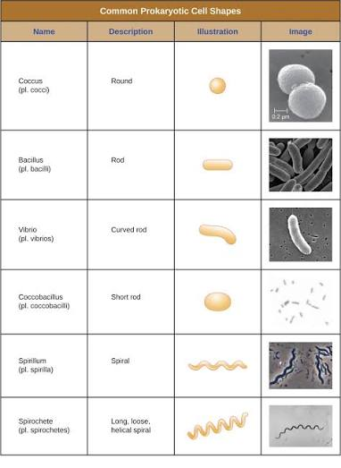
Describe common cell morphologies typical of prokaryotic cells and explain how cells maintain their morphology
Cells maintain morphology with a cytoskeleton
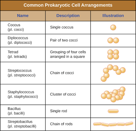
Describe common cellular arrangements typical of prokaryotic cells
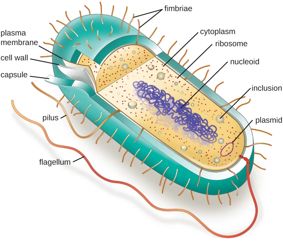
Describe internal and external structures of prokaryotic cells in terms of their physical structure, chemical structure, and function
Flagella
Pilus
Fimbriae
Nucleoid
Ribosome
Compare the distinguishing characteristics of bacterial and archaeal cells
Archaeal cells are more closely related to eukarya
They have different cell walls made of pseudomurein, more complex RNA, typically reside in extreme environments
Explain the distinguishing characteristics of eukaryotic cells
Circular, far more complex DNA, nuclear membrane, membrane bound organelles, cell wall made of cellulose or chitin
Describe internal and external structures of eukaryotic cells in terms of their physical structure, chemical structure, and function
Nucleus - stores DNA and site of transcription and replication
ER (rough and smooth) - Produces and folds proteins and lipids
Golgi complex - The mail center, modifies, packages, and sorts proteins and lipids for transport to different locations
Ribosomes - Make proteins that are inside the cell
Mitochondria - ATP production

Identify and describe symbiotic relationships
a close, long-term biological interaction between two different species.
Mutualism, parasitism, commensalism
Compare normal/commensal/resident microbiota to transient microbiota
Long term/life long residents of host
Transient microbiota only reside for so long.
Explain how prokaryotes are classified
16S RNA sequencing
Used to be more based on morphology, behavior, motility, etc.
Describe the unique features of each class within the phylum Proteobacteria: Alphaproteobacteria, Betaproteobacteria, Gammaproteobacteria, Deltaproteobacteria, and Epsilonproteobacteria
Alphaproteobacteria: Nutrient poor environments, Unusual cellular lifestyle, important symbiotic relationships
Betaproteobacteria: found in soil and water, metabolically diverse, some pathogenic
Gammeproteobacteria: very metabolically diverse, environmental organisms, normal microbiota, major pathogens
Epsilonproteobacteria: Found on animals and GI environments. Microaerophilic conditions
Deltaproteobacteria, zetaproteobacteria irrelevant
Describe the unique features of nonproteobacteria gram-negative bacteria
Includes Spirochetes, Chlamydiae, Bacteriodes
Chlamydiae does not have peptidoglycan, distinct life cycle
Bacteriodes includes fusobacteria. Important in decomposition and nutrient cycling
Several associated with human/animal microbiomes. Some can be opportunistic pathogens
Describe the unique features of phototrophic bacteria
Phototrophic bacteria capture energy from sunlight through photosynthesis or photophosphorylation.
ex. cyanobacteria
Describe the unique features of each category of high G+C and low G+C gram-positive bacteria
Actinobacteria: High GC content. Acid fast, partially acid fast, and filamentous branching bacteria.
Includes streptomyces and actinomycetota → Mycobacterium and Nocardia.
Firmicutes: Low GC content. Includes GPC, GPB, some endospore formers. Common human pathogens
Describe the unique features of deeply branching bacteria
Extremophiles, thermophiles, hyperthermophiles
First non-LUCA to be produced by evolution
Describe the unique features of each category of Archaea
Crenarchaeota - very diverse, all aquatice, mostly hyperthemophiles. Some alkaliphiles and acidophiles (pH).
Euryarchaeota - includes many methanogens, halophiles (salt)
Summarize the general characteristics of unicellular eukaryotic parasites
Some protozoa. Many many species, classified by motility: Amoeba (pseudopodia), Flagellates, ciliates, apicomplexans (apical complex; gliding motility)
No cell walls, chemotrophs and phototrophs, sexual or asexual reproduction, complex life cycles.
Describe the general life cycles and modes of reproduction in unicellular eukaryotic parasites
They are all very different but generally follow the same pattern
Somehow they get enveloped into the host (whether that be blood stream, ingestion, absorption, etc).
They multiply and divide into many different unicellular organisms
Find a way to infect another host and spread (sporulated oocyst, other things eat infected host, etc.)
Explain why we include the study of parasitic worms within the discipline of microbiology
Because where else would it go…
they cause diseases that are diagnosed by finding microscopic eggs and larvae in clinical specimens
Compare the basic morphology of the major groups of parasitic helminthes
Nematodes - round cross section
Cestodes - flat cross section, ribbon like tapeworms. Many segments.
Trematodes - flat cross section, one segment
Describe the characteristics of parasitic nematodes
Unsegmented. Have a complete digestive system. Most are dioecious.
Describe the characteristics of parasitic trematodes and cestodes, and give examples of each
Cestodes (tapeworms) - No mouth, digestive system, or vascular tracts. absorbs what it needs. Monoecious in each segment (proglottid). Scolex is the head region contains hooks/suckers to attach to wall of small intestine.
Trematodes (flukes) - have a ventral and oral sucker. most are monoecious. have complex lifestyles with intermediate hosts. Attach to inner walls of intestines, lungs, blood vessels, or liver.
Identify examples of the primary causes of infections due to nematodes, trematodes, and cestodes
Nematodes - ingestion or skin absorption of eggs. Pinworms
Cestodes - ingestion. Beef tapeworm
Trematodes - ingestion, skin penetration. Blood flukes.
Explain why the study of fungi such as yeast and molds is within the discipline of microbiology
Because the fungi domain includes mold, mushrooms,(multicellular) or yeast (unicellular). They are all eukaryotes
Describe the unique characteristics of fungi
Eukaryotes, multicellular or unicellular. Cell walls made of chitin. Asexual and sexual reproduction
Chemotrophs
Nonmotile
Potentially pathogenic
Grow in high sugar/ high salt concentrations, can grow in low moisture content, require less nitrogen than bacteria
Describe examples of asexual and sexual reproduction of fungi
A spore is how fungi reproduce asexually or sexually that detach from parent and grow.
Asexual reproduction - cell division and mitosis
-Conidiospore - not enclosed in a sac
-Arthroconidia - Fragmentation of septate hyphae
-Blastoconidia - Buds of the parent cell (budding yeast)
-Chlamydoconidia - Spore with a hyphal segment
-sporangiospore - enclosed in a sac
Sexual reproduction - fusion of nuclei from 2 opposite mating strains. 3 phases of sexual reproduction.
-Plasmogamy: + penetrates cytoplasm of -
-Karyogamy: + and - nuclei fuse to form a diploid zygote
-Meiosis: diploid nucleus produces haploid nuclei
Compare and classify the major groups of fungi
Classified by morphology: colonies, hyphae, and spores
Ascomycota, Basidiomycota, microsporidia, zygomycota
Explain why algae are included within the discipline of microbiology and describe unique characteristics of algae
Algae are multicellular or unicellular autotrophic protists
Chloroplasts contain pyrenoids that synthesize and store starches, boosting photosynthesis. Can be motile or non motile
Explain why lichens are included in the study of microbiology and describe the unique characteristics of lichen and the role of each partner in the symbiotic relationship of a lichen
A lichen is a combination of two organisms, a green alga or cyanobacterium and fungus, living in a symbiotic relationship.
They have large effects on the chemical composition of their environments.
Describe the general characteristics of viruses as pathogens
Infectious, acellular pathogens. Obligate intracellular parasites w/ host and cell type specificity
Describe viral genomes
Can have:
DNA or RNA
single stranded or double stranded
Envelope (some studded with glycoproteins) or naked
Lack genes for many products needed for successful reproduction, requiring exploitation of host-cell genomes to reproduce.
Describe the general characteristics of viral life cycles
Entry, replication, exit
Differentiate among bacteriophages, plant viruses, and animal viruses
Bacteriophages (phages) - infect bacteria. Attach to bacterium and usually only inject nucleic acid. Complex head and tail structure. Move from one bacterium to another after virions are released, often lyse the bacterial cell.
Plant viruses - Infect Plant cells. Usually enter through damaged tissue or transmitted by vectors like insects. Move between cells through plasmodesmata and throughout the plant vasculature tissue. Often spread without immediately lysing the plant cell. Commonly helical or icosahedral and usually lack an envelope
Animal Viruses - Enter through membrane fusion, endocytosis, or direct penetration. Commonly icosahedral, both naked and enveloped. Spread between cells through body fluids, nerves, blood, or respiratory secretions. Released through cell lysis, exocytosis, or budding.
Describe the characteristics used to identify viruses as obligate intracellular parasites
Acellular, no cell wall. No independent reproduction or metabolism, require a host cell to do that.
Describe the replication process of animal viruses
attachment - The phage attaches to the surface of the host
Penetration - the viral DNA enters the host cell
Biosynthesis - Phage DNA replicates and phage proteins are mage
Maturation - New phage particles are assembled
Lysis - The cell lyses, releasing the newly made phages
Describe unique characteristics of retroviruses and latent viruses
Latent viruses happens when a viral genome exists in a host genome as a provirus but virions are not produced within the cell: it is inactive. Proviruses insert themselves within the genome, so as the cell divides, the prophage DNA is passed onto daughter cells. Under stressful conditions, the prophage DNA is excised from the bacterial chromosome and enters the lytic cycle, replicating as normal. (ex. herpes)
Retroviruses are +ssRNA viruses that carry a reverse transcriptase within a capsid that synthesize a complementary ssDNA from the ssRNA template. This is made into dsDNA, and can integrate into the host chromosome and become a permanent part of the host (now a provirus). Then is now a latent virus until stressed into the lytic cycle (but doesn’t excise self from chromosome).
Discuss human viruses and their virus-host cell interactions
4 types:
Acute Infection - Replication of virions, host cell lysis. Acute signs/symptoms for host; often quick resolution
Chronic Infection - Slow/limited release of virions from host cell by budding (no lysis). Infected cell remains alive and continues to produce virus
Latent infection - Viral genome exists in host genome as a provirus, virions not produced. Infection asymptomatic until virus begins to replicate again
Transformation - Persistent/chronic infection that causes conversion of normal cell into tumor cell
Discuss why viruses were originally described as filterable agents
To isolate viruses, infected host cells can be obtained/grown. Virions can be separated from host cells by filtration: filters will remove anything above a certain size such as the host cell leaving the virions present in the medium to be collected below.
Describe the cultivation of viruses and specimen collection and handling (compare techniques)
Can be grown in vivo or in vitro (outside.)
In vivo could infect animals or egg embryos
In vitro can use many different type of cells, but cells are extracted and grown on a petri dish
Describe prions and their unique characteristics
PRIONS are misfolded, rogue forms of a normal protein in the cell. This misfolding can be due to many things like a genetic mutation, spontaneous. It can be infectious, stimulating other endogenous normal proteins to become misfolded leading to a whole clump of misfolded proteins. This is bad and can lead to cell death or whatever (ex. encephalopathy)