oat bio unit 7
1/53
Earn XP
Description and Tags
you are a digital video disc
Name | Mastery | Learn | Test | Matching | Spaced | Call with Kai |
|---|
No analytics yet
Send a link to your students to track their progress
54 Terms
chromatin
packaging of DNA around histone proteins.
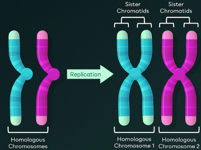
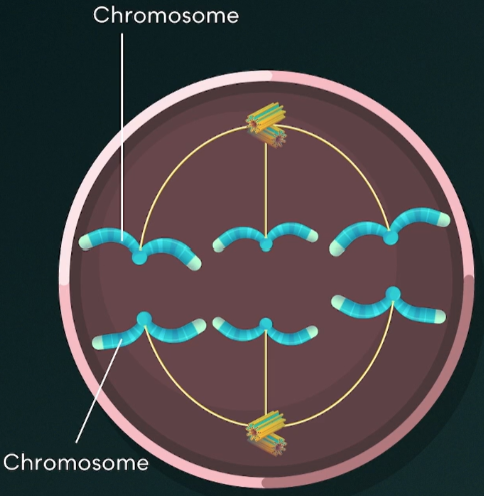
chromosomes
dense packaging of chromatin, existing during mitosis and meiosis.
chromatids
one half of a duplicated chromosome.
centromere
links two identical sister chromatids together.
haploid (n)
one set of chromosomes. n = number of chromosomes in a set (humans: n = 23).
diploid (2n)
two sets of chromosomes. n = number of chromosomes in a set (humans: 2n = 46).
homologous chromosomes
Found in diploid cells, it is two sets of every chromosome, one from each parent which form a pair.
They are similar in length, gene position, and centromere position.
Humans have 23 of them.
microtubule organizing centers (MTOCs)
in animal cells, they are called centrosomes, which are two centrioles.
spindle fibers
microtubules that emerge from the centrosome. They allow the chromosomes and chromatids to be separated during cell division.
kinetochores
proteins on the centromere of the chromosome where the spindle fibers attach.
somatic cells
bodily cells that aren’t used for reproduction that undergo only mitosis.
germ cells
cells that produce gametes that undergo both mitosis and meiosis.
5 phases of mitosis
Prophase
Prometaphase
Metaphase
Anaphase
Telophase and Cytokinesis
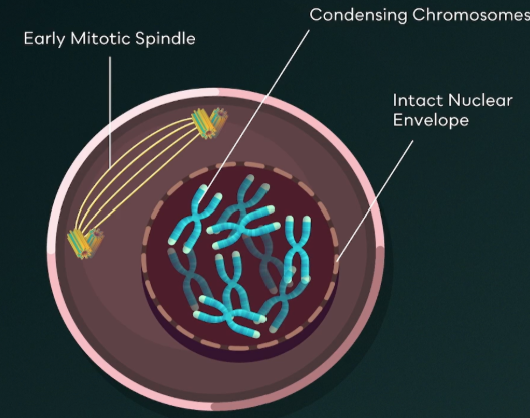
prophase (mitosis)
Chromatin condenses into chromosomes.
Nucleolus disappears.
Mitotic spindle begins to form.
Centrosomes begin to move upwards towards opposite ends of the cell.
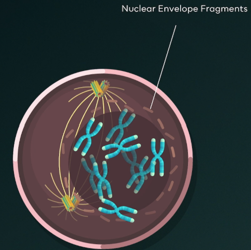
prometaphase (mitosis)
Nucleus disassembles.
Chromosomes condense even further.
Each chromatid is attached to a kinetochore.
Mitotic spindle further develops.
Spindle fibers begin to attach to kinetochores of chromosomes.
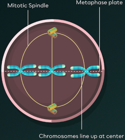
metaphase (mitosis)
Chromosomes are lined up at the center of the cell (metaphase plate).
Centrosomes have reached opposite ends of the cell.
Mitotic spindle is fully developed.
All chromosomes are attached to spindle fibers via kinetochores.
Karyotyping is performed here.
anaphase (mitosis)
Microtubules shorten.
Chromatids are pulled apart.
Each sister chromatid is now considered to be an individual chromosome.
Chromosomes are pulled to opposite ends of the cell.
telophase (mitosis)
Nucleoli reappear.
Two nuclear envelopes develop.
Chromosomes decondense back into chromatin.
Spindle fibers disappear.
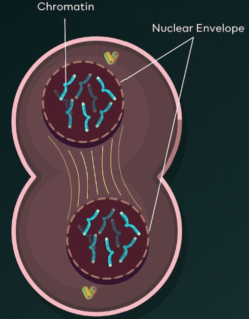
cytokinesis (mitosis)
Happens at the same time as telophase.
Division of cytoplasm to form two cells.
Animal cells: have cleavage furrow, which is a contractile ring formed by actin and myosin.
Plant cells: have a cell plate, which is an extension of the cell wall.
mitosis results
Two 2N daughter cells, where DNA sequence and amount is identical to the parent cell.
karyotyping
Viewing a complete set of chromosomes found during the metaphase stage. It can help identify genetic disorders.
gametes
An organism’s reproductive cells made by meiosis. They are haploid and carry only one copy of each chromosome.
meiosis I
Meiosis that involves separation of homologous chromosomes. Genetic recombination only occurs here.
meiosis II
Meiosis that involves separation of sister chromatids. It starts with two cells because that is the product of meiosis I.
prophase I (meiosis)
homologous chromosomes pair up to form tetrads, allowing for crossing over (genetic recombination). Also includes all steps of mitotic prophase.
chiasmata
region on chromosome where crossing over occurs, which creates genetic diversity in offspring.
telophase I and cytokinesis (meiosis)
Each daughter cell has a new nucleus with half the number of chromosomes. The daughter cells are haploid, and they are not genetically identical due to recombination.
meiosis results
four haploid daughter cells, each with half the amount of DNA compared to the parent cell, and with varied genetic makeup due to recombination.
genetic variation
happens in 3 places.
Recombination during prophase I
Independent assortment: genes are sorted independently relative to each other.
Random joining of gametes (sperm is random, egg is also random).
cell cycle
sequence of events that occur before and during the process of cell division.
interphase
sequence of that occurs before cell division that consists of cell growth, DNA replication, and protein synthesis. This is where cells spend the most time.
cell growth
aka the G1 (gap) phase. The cell increases in size, and protein synthesis begins in preparation for cell division.
DNA replication
aka the S phase. DNA is replicated OUTSIDE OF mitosis. Sister chromatids are formed and centrosomes replicate.
protein synthesis
aka the G2 (gap) phase. Organelles replicate and the cell continues to grow.
G0 phase
Outside of the cell cycle. Cells that are not replicating are here.
Permanently in Go = senescent
Temporarily in Go = quiescent
functional limitations of cells
surface to volume ratio and genome to volume ratio.
surface to volume ratio
volume increases faster than surface area, and the cell membrane can’t keep up with the needs of the rest of the cell.
genome to volume ratio
Genome size does not change based on cell size, so the cell can’t regulate enough processes if the volume is too large for the genome.
skeletal muscle cells
not capable of dividing, but grow larger in size. They combat functional limitations by having multiple nuclei.
cell specific regulations
Cell cycle checkpoints, density dependent inhibition, and anchorage dependence.
cell cycle checkpoints
Restriction point: End of G1 where cell checks for sufficient nutrients, necessary cell products, adequate cell size, and healthy DNA. Success = move to S phase.
End of G2: cell assesses accuracy and completion of DNA replication. Success = move into mitotic phase.
Spindle checkpoint: during metaphase, and the cell evaluates if sister chromatids are attached to the spindle fiber. Failure = mitosis stops.
density dependent inhibition
Cell stops dividing when density reaches a maximum.
anchorage dependence
Cells only divide when attached to an external surface, which prevents cells from multiplying in fluid.
cancer
caused by uncontrolled cell division. A mutated cell can disrupt cell regulations, and form a mass called a tumor.
malignant tumor
tumor where the cells break loose and travel to other parts of the body, called metastasis.
p53 gene
actively suppresses tumors. Mutation of the gene can cause the cell to divide uncontrollably.
cancer drugs
inhibit mitosis to stop uncontrolled growth.
labile cells
constantly dividing, like skin cells.
quiescent cells
cells that do not usually divide, but can be stimulated to as needed, like liver cells.
fixed/permanent cells
cells that have little to no capacity for cell division, like cardiac muscle cells.
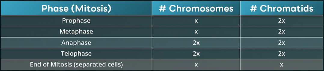
mitosis chromosome count
x = diploid number (humans = 46)
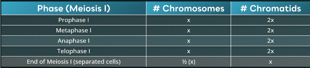
meiosis I chromosome count
x = diploid number (humans = 46)
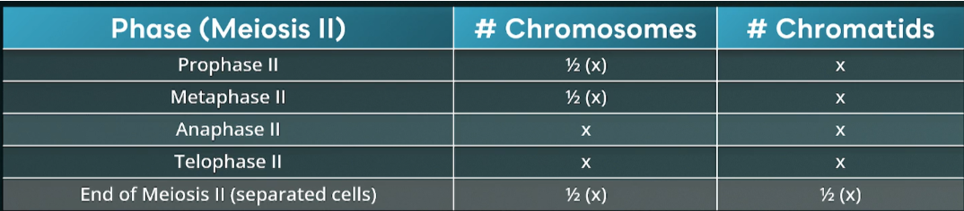
meiosis II chromosome count
x = diploid number (humans = 46)
CORRECT ANSWER
select the ““““correct answer”””” ← (hint)