The Fetal Genitourinary System
 Call Kai
 Learn
Learn Practice Test
 Spaced Repetition
 Match
Match Flashcards
 Knowt Play
1/82

There's no tags or description
Looks like no tags are added yet.
Name | Mastery | Learn | Test | Matching | Spaced | Call with Kai | Chat |
|---|
No analytics yet
Send a link to your students to track their progress
83 Terms
. If the kidneys fail to ascend into the normal position, the result is an
ectopic kidney (renal ectopica), most often located within the pelvis (pelvic kidney)
The most common renal anomaly is the
duplex collecting system, also referred to as a duplicated, duplex, or double collecting system

duplex collecting system consists of
two separate collecting systems, divided into what is termed an upper pole moiety and a lower pole moiety.
Horseshoe kidneys are kidneys that are attached at their
lower poles.

Initially , the bladder is continuous with the allantois, although eventually this channel closes and develops into a fibrous cord referred to as the
urachus
the urachus is located between the
apex of the bladder and the umbilicus.
The kidneys can be sonographically identified as early as
11 weeks with endovaginal imaging and by 12 weeks with transabdominal imaging
The fetal bladder can be seen as early as
12 weeks and should always be seen by 15 weeks and beyond
It is important to note that the fetal urinary bladder normally fills and empties once in every
30 to 45 minutes
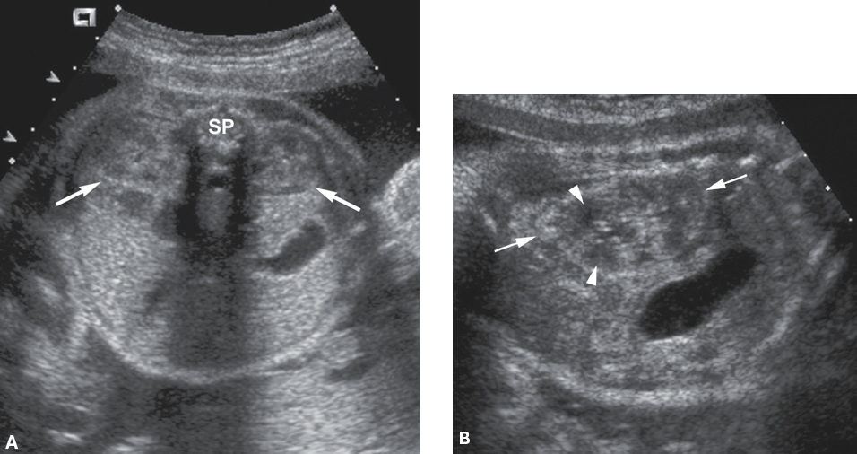
Sonographic image of kidneys
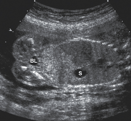
Sonographic image of bladder
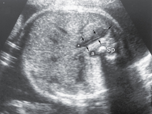
Sonographic image of adrenal gland
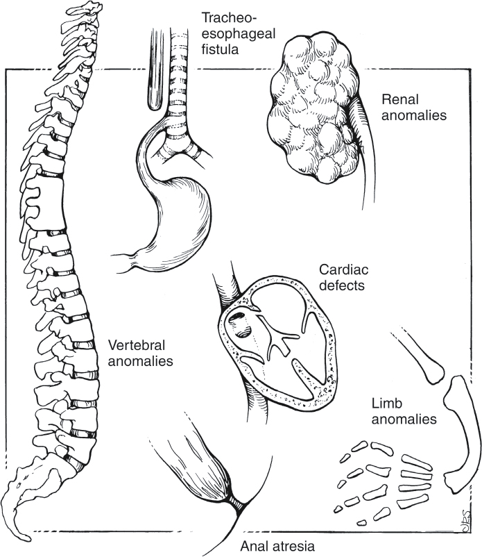
VACTERL stands for
vertebral anomalies, anal atresia, cardiac anomalies, tracheoesophageal fistula or esophageal atresia, renal anomalies, and limb anomalies; Patients are considered to have this association if three of the organ systems listed have abnormalities
Renal abnormalities are the most frequent cause of
oligohydramnios (after 9 weeks)
In circumstances in which the fetus has a renal abnormality, specifically those that are linked with bilateral renal agenesis, inadequately functioning kidneys, or obstruction of the urinary tract, oligohydramnios will be present, and, in some cases,
anhydramnios may occur
The most worrisome consequence of oligohydramnios is
pulmonary hypoplasia, or underdevelopment of the lungs.
Failure of a kidney to form is referred to as
renal agenesis
Bilateral renal agenesis, which results in
Potter syndrome or Potter sequence, is a fatal condition
Features of Potter syndrome or Potter sequence
Bilateral renal agenesis
Abnormal facial features (Potter facies)
Pulmonary hypoplasia (small chest)
Limb abnormalities (e.g., clubfeet)
Intrauterine growth restriction
Low-set ears
Oligohydramnios
Potter sequence is caused by any condition in which there is a significant lack of
amniotic fluid around the fetus as it develops
. Though bilateral renal agenesis can cause the typical Potter syndrome features, other conditions, such as an
obstructive defect of the urinary tract or amnion rupture, can cause the same issues.
Bilateral renal agenesis may be seen in conjunction with
sirenomelia and various cardiovascular malformations.
In the presence of unilateral renal agenesis, the contralateral kidney will enlarge, a condition known as
compensatory hypertrophy.
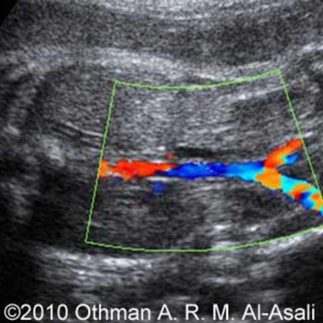
Sonographic Findings of Bilateral Renal Agenesis
Absent kidneys
Absent urinary bladder
Severe oligohydramnios or anhydramnios
Bilateral lying-down adrenal signs
Undetectable renal artery branches with color Doppler (bilateral)
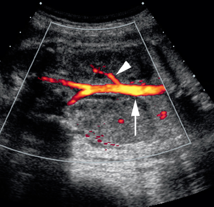
Sonographic Findings of Unilateral Renal Agenesis
Absent kidney
Compensatory hypertrophy of the contralateral kidney
Visible urinary bladder
Normal amniotic fluid volume
Unilateral lying-down adrenal sign
Undetectable renal artery branch with color Doppler (unilateral)
Most often, with unilateral renal agenesis, there is an
average amount of amniotic fluid, and the prognosis is good.
There are several distinct categories of fetal renal cystic disease that were formerly described by Potter
autosomal recessive polycystic kidney disease (ARPKD), autosomal dominant polycystic kidney disease (ADPKD), multicystic dysplastic kidney (MCDK) disease, and obstructive cystic dysplasia.
In order for an autosomal recessive disease to be passed to the fetus, both parents must be carriers of the disease. Each offspring of parents, who are both carriers of an autosomal recessive disorder, has a
25% chance of being affected and a 50% chance of being a carrier.
In the case of an autosomal dominant disease, at least one of the parents has to be the carrier of the disease, and the gene must be dominant. That means, the dominant gene is capable of overriding the normal gene from the parent who is not a carrier. Each offspring of a parent who is a carrier of an autosomal dominant disease has a
50% chance of receiving the gene from their parents
ARPKD may also be referred to as
autosomal recessive polycystic renal disease or infantile polycystic kidney disease
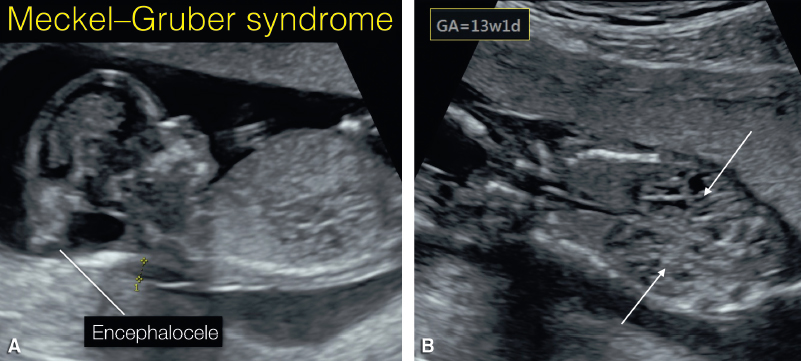
One condition associated with ARPKD is
Meckel–Gruber syndrome, which is a fatal disorder that includes renal cystic disease, occipital encephalocele, and polydactyly
. Fetuses with trisomy 13 and trisomy 18 may also have
polycystic kidney disease
It is significant to appreciate the differences in the sonographic appearance of ARPKD and MCDK disease. Cysts are typically not identifiable in
ARPKD but are evident with MCDK (multicystic dysplastic kidney)
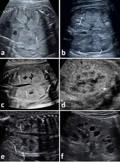
Sonographic Findings of Autosomal Recessive (Infantile) Polycystic Kidney Disease (ARPKD)
Bilateral, enlarged echogenic kidneys
Absent urinary bladder
Oligohydramnios
ADPKD may also be referred to as
autosomal dominant polycystic renal disease
If the kidneys do appear enlarged and echogenic, a distinguishing difference between the two diseases is that in the fetus with ADPKD, the urinary bladder is
often present and there is a normal amniotic fluid volume, whereas with ARPKD, the bladder is absent and there is oligohydramnios
Adult renal cystic disease is also associated with the development of
cysts within the liver, pancreas, and spleen (around 4th-5th decade of life)
Sonographic Findings of Autosomal Dominant (Adult) Polycystic Kidney Disease (ADPKD)
Normal-appearing or bilateral, enlarged echogenic kidneys
Visible urinary bladder
Normal amniotic fluid volume
Cysts often do not manifest until approximately the fifth decade of life
Multicystic dysplastic renal disease may also be referred to as
multicystic dysplastic kidney (MCDK) disease and multicystic renal dysplasia.
MCDK disease is thought to be caused by an
early, first-trimester obstruction of the ureter
There is typically no normal functioning renal tissue present in the kidney affected by MCDK disease. Therefore, MCDK disease is
fatal if bilateral (in most cases, it is unilateral)
Fetuses with MCDK disease can also have additional related anomalies, such as abnormalities of the
gastrointestinal tract and central nervous system, limb anomalies, and further renal abnormalities.
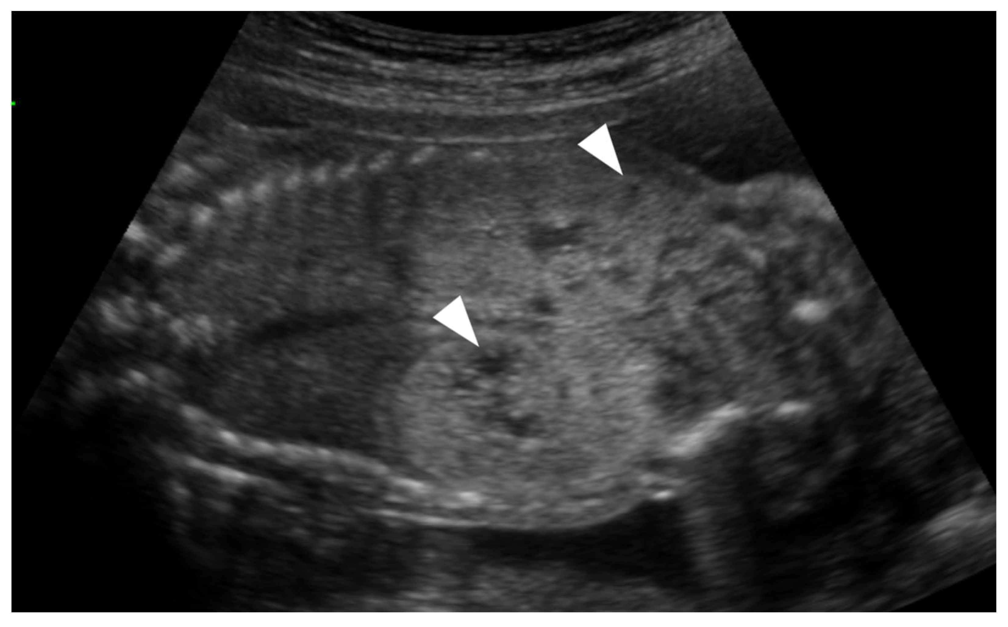
Sonographic Findings of Bilateral Multicystic Dysplastic Renal Disease
Bilateral, smooth-walled, noncommunicating cysts of varying sizes located within the renal fossae
Absent urinary bladder
Oligohydramnios

Sonographic Findings of Unilateral Multicystic Dysplastic Renal Disease
Unilateral, smooth-walled, noncommunicating cysts of varying sizes located within the renal fossae
Compensatory hypertrophy of the contralateral kidney
Visible urinary bladder
Normal amniotic fluid volume

Obstructive cystic dysplasia, like MCDK disease, is caused by an
early renal obstruction. It can be unilateral or bilateral
A ureterocele, or a severe bladder outlet obstruction, early in gestation, can lead to
bilateral obstructive cystic dysplasia, in which case oligohydramnios will be present.

Unilateral obstructive cystic dysplasia is most often caused by a
pelviureteral junction or vesicoureteral junction obstruction

. Bilateral cystic dysplasia may be associated with urethral atresia or
posterior urethral valves
Sonographic Findings of Bilateral Obstructive Cystic Dysplasia
Small, echogenic kidneys
Peripheral renal cysts
Bilateral hydronephrosis
Thick-walled urinary bladder
Oligohydramnios
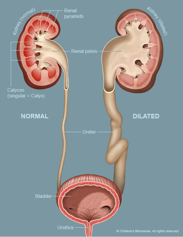
An obstruction of the fetal urinary tract can lead to distension of the
urethra, bladder, ureters, and renal collecting system, depending upon the level of obstruction
Urine is produced by the kidney; exits the kidney by means of the
renal pelvis; travels down the ureter, into the bladder; and exits the body via the urethra
if there is an obstruction at the region where the ureter meets the bladder, the ureterovesical junction (UVJ),
then those structures that are positioned proximal to the obstruction will be dilated ( entire ureter, the renal pelvis, and the renal calices will be eventually dilated and filled with urine.)
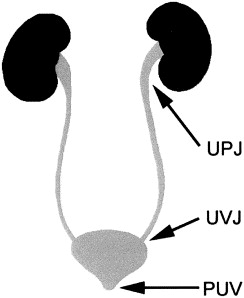
Conversely, if the obstruction level lies at the point at which the renal pelvis meets the ureter, the ureteropelvic junction (UPJ), t
then the ipsilateral renal pelvis and renal calices will be dilated, whereas the ureter and bladder will most likely remain normal, provided that contralateral urine flow is not obstructed in any way

what is the most common fetal abnormality noted during an obstetric sonogram
Hydronephrosis
Hydronephrosis, or pelvocaliectasis, may be described as
pelviectasis (pyelectasis) or caliectasis, depending on which part of the collecting system is dilated
Enlargement of the bladder is called
megacystis
whereas dilation of the ureter may be referred to as
megaureter or hydroureter.
Fetal pelviectasis, or dilation of the renal pelvis, can be established and measured with sonography by taking a
renal pelvic diameter

The measurement of the renal pelvis is made in the anteroposterior plane and should not exceed
7 mm before 20 weeks’ or 10 mm after 20 weeks’ gestation

what are the three most common areas where obstruction occurs.
The UPJ, the UVJ, and the urethra
It is also important to note that less common causes of hydronephrosis in the fetus include
ureterocele, ectopic ureter, vesicoureteral reflux, and urethral atresia.
UreteroPelvic junction obstruction, or UPJ obstruction, is the most common cause of
hydronephrosis in the neonate and the most common form of fetal renal obstruction (located at junction of renal pelvis and renal calices)
UPJ is usually
unilateral and more common in males
Sonographic Findings of UreteroPelvic Junction Obstruction
Hydronephrosis (dilated renal pelvis and calices)
Normal ureters (nonvisualization)
Normal bladder
A bladder outlet obstruction describes the condition in which there is a
blockage of the flow of urine out of the urinary bladder.
posterior urethral valves are a common cause of
bladder outlet obstructions in male fetuses.
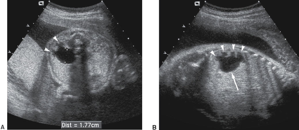
Sonographic Findings of Posterior Urethral Valves
“Keyhole” sign (dilated bladder and urethra)
Bilateral hydroureter
Bilateral hydronephrosis
Oligohydramnios
Thickened bladder wall
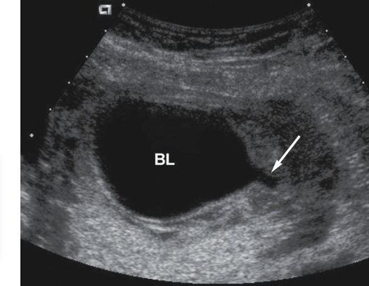
Prune belly syndrome (most seen in males) is typically caused by
megacystis, a massively dilated urinary bladder.
Sonographic Findings of Prune Belly Syndrome
Dilated bladder and, possibly, urethra (“keyhole” sign)
Absent abdominal musculature
Undescended testis
Urinary tract abnormalities (megacystis and hydronephrosis)
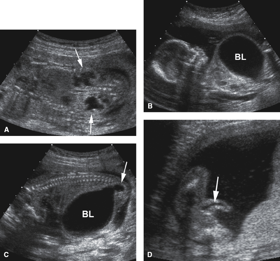
UreteroVesical junction obstruction, or UVJ obstruction, is the least
common cause of hydronephrosis in the fetus.
Sonographic Findings of Ureterovesical Junction Obstruction
Hydronephrosis
Dilated ureter
Normal bladder
Normal amniotic fluid (if unilateral)
Bladder exstrophy is an anomaly wherein the bladder is
located outside of the fetal pelvis.
After an extended amount of investigation, nonvisualization of the bladder in the presence of a normal amniotic fluid volume and normal kidneys should warrant a search for
bladder exstrophy
The cloaca is the embryonic structure that develops into the
rectum and urogenital sinus
With cloacal exstrophy, also referred to OEIS complex
there is an Omphalocele, bladder Exstrophy, Imperforate anus, and Spina bifida.
Sonographic Findings of Bladder Exstrophy
Lower abdominal wall mass inferior to the umbilicus
Absent urinary bladder
Normal kidneys

The most common solid fetal renal mass is the
mesoblastic nephroma (a hamartoma of the kidney. )
Sonographic Findings of The Mesoblastic Nephroma
Solid, homogeneous mass within the renal fossa and may completely replace the kidney.

Ambiguous genitalia is a birth defect in which the
sex of the fetus cannot be determined.
Findings of abnormal external genitalia in the male are
micropenis, hypospadias (opening on underside), hypospadias with chordee ((curvature of penis)), epispadias (opening on topside), and undescended testicles.
The most common female finding is
clitoromegaly
Fetal ovarian cysts may be noted in the fetal pelvis, secondary to
maternal hormone stimulation. These cysts are most often benign and resolve spontaneously, although if they continue to grow, they can lead to hemorrhage and, possibly, ovarian torsion.

The most common malignant adrenal pediatric tumor is the
neuroblastoma