COMP 2 Cards Anatomy (Blood Flow and ETC)
1/79
There's no tags or description
Looks like no tags are added yet.
Name | Mastery | Learn | Test | Matching | Spaced | Call with Kai |
|---|
No analytics yet
Send a link to your students to track their progress
80 Terms
Hesselbach’s triangle is bounded by:
rectus abdominis (medial), inferior epigastric vessels (lateral), inguinal ligament (inferior)
Gastro-omental arteries supply the
greater curvature of the stomach and the omental bursa
greater curvature of the stomach and the omental bursa are supplied by the
gastro-omental arteries
The lesser curvature of the stomach is primarily supplied by the
left gastric artery and the right gastric artery
left gastric artery and the right gastric artery supply the
Lesser curvature of the stomach
Pathway of blood for celiac trunk (origin to what it supplies)
The celiac trunk originates from the abdominal aorta —> left gastric artery —> the splenic artery —> common hepatic artery. This network of arteries supply blood to the foregut organs
The celiac trunk supplies the
Foregut (Esophagus, Stomach, Proximal Duodenum, Liver, Gallbladder, Pancreas, Spleen)
For “Every Slutty Drunk Loves Getting Pretty SLAMMMED.”
E – Esophagus
S – Stomach
D – (proximal) Duodenum
L – Liver
G – Gallbladder
P – Pancreas
S – Spleen
The splenic artery supplies (organs)
oxygenated blood primarily to the spleen, pancreas, and parts of the stomach
Short gastric aa. supply blood to the
stomach's fundus and near the greater curvature.
The common hepatic artery supplies (organs)
oxygenated blood to the liver, gallbladder, the pylorus region of the stomach, the duodenum, and the pancreas
The proper hepatic artery supplies (organs)
oxygenated blood to the liver and gallbladder.
The proper hepatic artery branches off the ….. and splits into
common hepatic artery and travels through the hepatoduodenal ligament before splitting into the hepatic arteries and right gastric artery
The left hepatic artery supplies
oxygenated blood to the left lobe and quadrate lobe of liver
The right hepatic artery supplies
oxygenated blood to the right lobe of the liver, the gallbladder, and the caudate lobe
The cystic artery supplies the
gallbladder, common hepatic duct, cystic duct and the proximal part of the (common) bile duct
The gastroduodenal artery supplies (organs)
blood to the pylorus, the proximal duodenum, and the head of the pancreas
The anterior superior pancreaticoduodenal (ASPD) artery is a terminal branch of the…. that supplies
branch of gastroduodenal artery. ASPD supplies blood to the head of the pancreas and the first and second parts of the duodenum
Splenic vein pathway
originates from Hilum of spleen
→ inferior mesenteric veins → joins superior mesenteric vein → forms portal vein.
“Spleen → runs behind Pancreas → meets IMV → joins SMV → Portal vein.”
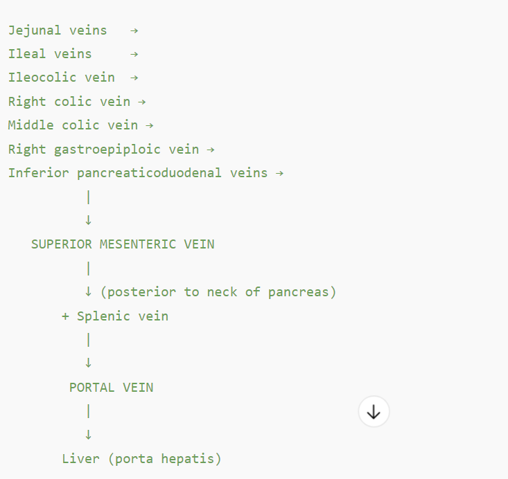
Superior Mesenteric Vein pathway
The SMV begins in the right iliac fossa then heads to mesentery of small intestines. It will receive tributaries of superior mesenteric artery.
Ileocolic vein
Ileal & Jejunal Vein
Right colic vein
Middle colic vein
Right gastro-omental vein (from stomach/greater omentum)
As it ascends, it joins with the splenic vein
Their union forms the portal vein, which then enters the liver at the porta hepatis.
Celiac Trunk branching pathway
→ Left gastric artery |
Splenic Artery branching pathway
→ Short gastric arteries
→ Left gastro-omental artery
→ Pancreatic branches
→ Hilum of spleen
Common Hepatic Artery Branching pathway
→ Proper hepatic artery
→ Gastroduodenal artery
Gastroduodenal artery Branching Pathway
→ Right gastro-omental artery
→ Anterior superior pancreaticoduodenal artery
Anterior superior pancreaticoduodenal artery Branching pathway
→ Head of pancreas, duodenum
Proper hepatic artery Branching Pathway
→ Right & Left hepatic artery
→ Right gastric artery (Lesser Curvature of the Stomach)
Right hepatic artery Branches to what?
→ Cystic artery (to gallbladder)
Left hepatic artery Branching Pathway
→ Left lobe of liver |
The gastroduodenal artery branches into the (arteries)
right gastro-omental artery and the Anterior superior pancreaticoduodenal a
Left Gastric Artery supplies the
lesser curvature of the Stomach and the esophageal branches.
What supplies the esophageal branches?
Left gastric artery
Hepatoduodenal Ligament:
Connects liver to the first part of the duodenum; contains portal triad:
Proper hepatic artery (left side, anterior)
Common bile duct (right side, anterior)
Portal vein (posterior, behind both artery and duct)
the Pringle maneuver is a surgical technique where the surgeon
clamps the portal triad within the hepatoduodenal ligament at the omental foramen to temporarily control bleeding from the liver.
The splenic artery travels
posterior to the stomach to supply blood to the spleen
The left gastro-omental artery is a branch off of the
splenic artery
The right gastro-omental artery is a branch off of the
gastroduodenal artery
The deep inguinal ring is formed by an
invagination of the transversalis fascia
The lateral umbilical folds are formed by the
peritoneum covering the underlying inferior epigastric arteries and veins
The medial umbilical fold is formed by the underlying
medial umbilical ligament, a remnant of the obliterated fetal umbilical artery
The median umbilical fold is formed by the
peritoneum covering the median umbilical ligament, which is the remnant of the urachus
Celiac Ganglia innervates
stomach, liver, pancreas, spleen, and parts of the small and large intestines
Hepatic portal vein carries ……. to where?
oxygen-poor blood to the liver
Left portal vein supplies the left functional lobe and flows to
hepatic sinusoids —> hepatic veins —> inferior vena cava.
Right portal vein supplies the right functional lobe and then blood flows into the
SMV + Splenic v. → Hepatic Portal v. → Liver → Portal vein branches into Left portal v. and Right portal v. → Hepatic sinusoids → Central v. → Hepatic vv. → IVC → Right atrium
Hepatic veins drain
oxygen-poor, nutrient-depleted, detoxified blood from liver directly into the inferior vena cava.
Inferior vena cava receives
hepatic veins and carries blood into the right atrium of the heart
Which nerve passes through the diaphragm’s esophageal hiatus?
vagus nerve
vagus nerve travels through what in diaphragm?
Esophageal hiatus
What passes through the diaphragm’s caval hiatus?
The inferior vena cava and parts of the right phrenic nerve
The aorta, the thoracic duct, and the azygos vein pass through the
aortic hiatus
What passes through the diaphragm’s aortic hiatus?
The aorta, the thoracic duct, and the azygos vein
lateral cutaneous nerve is a sensory nerve that supplies
the skin of the anterior and lateral (outside) thigh.
The iliohypogastric nerve supplies
sensation to the skin of the suprapubic (lower abdomen) and posterolateral gluteal regions
The obturator nerve provides
sensation to the medial thigh and helps adduct it.
The femoral nerve controls (actionwise)
hip flexion and knee extension
The genitofemoral nerve provides sensation to the
inner thigh, lower abdomen, and the external genitalia
The marginal artery is formed by the
anastomoses between branches of the superior mesenteric artery (SMA) and inferior mesenteric artery (IMA): (think colic arteries)
The superior suprarenal arteries arise from the
inferior phrenic artery
The superior suprarenal gland distribute blood to
the cortex and medulla
Caval Foramen is found at what level of vertebrae?
T8 (Think Inferior Vena cava and right phrenic nerve)
Remember I 8 10 eggs, At 12
The caval foramen is innervated by the
Phrenic Nerve (C3-5)
The esophageal foramen is formed in the
right crus of the diaphragm
The aortic hiatus attachments
median arcuate ligament, right and left crura, and T12 vertebrae
The lateral, medial arcuate ligament Attachments
Attached to TP L1, 12th rib
The lateral arcuate ligament arches over the
Quadratus Lumborum
The lateral arcuate ligament is innervated by the
phrenic nerve and intercostal nerve
The medial arcuate ligament passes over the
Psoas Major muscle
The median arcuate ligament arches over the
aorta and extends from the right crus to left crus
Subcostal Nerve innervates the
abdominal wall muscles
External oblique
Internal oblique
Transversus abdominis
Rectus abdominis
Pyramidalis (when present)
The iliohypogastric nerve supplies the
internal oblique and transversus abdominis muscles.
Provides Sensation to…. skin of gluteal region and skin above pubis.
The Ilioinguinal nerve innervates the
Internal oblique and transversus abdominis and skin over the penis, scrotum, labia majora
The genitofemoral nerve (L1-L2) supplies the
motor for cremaster muscle
Sensation to anterior thigh and scrotal skin, labia majora
The Femoral Nerve innervates what muscles?
Anterior compartment of thigh specifically,
Iliacus
Pectineus (partial, also obturator)
Sartorius
Quadriceps femoris (rectus femoris, vastus lateralis, vastus medialis, vastus intermedius)
These muscles mainly flex hip and extend knee
The intestinal Artery does what?
Forms arterial arcades which give rise to vasa recta
Ileocolic artery is a branch to what…. that leads to
SMA that gives way to appendicular artery
Difference between Hepatic Portal System and Caval Venous System
HPS: Formed by union of SMV and Splenic vein (some contributions from IMV)
This venous system drains blood and delivers nutrient rich, oxygen poor blood to the LIVER before returning to heart.
CVS: Deoxygenated blood drains directly into the SVC or IVC, bypassing the Liver. It will deliver directly to the heart.
Common iliac vein pathway of blood
Internal + External Iliac vein → Common iliac vein → Inferior vena cava → Right atrium
Right veins go
straight to inferior vena cava and then right atrium
Left veins go
straight to left renal vein → Inferior vena cava → Right atrium