A & P Practical 1
 Call Kai
 Learn
Learn Practice Test
 Spaced Repetition
 Match
Match Flashcards
 Knowt Play
1/86

There's no tags or description
Looks like no tags are added yet.
Name | Mastery | Learn | Test | Matching | Spaced | Call with Kai | Chat |
|---|
No analytics yet
Send a link to your students to track their progress
87 Terms
skull
Cranial
Face
Facial
Neck
Cervical
head
Cephalic
Armpit
Axillary
Arm
Brachial
Front of elbow
Antecubital
Wrist
Carpal
Palm
palmar
Fingers
Digital or phalangeal
thigh
Femoral
Anterior surface of knee
Patellar
Leg
Cural
Ankle
Tarsal
Toes
Digital or Phalangeal
Foot
Pedal
Big toe
Hallux
top of foot
Dorsum
Hand
Manual
Thumb
pollex
groin
inguinal
hip
Coxal
Shoulder
acromial
shoulder blade
scapular
spinal column
vertebral
Back of elbow
cubital
between hips
sacral
tail bone
coccygeal
buttox
gluteal
region between anus and external genitals
Perineal
Hollow behind knee
Popiteal
calf
sural
sole
plantar
heel
calcaneal
back of hand
dorsum
Frontal Plane
Sagittal Plane
Transverse Plane
Frontal Plane: divides anterior and posterior axis
Sagittal Plane: medial and lateral axis
Transverse Plane: Superior and inferior axis
Ventral
Dorsal
Anterior
Posterior
Cephalic
Rostral
Caudal
Superior
Inferior
Medial
Lateral
Proximal
Distal
Ipsilateral
Contralateral
Superficial
Deep
Ventral- towards the front or belly
Dorsal-towards spine
Anterior-towards ventral side
Posterior-towards dorsal side
Cephalic-towards head
Rostral-toward the forehead or nose
Caudal- toward the tail or inferior end
Superior-above
Inferior-below
Medial-toward median plane
Lateral-away from median plane
Proximal-closer to the point of attachment
Distal-farther from the point of attachment
Ipsilateral-same side
Contralateral-opposite sides
Superficial-closer to body surface
Deep-farther from body surface
Quadrants
Right Upper Quadrant
Left Upper Quadrant
Right Lower Quadrant
Left Lower Quadrant
9 sections of abdomen
Right Hypochondriac | Epigastric | Left Hypochondriac
Right Lumbar | umbilical | left lumbar
Right Inguinal | Hypogastric | Left inguinal
Cavaties
Cranial
Vertebral
Thoracic
Diaphragm
Abdominal
Pelvic
Pericardial
Thoracic Cavity
Divided from Abdominopelvic cavity by diaphragm
Thoracic cavity divided by mediastinum
Mediastinum houses the heart
pleurae lines the lungs
pericardium lines the heart
Abdominal cavity
Contains most digestive organs, spleen, kidneys, ureters
Epithelial tissue functions
protections
secretion
excretion
absorption
filtration
sensation

Simple Squamous-allows rapid diffusion of gasses
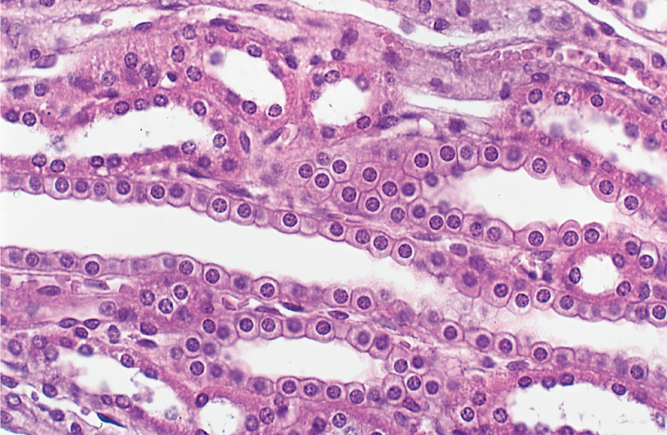
Simple Cuboidal-absorption and secretion

Simple Columnar-absorption and secretion of mucus

Stratified Cuboidal-swear secretion; secretes ovarian hormones

Pseudostratified Columnar/ Goblet-secretes and produces mucus

Cilla

Microvilli

Transitional Epithelium/urothelium
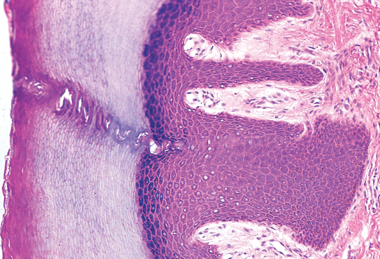
Keratinized Stratified Squamous

nonkeratinized Stratified Squamous
Connective Tissue Function
Binding of organs
Support
physical protection
immune protection
movement
storage
heat production
transport
Fibrous CT fiber types
Collagenous fibers-made of collagen, tough and flexible, resists stretching
reticular fibers-thin collagen fibers, form spongelike network for such organs and constitute part of basement membranes
elastic fibers-thinner than collagenous fibers, allows stretch and recoil

Areolar Tissue (loose)

Fibroblasts

Adipocytes

Reticular (loose)
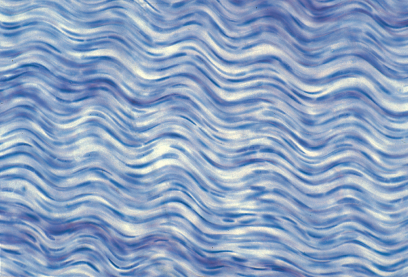
Dense regular connective tissue
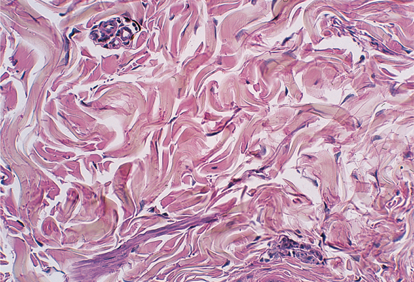
Dense Irregular Connective Tissue
Cartilage
Stiff and rubbery material, made of chondroblasts, which then encase themselves into a cavity called a lacunae, then they are called chondrocyte

Hyaline Cartilage
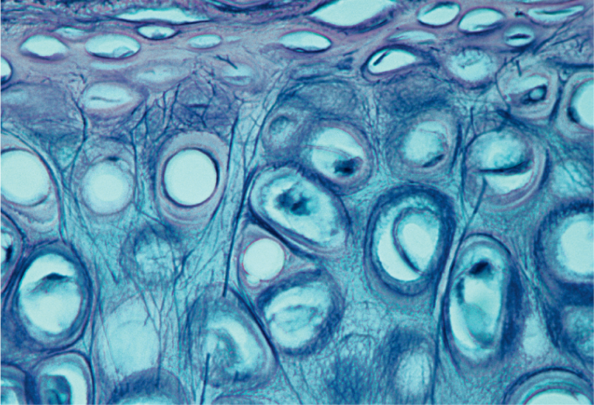
Elastic Cartilage

Fibrocartilage

Bone
3 main Skin layers
Epidermis-outer epithelial
Dermis-middle layer comprised of CT, skin appendages, off shoots of nervous tissue and blood vessels
hypodermis-fat, nervous tissue, blood vessels
Cells of epidermis
Keratinocytes: produce keratin, majority of cells in epithelium
Stem cells:cells involved in cell production reside in stratum basale
melanocytes:in stratum basale, produce melanin and protect DNA from UV rays
Tactile cells: initiate touch perception, found in basal and linked with nerve endings in dermal layer
Dendritic: in stratum spinosum and granulosum, immune cells
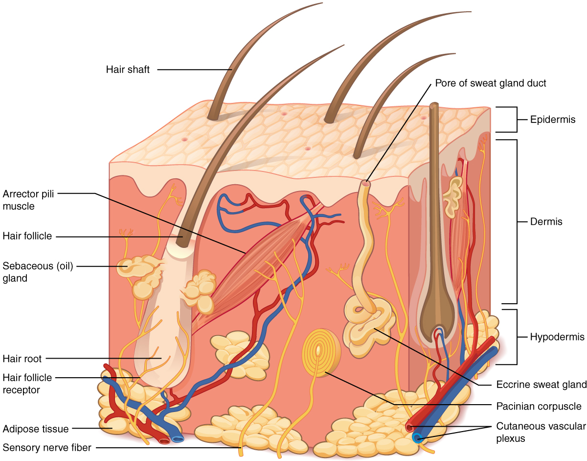
Know cube
Layers of epithelium (deep to superficial)
Stratum basale: single layers of simple cuboidal or columnar keratinocytes. Also home to stem cells, melanocytes, and tactile cells. stem cells divide into keratinocytes an migrate superficially
stratum spinosum: several layers of keratinocytes. Thickest portion on histological section (except palms and soles its stratum corneum)
Stratum granulosum: 3-4 layers of keratinocytes, appear as darker stain
stratum lucidum: thin layer which appears clear
Stratum corneum: thick superfifical layer of dead keratinized cells.

know diagram
Dermis
CT Layer comprised mostly of collagen, but also some reticular and elastic fibers
Good blood supply, cutaneous glands, nerve endings
dermal papilae are finger like projections which act as a locking mechanism
2 zones of dermis: papillary and reticular
Papillary layer composed of areolar CT near dermal papillae rich in leukocytes and small blood vessels
Reticular layer thicker layer comprised of dense irregular CT tissue
Hypodermis
Fat layer rich in blood vessels and areolar CT
Hair
Also known as pilus, grow from hair follicle
portion of hair above skin is shaft, beneath skin is the root, bulb is base of hair grown around highly vascularized CT called dermal papilla
directly above papilla is the hair matrix (growth center)
hair medulla is the inner layer while bulky outer layer is cortex
each hair has an arrector muscle controlled by ANS

know diagram
Glands
5 kinds of glands: eccrine, apocrine, sebaceous, ceruminous, mammillary
Eccrine glands
Stratified cuboidal epithelium throughout body use for cooling
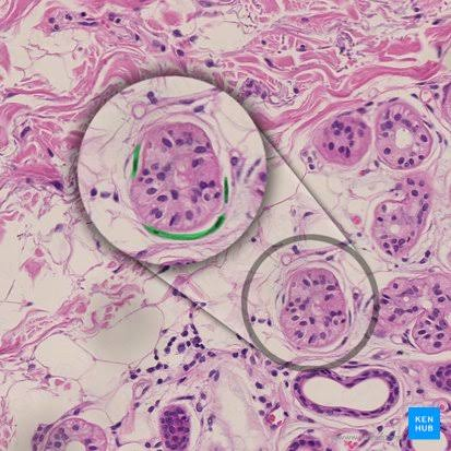
Apocrine Gland
Located in anus, groin, beard, axilla, and areola regions
ducts of these glands lead to nearby hair follicles, not directly to skin

Sebaceous Gland
Produce sebum, short ducts that also lead into hair follicles. Oily secretion that prevent hair and skin from drying out

Ceruminous Glands
Produce cerum (earwax)

Mammary gland
modified apocrine gland, produces milk found in females only

Nails
Hard derivatives of stratum corneum
hard part of nail is nail plate
nail plate consists of: free edge(portion that hangs over the finger), nail body (visible part attached to finger), nail root(continuation of nail under skin)
Skin that rises above the nail root is the nail fold
nail groove is space around the nail body
skin under the nail plate is the nail bed
epidermal layer of the nail plate is called hyponychium
Nails Cont
Stratum basale at the proximal end of the nail hardens and is called nail matrix
apaque clear base of the nail is called the lunule
cuticle overhands the nail at the proximal end

Know diagram
Features of Bone
Flat bones: thin curved bones found in cranium, sternum, scapula, ribs, pelvis
long bones: limbs, comprised of a dense over layer called compact or cortical bone
Cortical none encompasses the marrow cavity
spongy bone fills the central space along the ends of the bones
Shaft of the long bone is the diaphysis with an expanded head on each end called the epiphysis
Epiphyseal line is the location of fusion between the epiphysis ad diaphysis in mature bone. This epiphyseal plate in developing bone
articular cartilage (hylaline) lines the ends of space where movement occurs
bones are covered by a periosteum externally

Know Diagram
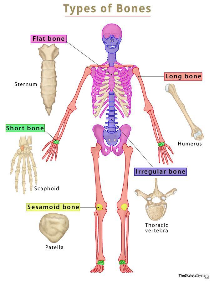
know difference and where each type of bone can be found