MSK TBL 11: Wrist & Hand Functional Anatomy
 Call Kai
 Learn
Learn Practice Test
 Spaced Repetition
 Match
Match Flashcards
 Knowt Play
1/49

There's no tags or description
Looks like no tags are added yet.
Name | Mastery | Learn | Test | Matching | Spaced | Call with Kai | Chat |
|---|
No analytics yet
Send a link to your students to track their progress
50 Terms
palmar aponeurosis
thick, fibrous central fascia
proximal end [apex] of triangular aponeurosis is continuous w/ flexor retinaculum & palmaris longus tendon
wrist joint
radiocarpal joint
articulation
btwn radius & articular disc & scaphoid, lunate & triquetrum
ulna does NOT participate
proximal articular surface forms concave surface
distal convex surface
capsule
encloses joint & is attched above to distal ends of radius & ulna & below to the proximal row of carpal bones
synovial membrane
lines capsule & is attached to margins of articular surfaces
joint cavity does NOT communicate w/ distal radioulnar joint or w/ joint cavities of intercapal joints
nerve supply
anterior interosseous nerve [from median nerve] & deep branch of radial nerve
wrist [radiocarpal] joint ligaments [4]
anterior [palmar radiocarpal] ligament
posterior [dorsal radiocarpal] ligament
1&2 strengthen capsule
ulnar collateral [medial] ligament
attached to styloid process of ulna & to triquetral bone
radial collateral [lateral] ligament
attached to styloid process of radius & to scaphoid bone
movement that’s not possible at wrist joint
rotation not possible [achieved by radioulnar joint]
pronation/supination compensate for this lack of rotation
[flex/extend, ab/aduct, & circumduction possible]
flexors attach to
metacarpal bones
abduction of wrist is performed by which muscles
flexor carpi radialis
extensor carpi radialis longus & brevis
adduction of wrist is performed by which muscles
flexor carpi ulnaris
extensor carpi ulnaris
carpal tunnel
carpal bones form arch that convex on dorsal side, concave on palmar side
groove of palmar side called sulcus carpi
covered by sheath of tough connective tissue = flexor retinaculum [transverse carpal ligament]
attached radially to trapezium & ulnar side to pisiform & hook of hamate
9 flexor tendons passing thru carpal tunnel
flexor digitorum profundus [4 tendons]
flexor digitorum superficialis [4 tendons]
flexor pollicus longus [1 tendon]
in carpal tunnel, median nerve courses btwn
FDP & FDS
which nerve innervates the thenar muscles?
recurrent branch of median nerve
where does median nerve bifurcate?
typically, after nerve exits carpal tunnel
[in 5-10% of pple, happens more proximally]
guyon’s canal
AKA ulnar canal or ulnar tunnel
space btwn pisiform & hamate
ulnar artery & ulnar nerve travel thru into hand
anatomical snuffbox borders
medially: extensor pollicus longus
laterally: extensor pollicus brevis & abductor pollicis longus
floor: scaphoid & trapezium
radial artery cross these bones as it passes diagonally from anterior surface of radius to dorsal surface of hand
most frequently fractured carpal bone
scaphoid [fracture along waist]
from fall on palm when hand abducted
synovial flexor sheaths
in hand, tendons of flexor digitorum superficialis & profundus invaginate a common synovial sheath
sometimes called ulnar bursa
medial part extends distally w/o interupption on tendons of little ginger
lateral part of seath ends on middle of palm
distal ends of long flexor tendons of index, middle, & ring fingers acquire digital synovial sheaths as they enter the fingers
*pattern of infection spread
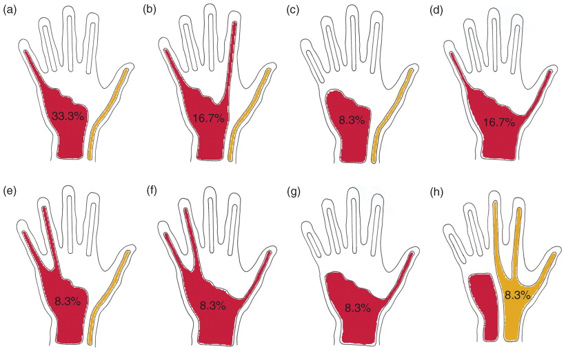
Flexor pollicus longus sheath
FPL has its own synovial sheath that passes into thumb
sometimes called radial bursa
communicates w/ common synovial sheath at level of wrist in 50% of subjects

oblique intertendinous connections
[3]
linked proximal to MCP joints
resist independent extension of 4 medial digits
esp ring finger
extensor expansion
on posterior surface of each finger, extensor tendon joins this fascial expansion
near PIP joint, splits into 3 parts
central part inserted into base of middle phalanx
2 lateral parts converge to insert into base of distal phalanx
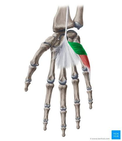
palmaris brevis
small superficial muscle over hypothenar eminence
origin
flexor retinaculum & palmar aponeurosis
insertion
skin on medial border of palm of hand
innervation
superficial branch of ulnar nerve
action
corrugates skin of hypothenar eminence to improve grip of palm
fascial spaces of palm: medial fibrous septum
passes from ulnar border of palmar aponeurosis & attchs to anterior border of 5th metacarpal bone

fascial spaces of palm: lateral fibrous septum
passes from lateral border of palmar aponeurosis & attchs to anterior border of 3rd metacarpal bone

fascial spaces of palm: oblique fibrous septum
btwn 1st & 3rd metacarpal
palm of hand is divided into how many compartments?
4
medial
mid-palmar space
thenar space [misnomer]
lateral
medial compartment of palm
contains hypothenar muscles
mid-palmar space compartment of palm
lies btwn medial & oblique fibrous septum
contains 2nd, 3rd, & 4th lumbrical muscles
lies posterior to long flexor tendons to middle, ring, & little fingers
lies superficial to interosseous muscles & the 3rd, 4th, & 5th metacarpal bones

thenar space compartment of palm
lies btwn oblique & lateral septa
contains 1st lumbrical muscle
lies deep to long flexor tendons to index finger & superficial to adductor pollicus

lateral space compartment of palm
contains muscles of thenar
which compartments of palm are closed off from forearm?
thenar & mid-palmar space closed off by carpal tunnel
[distally, continuous w/ appropriate lumbrical canals]
thenar muscles / muscles of thumb [4]
abductor pollicis brevis
flexor pollicis brevis
opponens pollicis
1-3 median nerve
adductor pollicis
ulnar nerve
hypothenar muscles [3]
abductor digiti minimi
flexor digiti minimi
opponens digiti minimi
all innervated by deep branch of ulnar nerve
intrinsic hand muscles
4 lumbricals
2 median [1st & 2nd], 2 ulnar [3rd & 4th]
3 palmar interossei
ulnar
4 dorsal interossei
ulnar
lumbrical muscles originate from
tendons of flexor digitorum profundus
lumbrical muscles insert onto
extensor expansions of corresponding finger
action of lumbricals
flexion at MCP joints [not that imp]
extension of IP joints [v imp!]
only extensors of fingers [pretty much]
palmar interossei muscles [3]
act on index, ring, & little fingers
origin
metacarpal of corresponding finger
insertion
proximal phalanx
extensor expansion
innervation
deep branch of ulnar nerve
action
adduct at MCP joints
flex at MCP joints
extend at IP joints
PAD
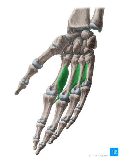
dorsal interossei muscles [4]
act on index, middle [2 muscles], & ring fingers
origin
2 heads from adj. sides of 2 metacarpal bones
insertion
side of base of proximal phalanx
extensor expansion
innervation
deep branch of ulnar
action
abduct at MCP joints
flex at MCP joints
extend at IP joints
DAB
![<p>act on index, middle [2 muscles], & ring fingers</p><ul><li><p>origin</p><ul><li><p>2 heads from adj. sides of 2 <strong>metacarpal </strong>bones</p></li></ul></li><li><p>insertion</p><ul><li><p>side of base of proximal phalanx</p></li><li><p>extensor expansion</p></li></ul></li><li><p>innervation</p><ul><li><p>deep branch of ulnar</p></li></ul></li><li><p>action</p><ul><li><p><strong>abduct</strong> at MCP joints</p></li><li><p>flex at MCP joints</p></li><li><p>extend at IP joints</p></li></ul></li></ul><p><strong>DAB</strong></p>](https://assets.knowt.com/user-attachments/ffdd4a2f-7b41-48ca-a10c-8358e1a01cd5.png)
FDS attaches to
middle phalanx
FDP attaches to
distal phalanx
5 synovial joints of hand & fingers
midcarpal
carpometacarpal
intermetacarpal
metacarpophalangeal [MCP]
interphalangeal [IP]
ligaments of MCP joints
palmar metacarpal ligaments
strong w/ some fibrocartilage
palmar ligaments
united by deep transverse metacarpal ligaments, which hold heads of metacarpal bones together
collateral ligaments
on each side of joints
taut when joint in flexion
finger pulp space
deep fascia of pulp of each finger fuses w/ periosteum of terminal phalanx
this closes off a fascial compartment known as pulp space
terminal branch of digital artery runs thru here
filled w/ fat
blood supply of hand: 3 anastomotic arterial arches from branches of radial & ulnar arteries
superficial palmar arterial arch
deep palmar arterial arch
dorsal carpal arch
collateral circulation of hand: ulnar artery
superficial branch is main supply
collateral circulation of hand: radial artery
dorsal carpal branch is main supply
digit arteries
from dorsal, deep, & superficial arches
supply fingers
every digit gets 4 arteries
2 dorsal digital from dorsal carpal arch
2 proper palmar digital from superficial arterial arch
princeps pollicis
supplies blood to thumb
from radial artery
radialis indicis
supplies blood to index finger
from radial artery
radial tubercle [Lister’s tubercle]
distal radius prominence on wrist dorsum
palpate radius dorsum while pt flexes wrist
lines up w/ 3rd metacarpal

most common carpal dislocation
scapholunate joint or interval