Bio 314 - Exam 1
 Call Kai
 Learn
Learn Practice Test
 Spaced Repetition
 Match
Match Flashcards
 Knowt Play
1/97

There's no tags or description
Looks like no tags are added yet.
Name | Mastery | Learn | Test | Matching | Spaced | Call with Kai | Chat |
|---|
No analytics yet
Send a link to your students to track their progress
98 Terms
Prokaryotic cells
small, bacteria + archaea, no membrane bound organelles, circular chromosomes in cytoplasm (nucloid)
Eukaryotic cells
large, animals, protists, fungi, plants, membrane bound organelles, chromosomes in nucleus
What do prokaryotic and eukaryotic cells have in common?
Both bound by a plasma membrane, both with DNA and ribosomes for gene exp.
plasma membrane
Controls what enters or leaves a cell, functions in cell-cell signaling and cell adhesion
mitochondria
Surrounded by a double membrane, generates ATP by oxidation of glucose and fatty acids
Lysosomes
Have acid lumen, degrade material internalized by the cell, worn-out cellular membranes, and organelles
nuclear envelope
A double membrane that encloses contents of the nucleus, outer nuclear membrane is continuous with rough ER
nucleolus
A nuclear subcompartment where most of cells rRNA is synthesized
nucleus
Contains DNA and proteins in eukaryotic cells, site of mRNA and tRNA synthesis
smooth ER
Synthesizes lipids and detoxifies certain hydrophobic compounds
rough ER
Synthesizes, processes, and sorts secreted proteins, lysosomal proteins, and certain membrane proteins
golgi complex
Processes and sorts secreted proteins, lysosomal proteins, and membrane proteins synthesized on the rough ER
secretory vesicles
Store secreted proteins and fuse with the PM to release their contents
peroxisome
Detoxifies molecules and breaks down fatty acids to produce acetyl groups for biosynthesis
cytoskeletal fibers
Form networks and bundles to support cellular membranes, organize organelles, and for cell movement
microvilli
Increase surface area for absorption of nutrients from surrounding medium
cell wall
Composed of cellulose, maintains plant cells shape and provides protection against mechanical stress
vacuole
Stores water, ions, nutrients, degrades macromolecules, and functions in cell elongation during growth
chloroplasts
Carry out photosynthesis, surrounded by a double membrane and contain a network of internal membrane-bounded sacs
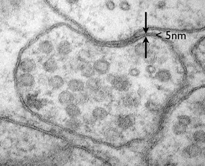
plasma membrane

mitochondria

lysosomes
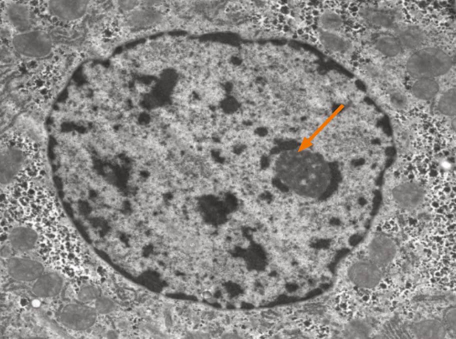
nucleolus
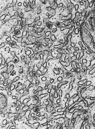
smooth ER

rough ER

golgi complex

secretory vesicles
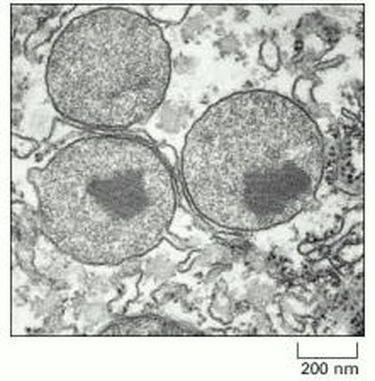
peroxisomes

microvilli
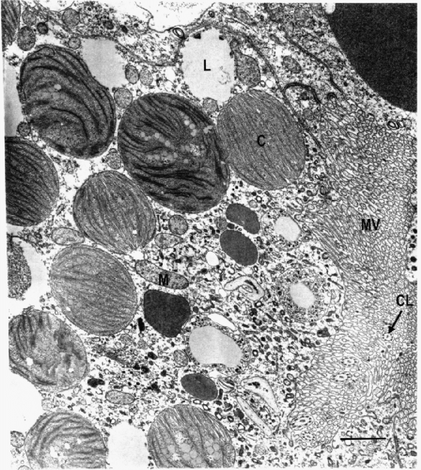
chloroplasts

phosphatidylethanolamine
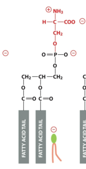
phosphatidylserine

phosphatidylcholine

sphingomyelin
visible light
radiation source and most limiting factor in a light microscope
limit of resolution
the separation that occurs at which the specimen becomes blurry, depends on wavelength and numerical apperature
bright field microscopy
light passes through a cell and forms the image directly
dark field microscopy
Oblique light is angled to not pass through the objective. Low density areas (cytosol) don’t scatter light, high density areas do and appear bright
Phase contrast and differential-inference contrast
Low density areas remain in phase to appear brighter, high density areas deviate waves to appear darker
fluorescent molecules
absorb light at a specific wavelength and emit it at a lower frequency. Used to detect specific proteins and target molecules
Indirect immunohistochemistry
Primary antibodies bind to a specific antigen, and secondary antibodies tagged with fluorescence amplify the signal by binding to primary antibodies
confocal microscopy
Focuses a laser onto a specific point at a specific depth to photograph a given plane, software compiles images to produce a clear “optical section”
Transmission electron microscope
Electrons pass through specimen to view intracellular structures, samples exposed to vacuum, samples dehydrated, permeated with monomeric resin, sliced with ultramicrotome, and covered with heavy metal salts or immunogold
scanning electron microscope
Electrons reflect off specimen to produce 3D structure of specimen surface, specimens are dried and prepped with thin layer of heavy metals. Smaller, simpler, and cheaper than TEM
glycero-phospholipids
p-ethanolomine, p-serine, p-choline
sphingolipids
sphingomyelin
glycolipid
a sugar containing lipid found facing the ECF, involved in cell recognition like blood type
cholesterol
Found 1:1 with phospholipids in PM, amphiphilic and rigid rings decrease fluidity
Why is it important for the plasma membrane to be asymmetrical?
The intracellular layer has more negatively charged lipids, and during apoptosis the bilayer flips to move (-) charges outside the cell
lipid raft
concentrated areas of specific lipids and proteins used for signaling
lipid droplet
stored excess neutral lipids formed from the smooth ER that sit in the cytoplasm until needed
What makes an amino acid?
R-group, amino group (H3N), carboxyl group (COO-), hydrogen around an alpha carbon
peptide bond
dehydration rxn that bonds AAs
What bonds do AA side chains form?
Noncovalent bonds- H bonds, electrostatic attraction, or Van der Walls forces
Main protein folding patterns
a helix and b-sheet, bonds are between AA backbone
transmembrane proteins
amphiphilic and extend through bilayer, single pass, multi-pass, beta barrel
integral membrane proteins
Don’t pass through the bilayer, anchor in membrane via a-helix, GPI, or lipid anchor (covalent bond w/lipid)
peripheral membrane proteins
Don’t enter membrane, non-covalent interactions
hydropathy phot
Use protein sequences to predict hydrophobic membrane spanning regions, only works for a-helices
hydropathy index
How much it takes something hydrophobic to go into water, plotted against AA #
glycosylation
Sugar residues get attached to proteins to protect from chemical/mechanical damage or prevent cell-cell interaction
detergents
Disrupt hydrophobic interactions to degrade the PM and allow proteins to be solubilized and purified
Membrane transporters
Exposed to one side of the membrane at a time
strong interaction with substrate
energetically active/passive transport
Channel proteins
Exposed to both sides of membrane
weak interaction with substrate
only passive transport (faster)
What drives passive transport?
The solutes electrochemical gradient
What cells have the highest permeability in the membrane?
hydrophobic molecules and small uncharged polar molecules
What cells have the lowest permeability in the membrane?
large uncharged polar molecules and ions
What factor influences solute transport?
Membranes have electrical potentials that favor influx of (+) ions and oppose (-) ions
transporter saturation (Vmax)
max rate of transport, speed transporter can flip b/t conformation states
transporter affinity (Km)
affinity of transporter to a solute
transporter abundance
alterations in the amount of transporters in the membrane
coupled transporter
Use energy from electrochemical gradient to transport a solute actively, only change conformation when both binding sites are full
ATP-driven transporters
Pump solutes across membranes through hydrolysis of ATP, p-type pumps and ABC transporters
Pathway of p-type pumps
Substrate binds to open transporter
ATP transfers P, forms ADP
2nd conformation change
Substrate leaves transporter
Removal of P resets pump to original conformation
P-type: Ca2+ pump
Actively holds Ca concentrations low in the cytoplasm to allow for rapid influx of Ca from ECF
P-type: Na+/K+ pump
Maintain steep concentration gradients of Na and K, is electrogenic (allows uneven mov’t of charges)
ABC transporter
Transport a huge variety of substrates, have 2 ATP binding cassettes on the cytosolic side of membrane
ion channels
Only passive transport, control ion specificity with selectivity filter (accounts for ion size and charge)
membrane potential
Difference in electrical charge on two sides of a membrane (mV), created by Na/K pumps
Measuring membrane potential w/whole cell
Record electrical currents through multiple ion channels
Measure membrane potential w/patch clamp
Record electrical current from ion flow through one singular ion channel
Mechanically gated channels
Respond to physical touch. Ex. in animals a sensation or change in blood pressure, in plants respond to mechanical stretching for turgor pressure regulation
Ligand and voltage gated channels
A ligand gated channel is amplified by a voltage gated channel until the ligand is released
voltage gated channel
generates action potentials, a traveling wave of electrical excitement triggered by depolarization
Mechanism of voltage-gated channel
channel is closed, membrane depolarization is detected via voltage sensors
a local change occurs and gate opens
Na+ flows in causing changes in other channels until inactivation gate closes
membranes repolarize through voltage-gated K+ channels
return to resting membrane potential
Charge of ions in ECF
Na+=145mM, Mg2+=1-2mM, Ca2+=1-2mM, Cl=110
Charge of ions in ICF
K+=140mM
cytosol
The site of protein synthesis and degradation
Proteins: Gated transport
cargo moves through gates b/t two spaces w/the same composition
Protein translocation
Proteins move from cytosol into something w/a distinct composition (crossing a membrane)
vesicular protein transport
Proteins start in ER and go to other parks of the endomembrane system
How do proteins get sorted?
Receptors recognize a sorting signal (AA sequence) and guides proteins to their target
nuclear pore complexes (NPCs)
Gates in the nuclear envelope made of nucleoporins, lined with FG motifs that regulate what goes through
Ran GTPase
Molecular switch localized in the nucleus that changes form when bound to GTP or GDP nucleotides
Function of Ran GTPase
Nuclear protein in cytosol binds to importin receptor and moves through nuclear pore
Ran GTP binds to importin receptor to cause unloading of protein
Protein falls off and is discharged in the nucleus
Importin-Ran GTP complex goes back out to the cytoplasm
Cytosolic GAP hydrolyzes Ran GTP to Ran GDP
Ran GDP and importin dissociate
GTPase activating protein (GAP)
Transforms GTP→GDP
Guanine exchange factor (GEF)
Transforms GDP→GTP