BIOL 2220: Unit 4 Test
 Call Kai
 Learn
Learn Practice Test
 Spaced Repetition
 Match
Match Flashcards
 Knowt Play
1/268

There's no tags or description
Looks like no tags are added yet.
Name | Mastery | Learn | Test | Matching | Spaced | Call with Kai | Chat |
|---|
No analytics yet
Send a link to your students to track their progress
269 Terms
what mesenchymal layer becomes the brain and CNS
ectoderm
sympathetic preganglionic neurons branch off of...
thoracic and lumbar spinal cord
parasympathetic preganglionic axons branch off of...
cervical and sacral spinal cord
steps of embryological development
1. ectoderm thickens along midline to form neural plate
2. neural plate invaginate to for neural groove and folds
3. folds fuse to form tube
4. neural crest develops as offshoot of neural folds, becomes ganglia
primary brain vesicles
prosencephalon, mesencephalon, rhombencephalon
prosencephalon
forebrain
mesencephalon
midbrain, does not develop into secondary vesicles
rhobencephalon
hindbrain
the proencephalon gives rise to...
telencephalon and diencephalon
the rhombencephalon gives rise to...
metencephalon and myelencephalon
cerebrum
from telencephalon, undergoes greatest structural changes
diencephalon
hypothalamus, thalamus, epithalamus, retina
Metencephalon
pons and cerebellum
Mylencephalon
medulla oblongata
brain ventricles
cerebrospinal fluid filled cavities within the brain, connect with spinal cord, expansions of lumen of neural tube
lateral ventricles
left and right, do not directly connect
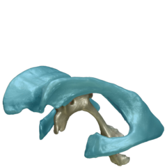
third ventricle
the ventricle located in the center of the diencephalon
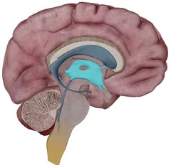
fourth ventricle
small triangular chamber between pons and cerebellum

interventricular foramen
connects lateral and third ventricles

cerebral aquaduct
connects the third and fourth ventricles
lateral apertures
Two openings in the side walls of the fourth ventricle that connect to the subarachnoid space
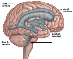
median apertures
holes by cerebellum that connect ventricle CSF to CSF in the subarachnoid space

aperture function
allows fluid to go out of brain to meninges
insula lobe
processes sensory information, emotional states and self awareness
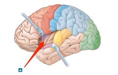
frontal lobe
motor control and planning, decision making, personality, language
gyri and sulci function
allow for more cerebrum to fit in skull
parietal lobe
sensory perception, spacial awareness
temporal lobes
auditory processing, language, memory
occipital lobe
visual processing and recognition
white matter
deep to cerebral cortex, made of myelinated axons
- commissures, association fibers, projection fibers
commissures
connect cerebral cortex of both hemispheres
association fibers
connect different parts of the same hemisphere
projection fibers
two way path, ties cerebral cortex to res of nervous system, link between sensory and motor
basal nuclei
islands of gray matter buried within the white matter
- caudet nucleus, putamen, globus pallidus, amygdala
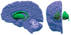
functions of basal nuclei
- similar to cerebellum
- refine skeletal muscle activity
- regulate stereotype movement (ie; eating)
- attention, cognition, emotion
- amygdala = fight or flight
cerebral cortex
differing degrees of thickness and structure linked to function, gray matter, mostly cell bodies, each hemisphere controls opposite side of body, no area acts alone
lateralization of function
right hemisphere -> visual/spacial tasks, art, music emotion
left hemisphere -> logic, language, math, science
functional areas of cerebral cortex
motor, association, and sensory areas
motor areas of cerebral cortex
primary motor cortex, premotor cortex, frontal eye field, broca's area
primary motor cortex
contracts muscles for skeletal muscle movements, pyramidal cells fine tune movement
premotor cortex
active during the planning of a movement, cannot directly control skeletal muscle, signals sent to primary motor cortex
Broca's area
left hemisphere only, motor area connected to speech, forming mouth shape and movement for words
frontal eye field
controls voluntary eye movements
sensory areas
-primary somatosensory cortex
-somatosensory association cortex
-visual and auditory areas
-olfactory, gustatory, and vestibular cortices
primary somatosensory cortex
receives signals from sensory receptors on the skin and proprioceptors, telling us where we are in space, figures out which part of the body is being stimulated
Somatosensory association cortex
integrates the information from primary somatosensory cortex
primary visual cortex
receives information from the retina of the eye
ex. football did not go through uprights
visual association area
interprets information from retina using prior experiences
primary auditory cortex
receives information from the inner ear
auditory association area
interprets information from inner ear
olfactory cortex
receives information from the nose
gustatory cortex
associated with the reception of taste
vestibular cortex
associated with sensations related to balance, located in the insula
association areas
-prefrontal cortex
-broca's and wernicke's
- general interpretation area
- visceral association area
prefrontal cortex
most evolved in humans
- associated with intellect, complex learning, recall, personality
-develops slowly, dependent on social feedback
wernicke's area
-left side of cerebrum
-used to sound out words
general interpretation area
- only on left side of brain
- integrates information from all other association areas into a single thought
visceral association area
-located in the cortex of the insula
-involved in conscious perception of visceral sensations (bladder full, upset stomach)
Cortex vs. Association area
association areas are specific parts of the cortex that aren't primary sensory or motor zones but instead integrate information
- if it doesn't start with primary it is an association area
thalamus
- all sensory information except for spinal reflexes travels through thalamus
- filters and codes sensory impulses, sending them to the correct cortex
hypothalamus
- internal organ control center
- associated with the autonomic nervous system
- controls food intake, water balance, sleep, hormone release
- daily maintenance
epithalamus
contains the pineal gland, secretes melatonin and produces CSF
brain stem
pons + medulla
- deep gray matter surrounded by white matter
- autonomic behaviors for survival: digestion, breathing, heartbeat
- connects higher and lower brain function
midbrain
- connects to both spinal cord and cerebellum
- pain suppression, physical reaction to emotion (links amygdala to ANS)
- visual reflex center (neck movement to eyball movement)
- auditory impulses travel through
pons
pathway between cortices and spinal cord
- connects cerebellum and motor cortex
medulla oblongata
- controls heart rate and blood vessel diameter
- rate and depth of breathing
- vomiting, swallowing, hiccuping
cerebellum
- refines skeletal muscle contractions
p plays role in cognition and problem solving
- subconscious activity
- gray cortex with internal white matter (arbor vitae)
steps of cerebellar processing
visual receptors, equilibrium receptors, motor association area of cerebral cortex --> cerebellar cortex calculates best path for muscle movement --> sends message to muscle --> muscle contracts
limbic system
emotional brain
- closely related to odor and smell
- output relayed through hypothalamus, resulting in gestures
- strong link between feelings and thoughts (feelings always override thoughts)
reticular formation
- loose cluster of neurons around midbrain and brainstem
- filters sensory information and sends to cerebral cortex
- linked to pineal gland and melatonin (wakes brain up)
EEG
recording of the waves of electrical activity that sweep across the brain's surface
alpha waves
slow brain waves of a relaxed, awake state
beta waves
awake and alert, concentrating
theta waves
common in children, vegetative, ignoring surroundings
delta waves
the large, slow brain waves associated with deep sleep
continuum of consciousness
alert --> drowsy --> lethargic --> stupor --> coma
NREM sleep
non-rapid eye movement sleep; encompasses all sleep stages except for REM sleep
- restorative sleep
REM sleep
- go from alpha wave to beta wave
- rapid eye movements
- dreaming
- NREM-->REM 4 times , 4th time being the longest and the one we usually remembers
REM function hypothesis
- analyzing our day
- reverse learning , forgetting unimportant things and remembering important
memory
created by repeated use of neuronal pools/networks
- stimulus--> temporary buffer (filters) --> short term/working--> goes into long term with reppitition and association
meninges
cover CNS, protect blood vessels, contain CSF
dura mater
two layered sheet of fibrous connective tissue that covers the brain
- one layered around spinal cord
- strongest, outermost covering of meninges
arachnoid mater
middle meningeal layer
- creates a space as it is filled with fluid and blood vessels
pia mater
innermost meninx, immediately covers brain and spinal cord
cerebrospinal fluid
reduces brain weight, protects and cushions brain and spine, nourishes brain
- formed by leaky capillaries in brain ventricles
blood-brain barrier
Blood vessels (capillaries) that selectively let certain substances enter the brain tissue and keep other substances out
where is the BBB absent?
vomiting center (poison triggers vomiting)
hypothalamus (assesses blood temperature and water levels)
What substances can pass through BBB?
glucose (ATP), amino acids (building), electrolytes (APs)
spinal cord development
neural tube develops an alar plate (interneurons) and basal plate (motor neurons)
- plates become grap matter, everything surrounding becomes white matter
conus medullaris
end of CNS
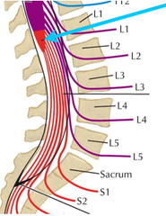
filum terminale
fibrous extension of the pia mater; anchors the spinal cord to the coccyx
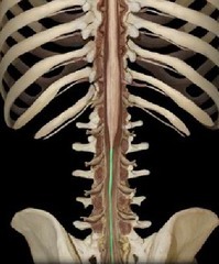
denticulate ligaments
extensions of pia mater that secure cord to dura mater

cauda equina
collection of spinal nerves below the end of the spinal cord
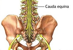
gray matter of spinal cord
nerve cell bodies arranged in a butterfly shape with anterior and posterior "horns"
white matter of spinal cord
myelinated axons "columns"
lateral horns of spinal cord
cell bodies of autonomic motor neurons
anterior horns of spinal cord
cell bodies of somatic motor neurons
posterior horns of spinal cord
cell bodies of somatic and visceral sensory neurons
dorsal root ganglion
pseudounipolar sensory neuron that synapes with PNS and interneurons of posterior hors
spinal cord tracts
bundles of axons that run in the white matter of the spinal cord
- stem from cell bodies in gray matter
- allow for signal amplification and transport to mulriple pathways
- most exhibit decussation and somatotopy
- have left and right pairs
- made up of x order neurons