Lab 1 SCB 204 Lagcc
 Call Kai
 Learn
Learn Practice Test
 Spaced Repetition
 Match
Match Flashcards
 Knowt Play
1/83

Earn XP
Description and Tags
3 sets combined
Name | Mastery | Learn | Test | Matching | Spaced | Call with Kai | Chat |
|---|
No analytics yet
Send a link to your students to track their progress
84 Terms
Which chamber receives deoxygenated blood before it is pumped into the pulmonary circulation?
Right atrium
Which valve with two cusps prevents backflow of oxygenated blood into the left atrium during left ventricular contraction?
Bicuspid (mitral) valve
Which chamber is responsible for pumping oxygenated blood into the aorta?
left ventricle
what are the muscular projections inside the ventricles that help secure the atrioventricular valves?
papillary muscles
which vessel receives deoxygenated blood from the right ventricle to carry it toward the lungs?
pulmonary trunk
Which intermediate muscular layer of the heart wall is responsible for contraction?
Myocardium
What is the muscular wall that separates the right and left ventricles?
interventricular septum
Which valve with three cusps is located between the right ventricle and the pulmonary trunk?
pulmonary semilunar valve
Which valve prevents the backflow of blood into the right atrium during right ventricular contraction?
tricuspid valve
What fibrous, thread-like structures anchor the AV valve cusps to the papillary muscles?
chordae tendineae
What is the percentage of pacemaker cells in cardiac myocytes?
1%
What is the percentage of contractile cells in cardiac myocytes?
99%
What is the primary function of pacemaker cells?
spontaneously and rhythmically generates action potentials to set the pace of the heart
What is the primary function of contractile cells?
depolarize in response to pacemaker action potentials and contract to pump blood
What are the three populations of the cardiac conduction system?
Sinoatrial (SA) node
Atrioventricular (AV) node
Purkinje fiber system
Where is the Sinoatrial (SA) node located?
upper right atrium
What is the intrinsic rate of depolarization of the SA node?
60 times per minute or more
What is the normal pacemaker of the heart?
Sinoatrial (SA) node
What is the anatomical location of the Atrioventricular (AV) node
posterior and medial to the tricuspid valve
What is the intrinsic rate of depolarization of the AV node?
40 times per minute
What are the components of the Purkinje fiber system in order of the signal flow?
Atrioventricular bundle
right and left bundle branches
terminal branches
What is the intrinsic rate of depolarization of the Purkinje fiber system?
20 times per minute
Explain the path of the Atrioventricular bundle
Penetrates the heart’s fibrous skeleton in the inferior interatrial septum and superior interventricular septum
explain the path of the right and left bundle branches
course along the right and left sides of the interventricular septum toward the apex
What is the function and endpoint of terminal purkinje fibers?
spread from the apex to penetrate ventricles and directly contact ventricular contractile cardiac muscle cells
What is step 1 in action potential propagation?
The SA node generates an action potential that spreads through atrial contractile myocytes via gap junctions
What is step 2 in action potential propagation?
Atrial contractile cells depolarize and contract, while the signal travels to the AV node
What is step 3 in action potential propagation?
signal reaches the AV node, where conduction slows slightly to allow ventricles to fill with blood
what is step 4 in action potential propagation?
the signal travels through the AV bundle, crossing the fibrous skeleton into the interventricular septum
what is step 5 in action potential propagation?
the signal splits into the right and left bundle branches, descending along the septum toward the apex
what is step 6 in action potential propagation?
terminal Purkinje fibers conduct the action potential rapidly upward through the ventricular wall muscle
what is step 7 in action potential propagation?
ventricular contractile cells depolarize and contract in unison, pumping blood to the lungs and body
what mechanism allows fast electrical speed between cardiac cells?
gap junctions within intercalated discs
what is the functional purpose of the AV nodal delay?
allows the atria to complete contraction and fill the ventricles before ventricular contraction begins
what is the direction of ventricular depolarization vector?
spreads from the apex upward through the ventricular walls toward the base
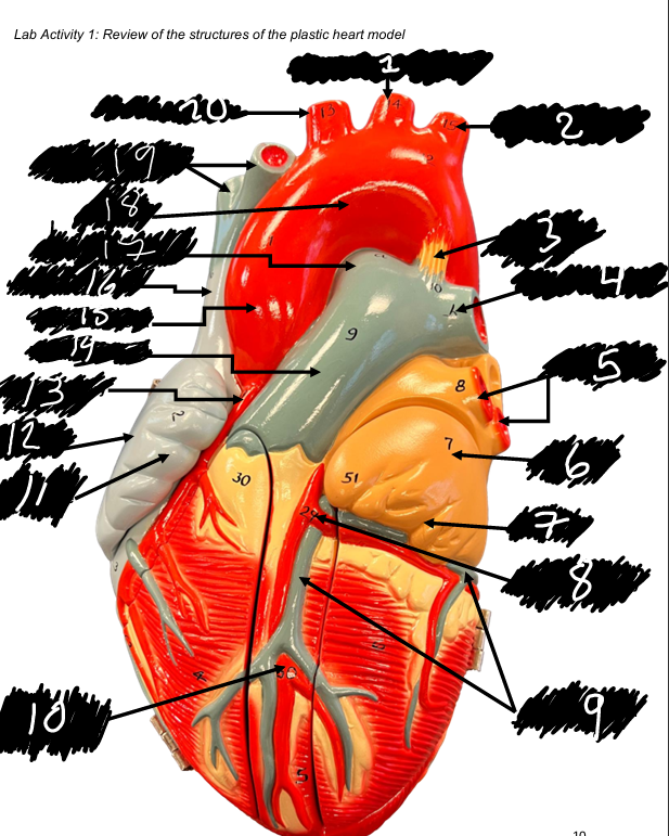
Name structure 1
Left common carotid artery
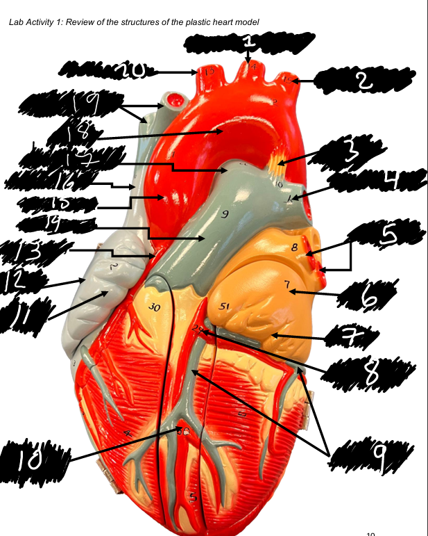
name structure 2
Left subclavian artery
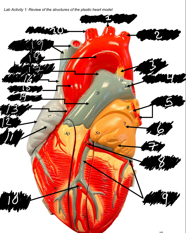
name the structure 3
Ligamentum arteriosum
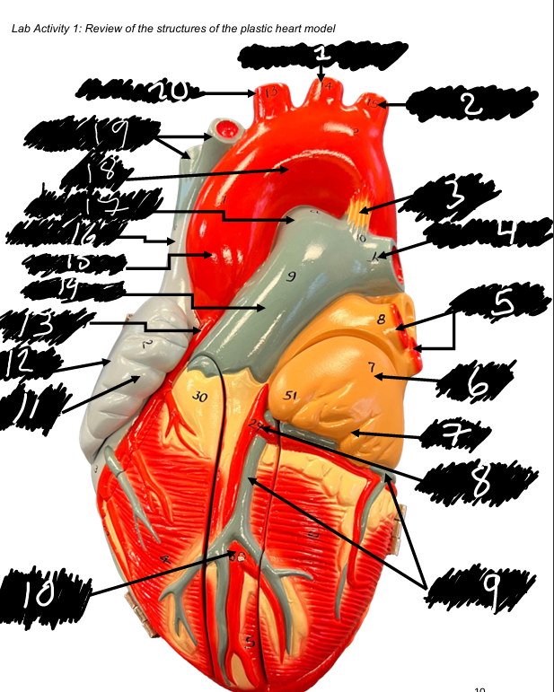
name the structure 4
Left pulmonary artery
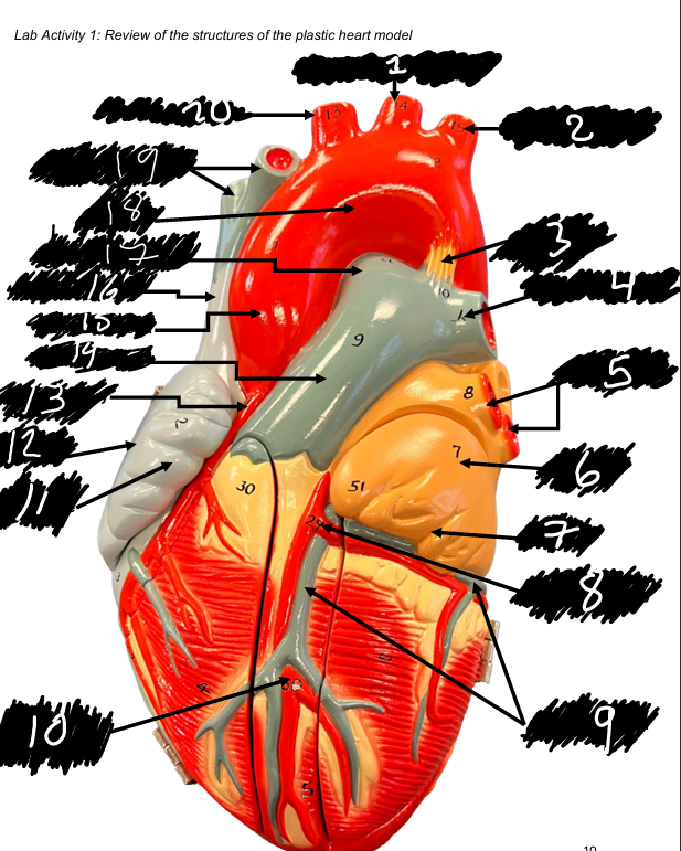
name the structure 5
Pulmonary veins
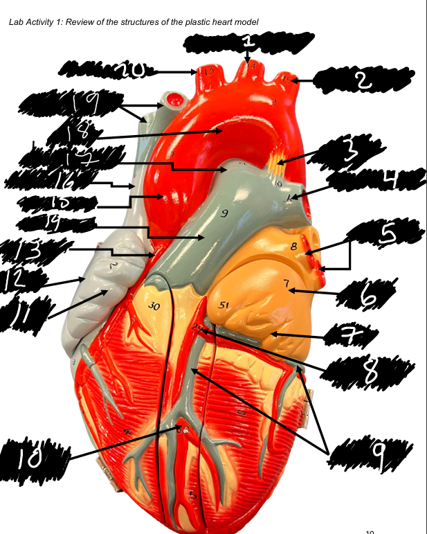
name the structure 6
Left atrium
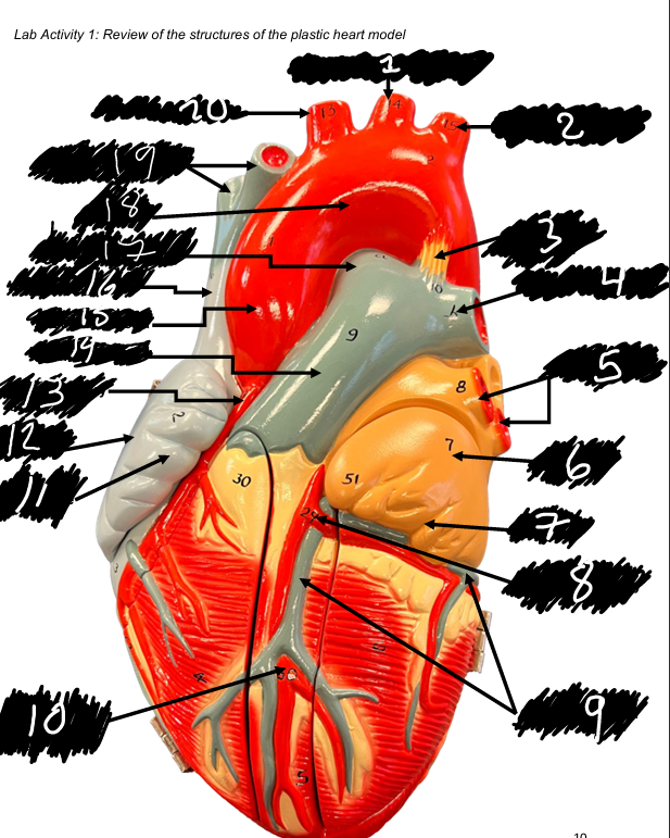
name the structure 7
left auricle
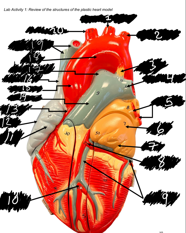
name the structure (8)
left coronary artery
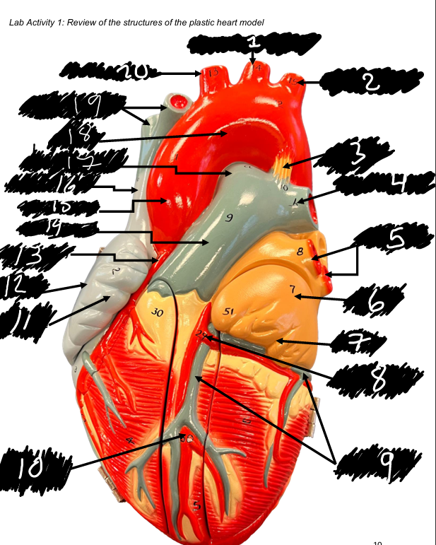
name the structure (9)
Great cardiac vein
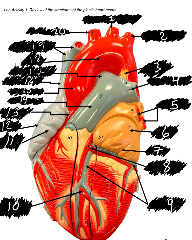
name the structure (10)
Anterior interventricular artery
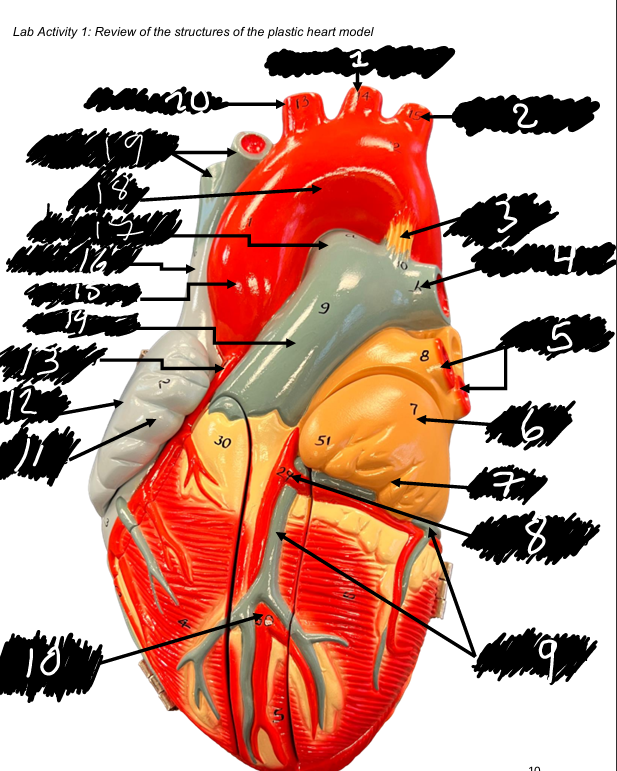
name the structure (11)
Right auricle
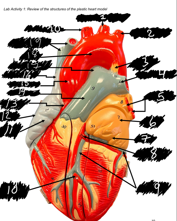
name the structure (12)
right atrium
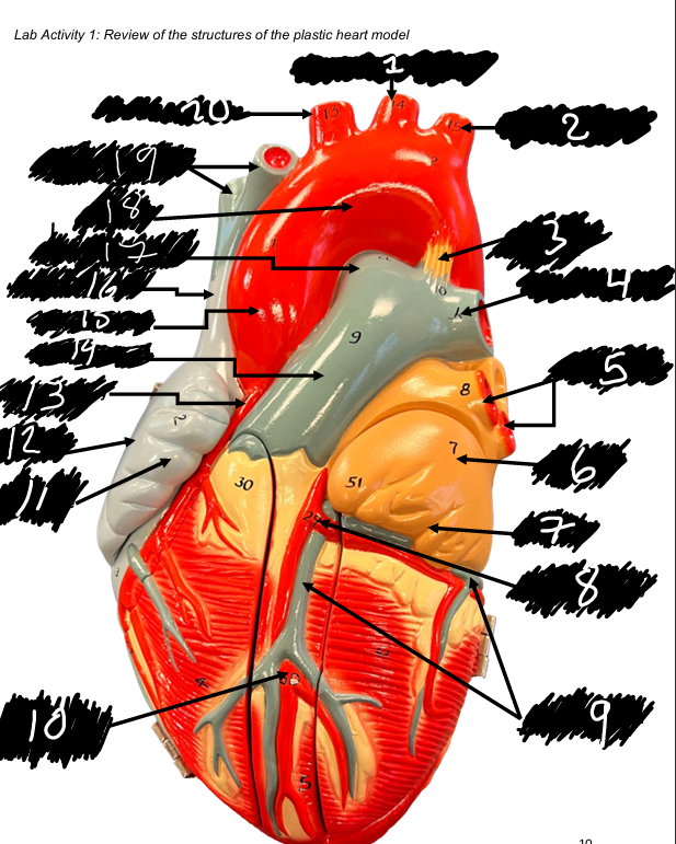
name the structure (13)
Right coronary artery
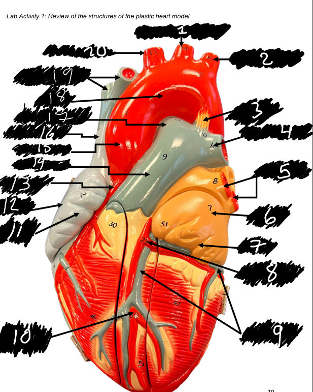
name the structure (14)
Pulmonary trunk
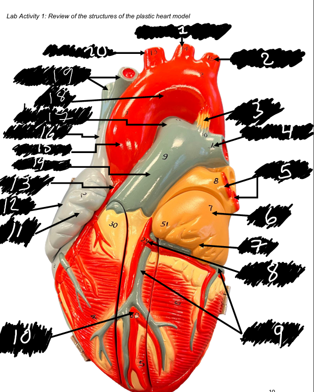
name the structure (15)
Ascending aorta
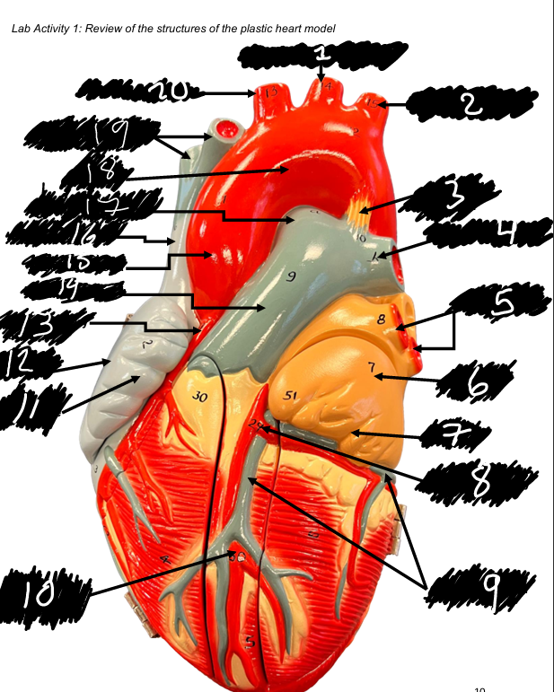
name the structure (16)
Superior vena cava
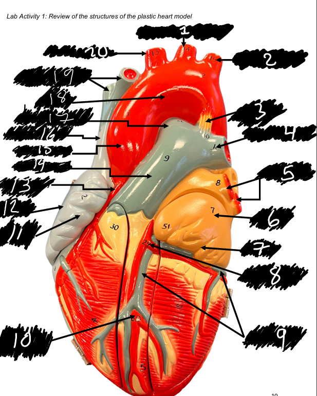
name the structure (17)
Right pulmonary artery
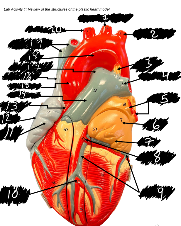
name the structure (18)
Aortic arch
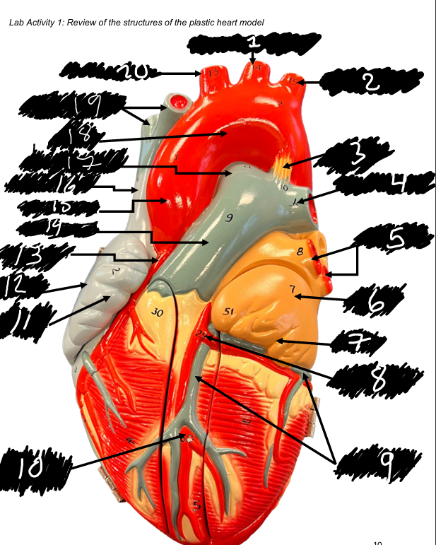
name the structure (19)
Brachiocephalic veins
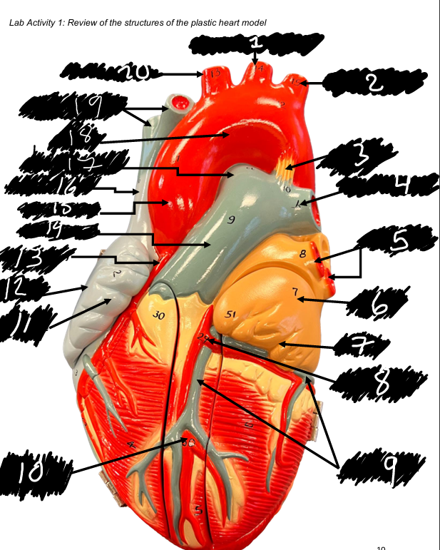
name the structure (20)
Brachiocephalic trunk
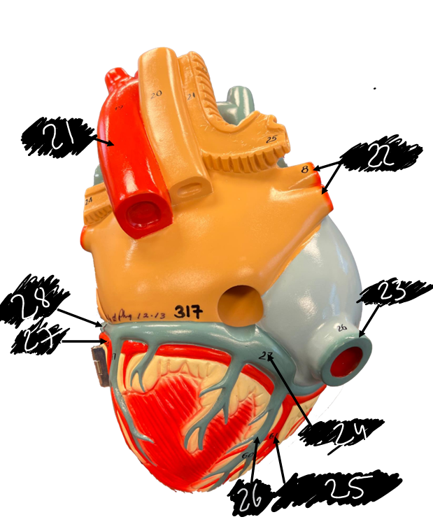
name the structure (21)
Descending thoracic aorta
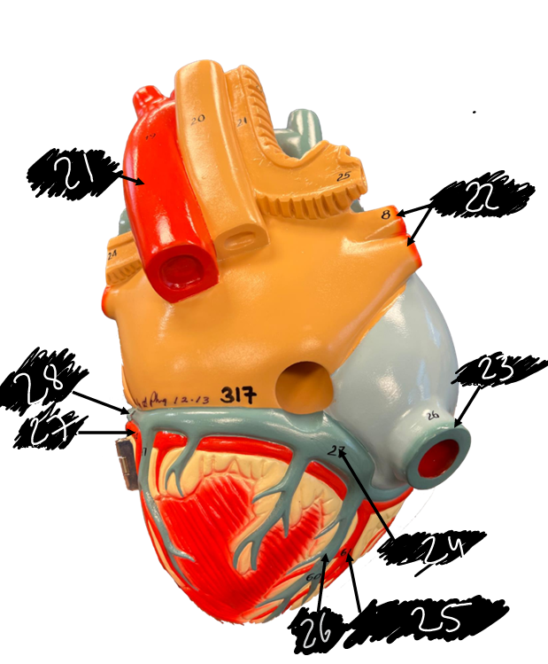
name the structure (22)
Pulmonary veins
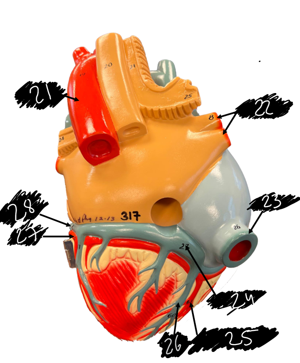
name the structure (23)
Inferior vena cava
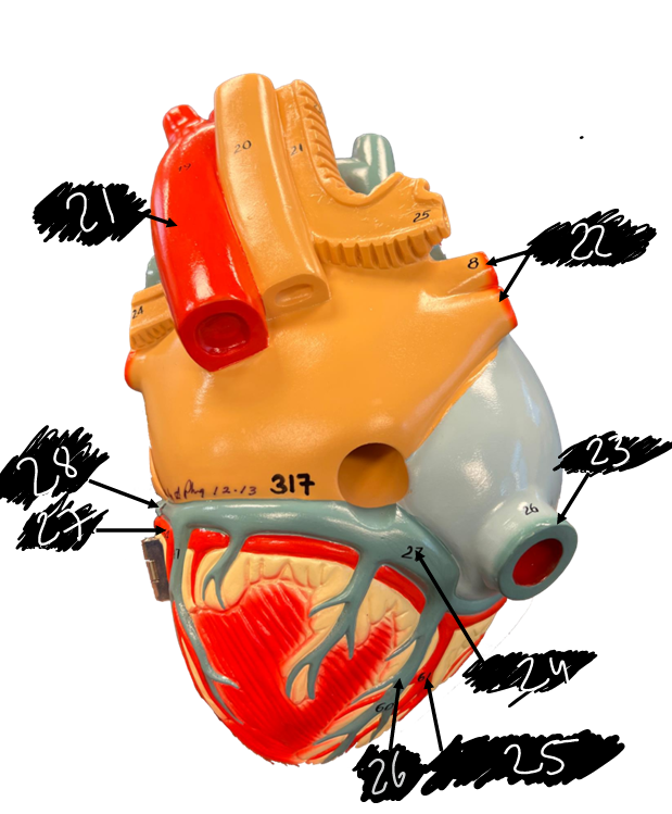
name the structure (24)
Coronary sinus
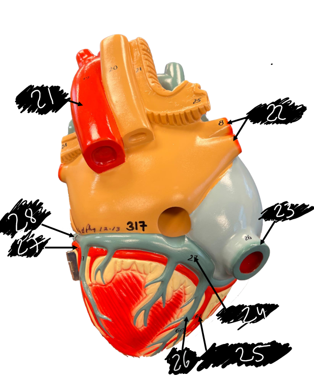
name the structure (25)
Posterior interventricular artery
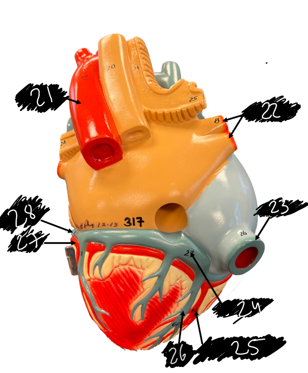
name the structure (26)
Middle cardiac vein
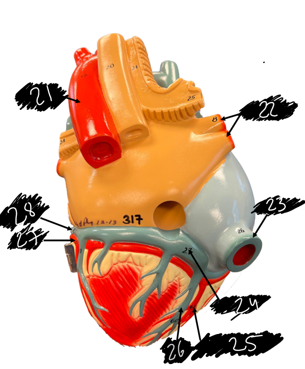
name the structure (27)
Circumflex artery
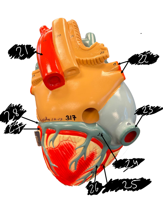
Name the structure (28)
Great cardiac vein
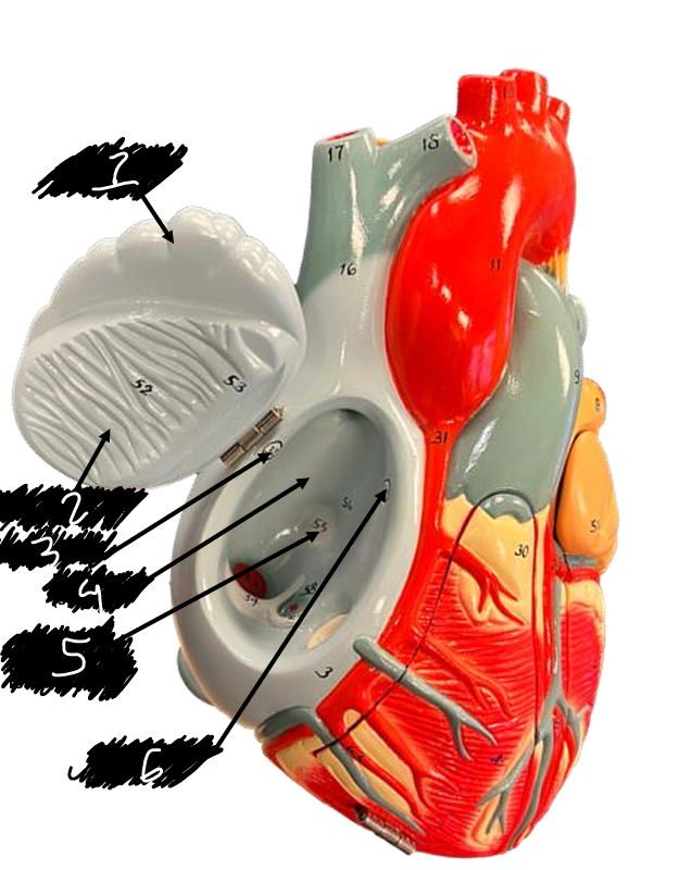
name the structure (1)
Right auricle
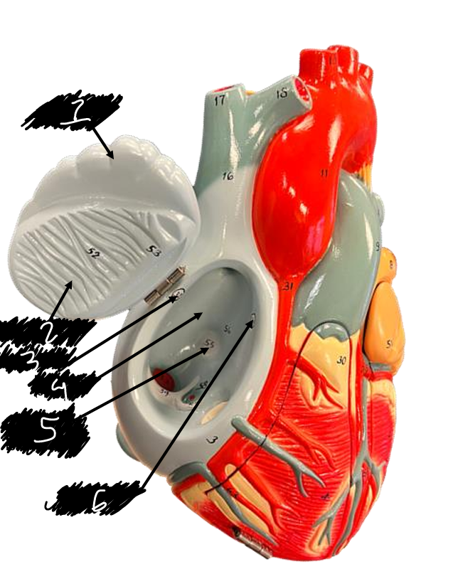
name the structure (2)
Pectinate muscles
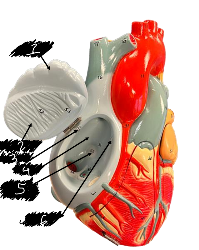
name the structure (3)
Sinoatrial node
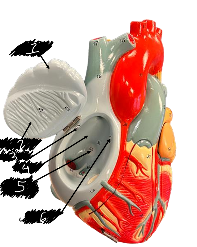
name the structure (4)
Right atrium
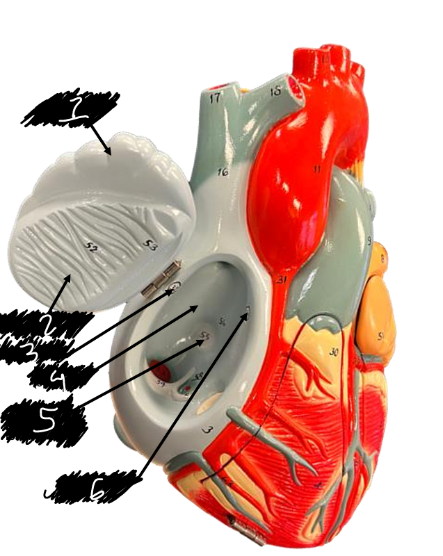
name the structure (5)
Fossa ovalis
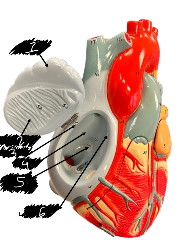
name the structure (6)
Atrioventricular node
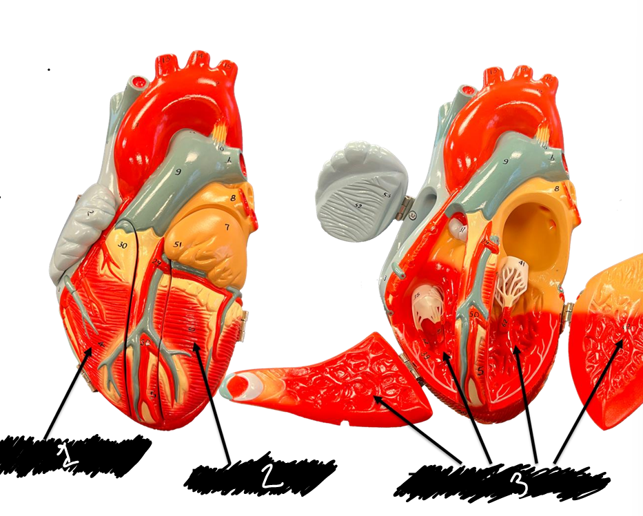
name the structure (1)
Right ventricle
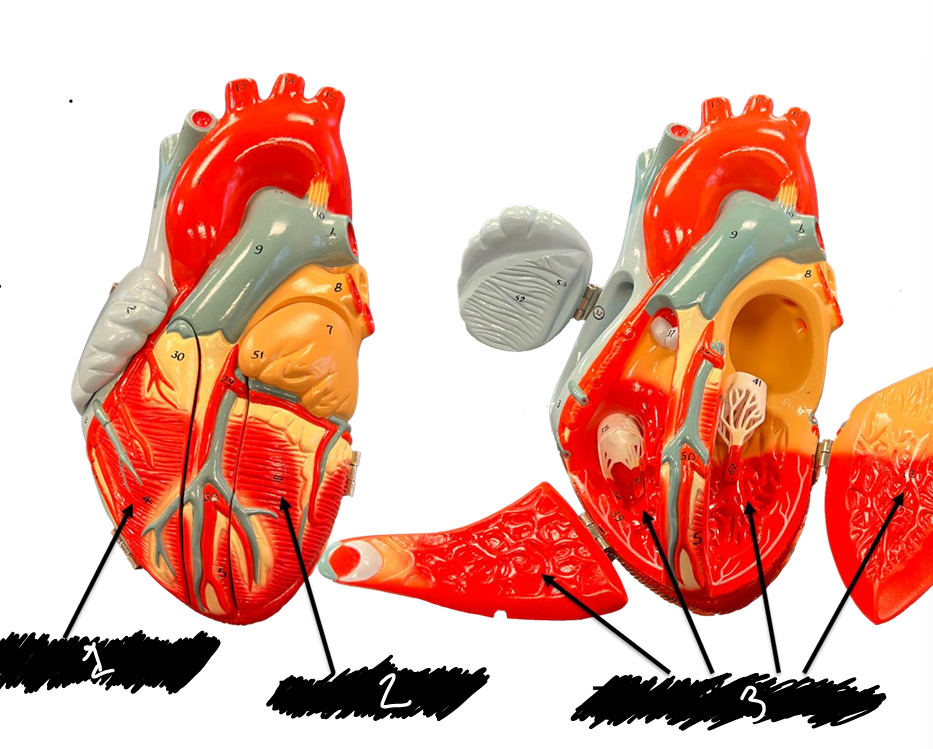
name the structure (2)
Left ventricle
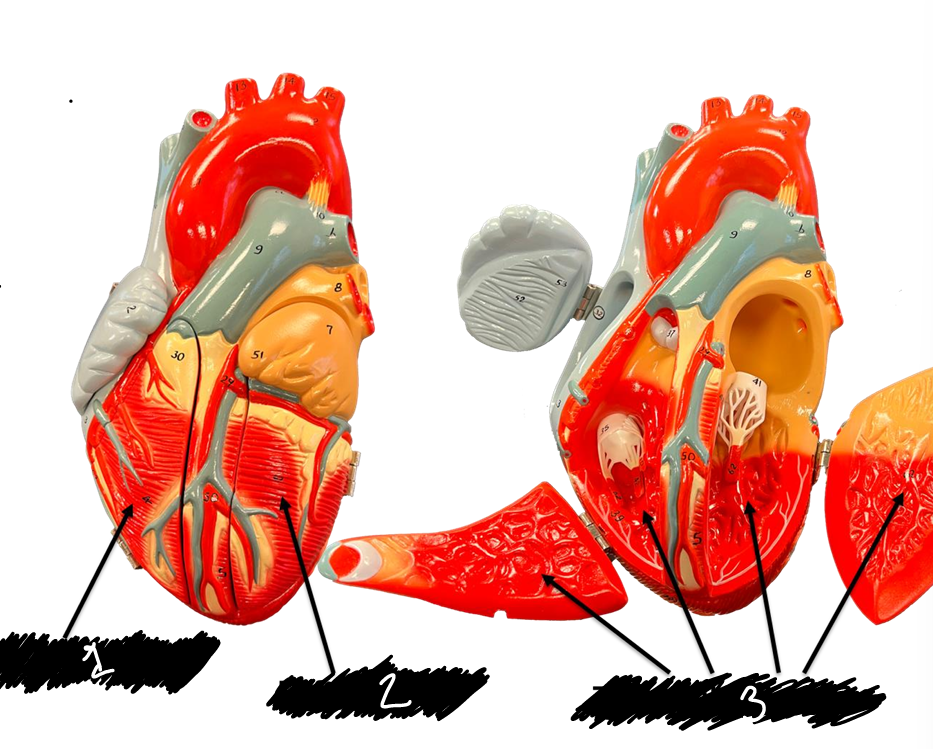
name the structure (3)
Trabecule carneae
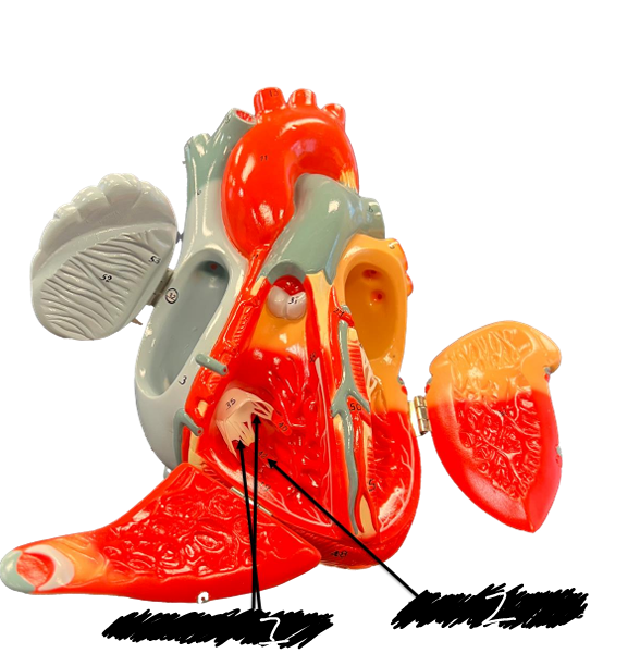
name the structure (1)
Chordae tendineae
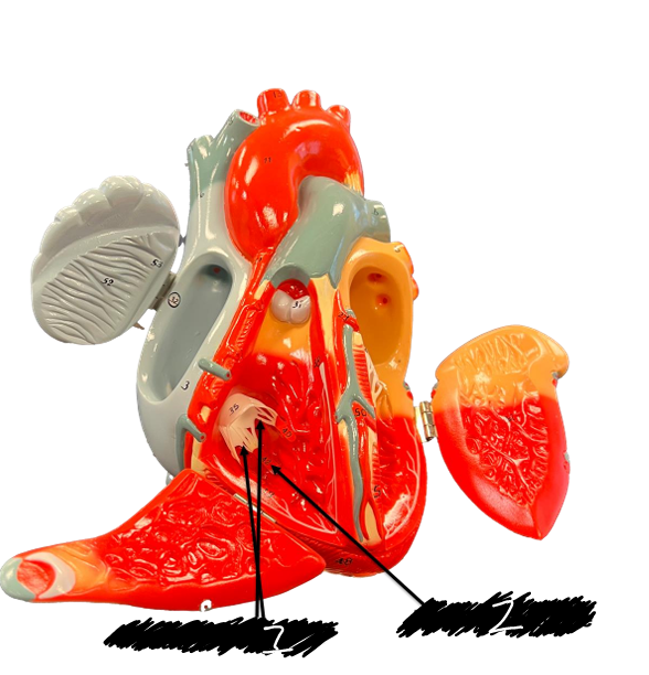
name the structure (2)
Papillary muscle
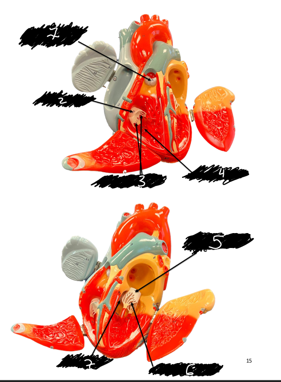
name the structure (1)
Pulmonary valve
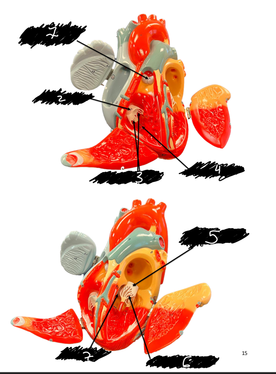
name the structure (2)
Tricuspid valve
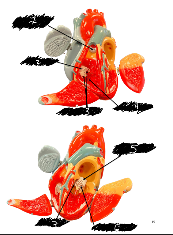
name the structure (3)
Chordae tendineae
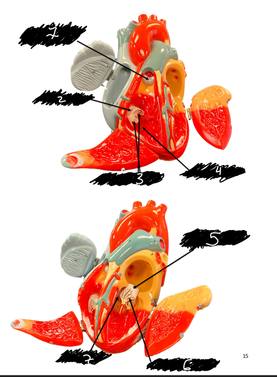
name the structure (4)
Papillary muscle
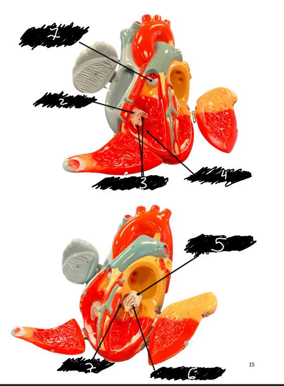
name the structure (5)
Bicuspid valve
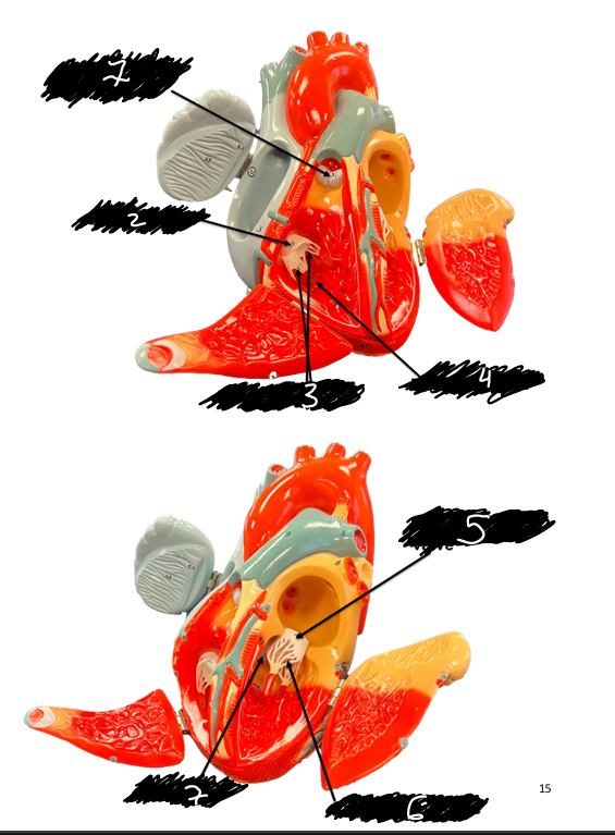
Name the structure (6)
Chordae tendineae
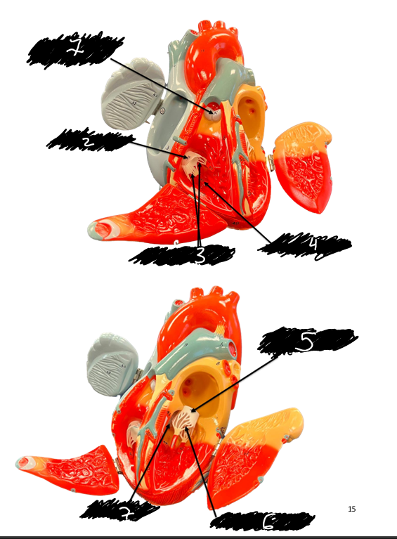
name the structure (7)
Aortic valve
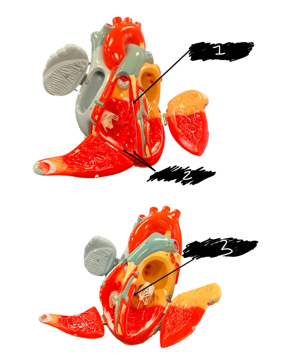
name the structure (1)
Right branch of bundle of His
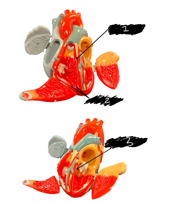
name the structure (2)
Purkinje fibers
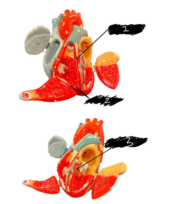
name the structure (3)
Left branch of bundle of His