Anatomy Lab 2: Trunk Musculature, Vertebral Column, Meninges and Spinal Cord
1/23
There's no tags or description
Looks like no tags are added yet.
Name | Mastery | Learn | Test | Matching | Spaced | Call with Kai |
|---|
No analytics yet
Send a link to your students to track their progress
24 Terms
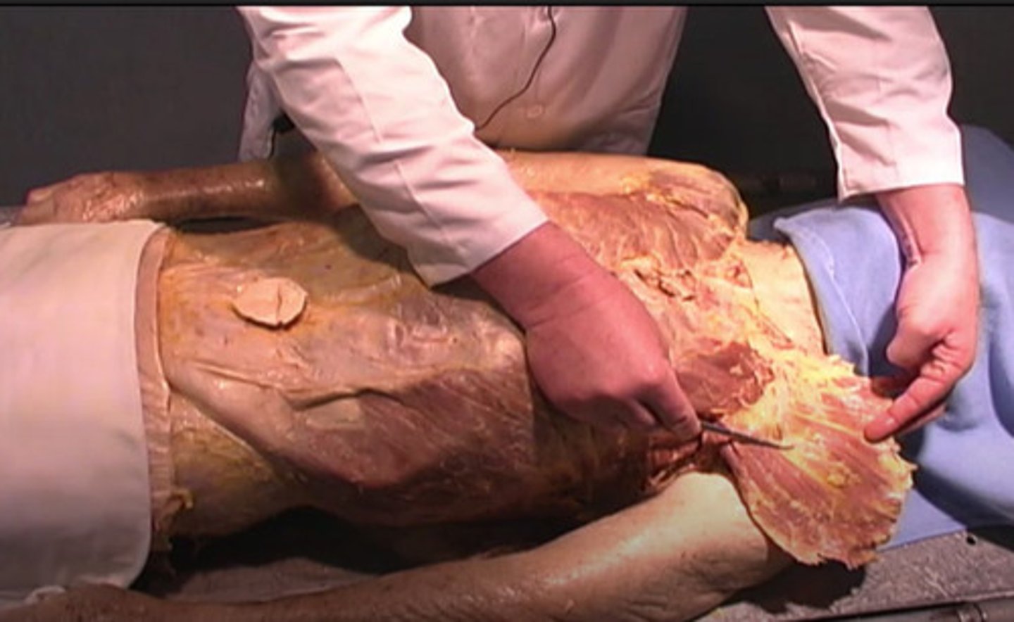
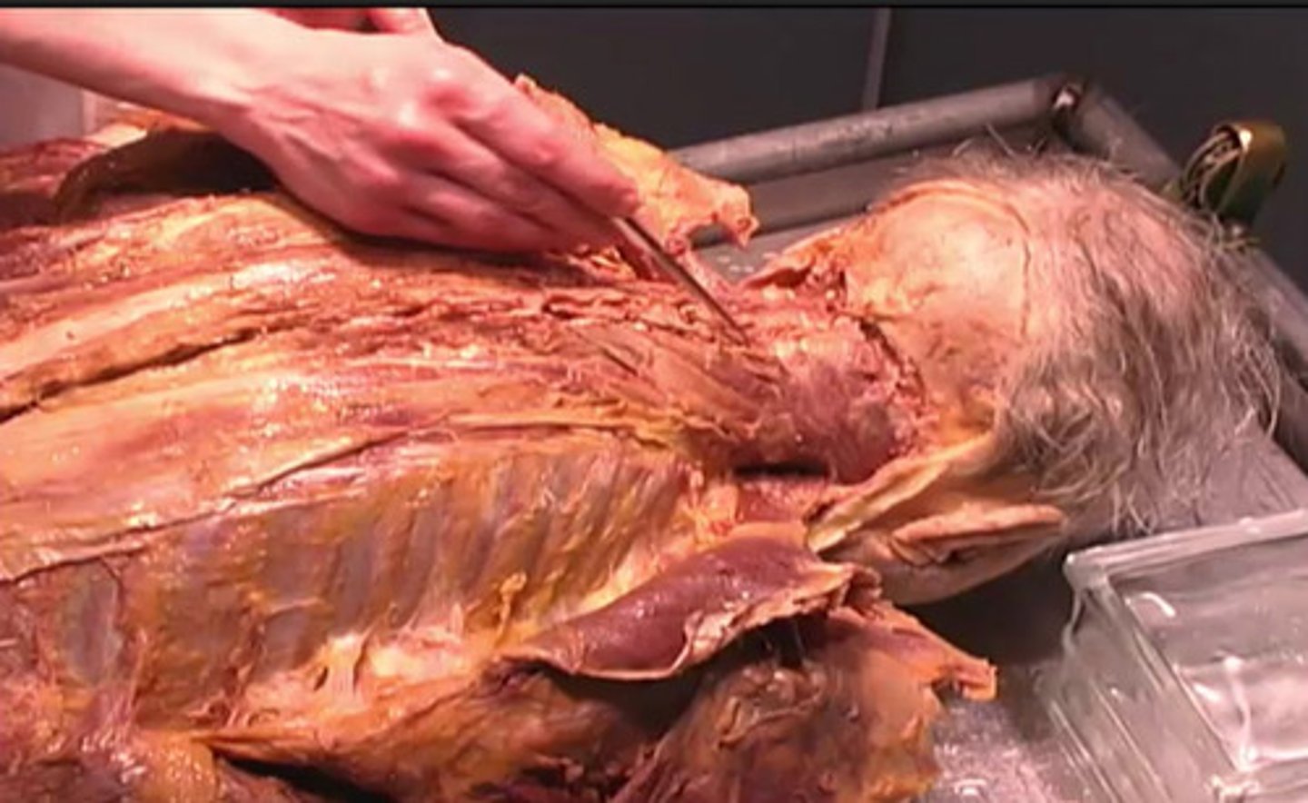
pectoralis major
What is the structure being shown here?
pectoralis major
inferior margin of the muscle
spine
sternum
Cutting off the muscle from the underlying ribs
What is the purpose of removing the pectoralis major?
cutting off the ribs
detaching the xiphoid process
cutting off the muscle from the underlying ribs
removing the spine
reflected
This muscle is being:
reflected
refracted
insized
excized
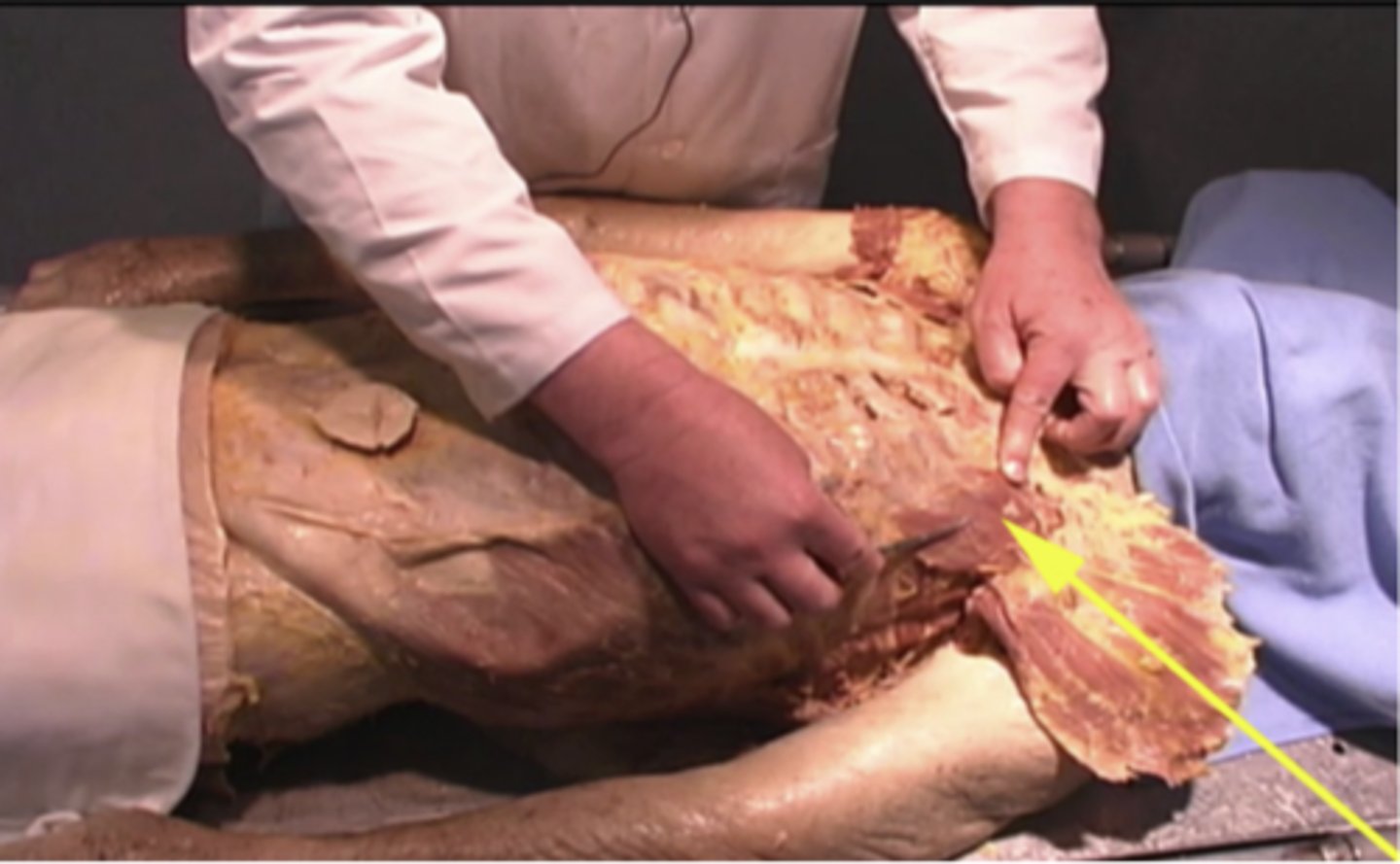
Due to their location on the underside of the muscle
Why do you not need to worry about destroying the nerves and arteries during this incision?
due to their location on the inferior spine
due to their location on the underside of the muscle
due to their location on the underside of the spine
due to the epidermis
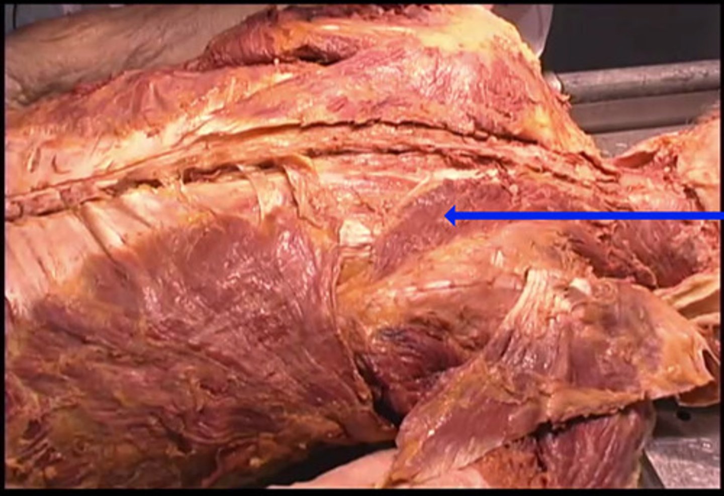
Pectoralis minor
What structure is being shown here?
nerve bundle
pectoralis minor
pectoralis major
spine
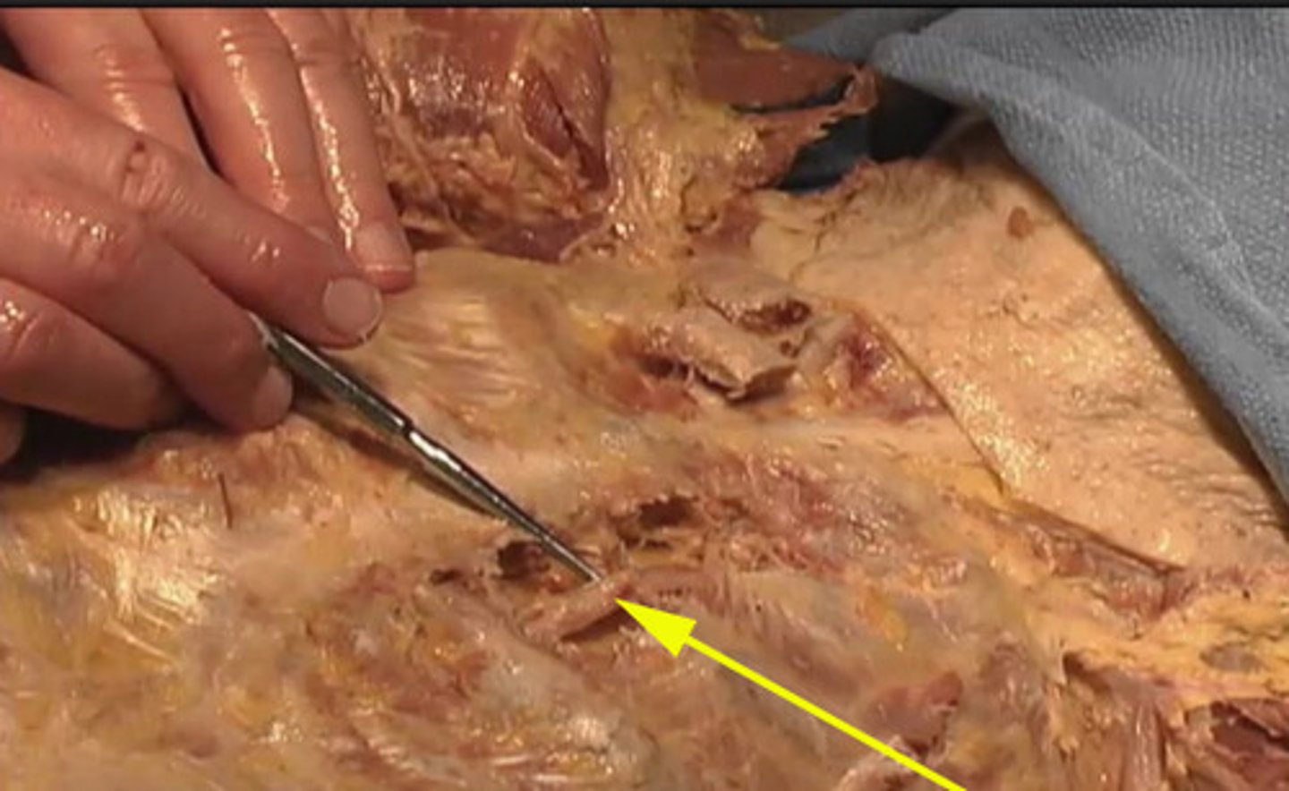
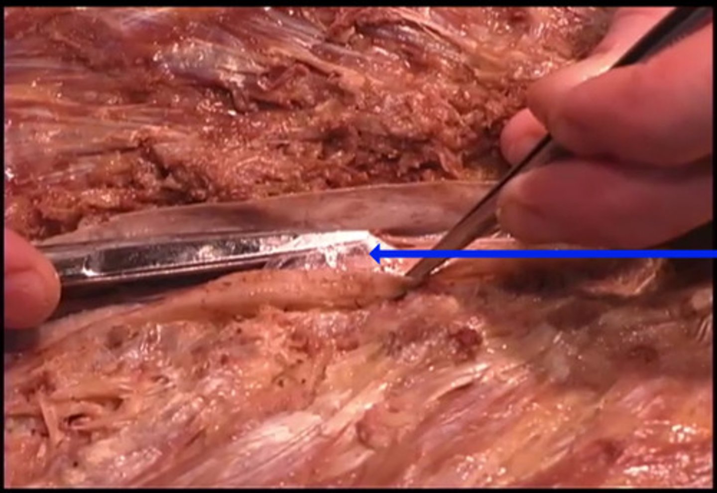
internal thoracic artery
What is this structure is being shown here?
epidermal artery
mylin sheath
internal thoracic vein
internal thoracic artery
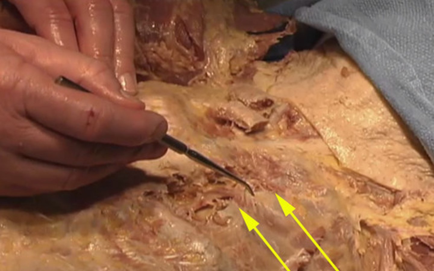
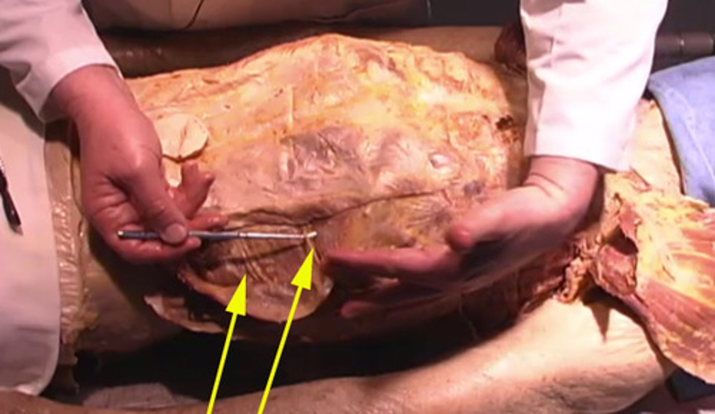
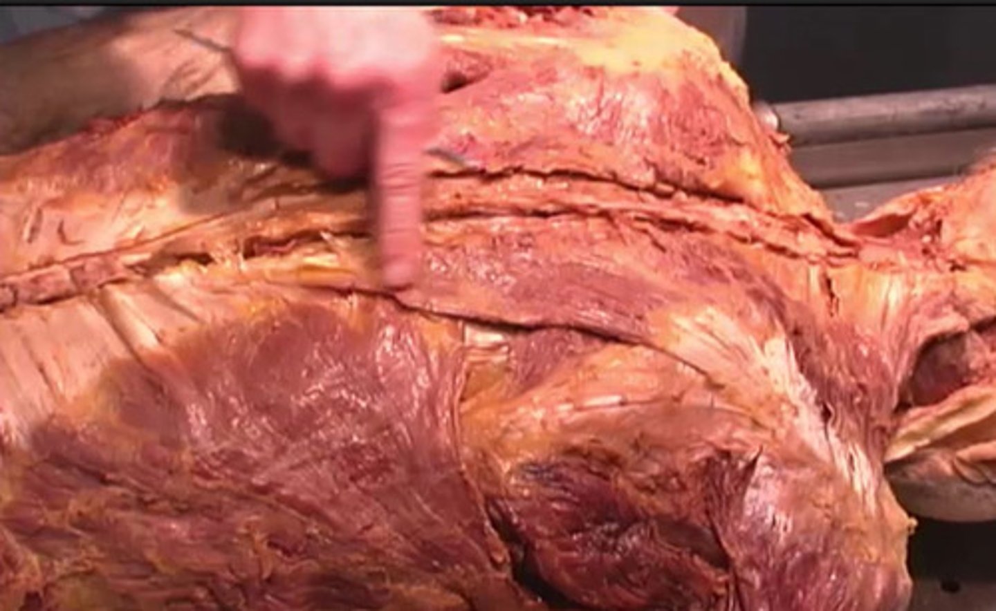
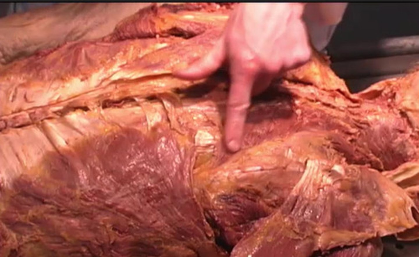
Intercostal spaces
To where are these arrows pointing?
intercostal spaces
pectoralis major
spine
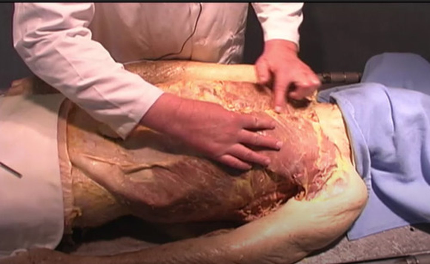
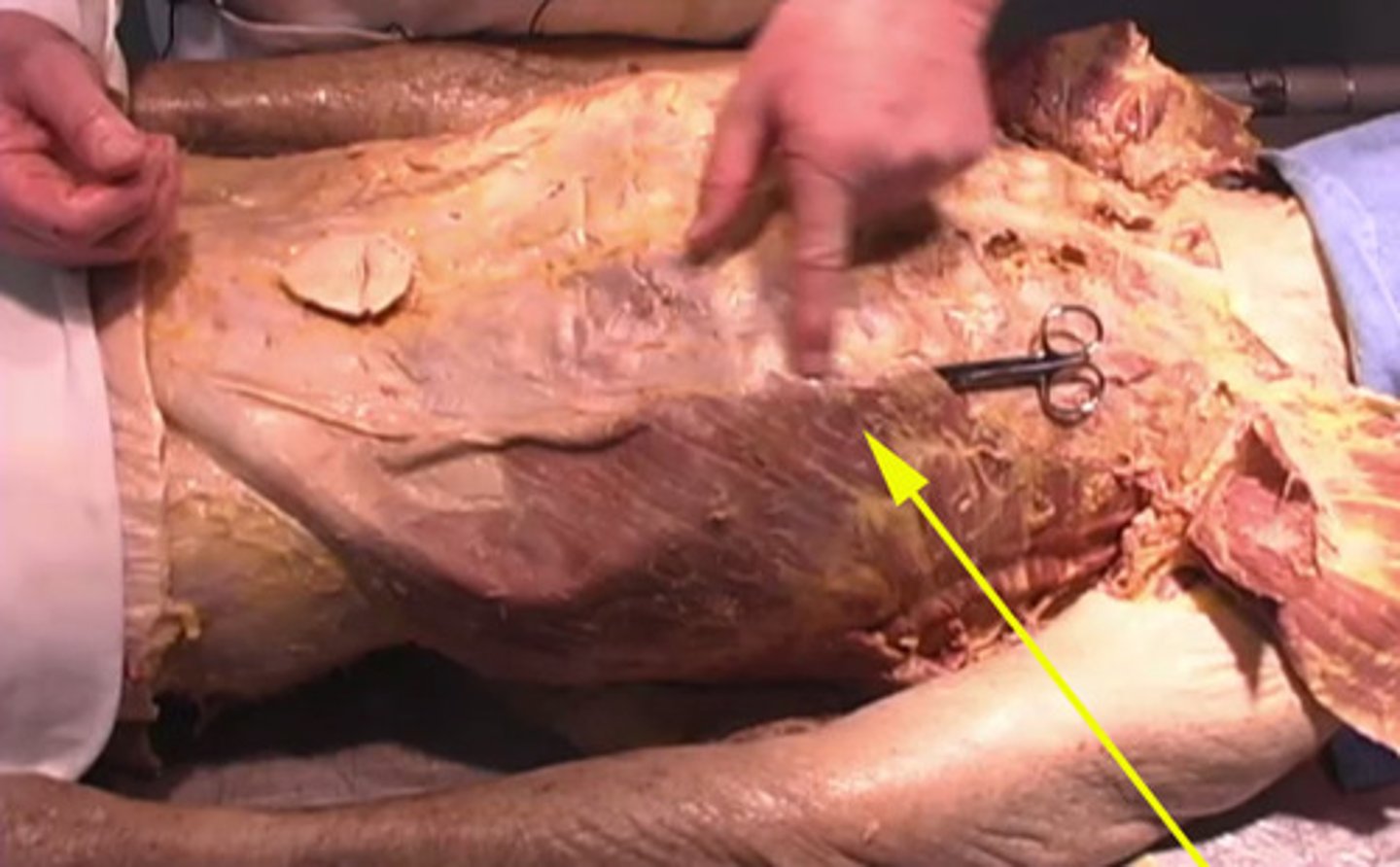
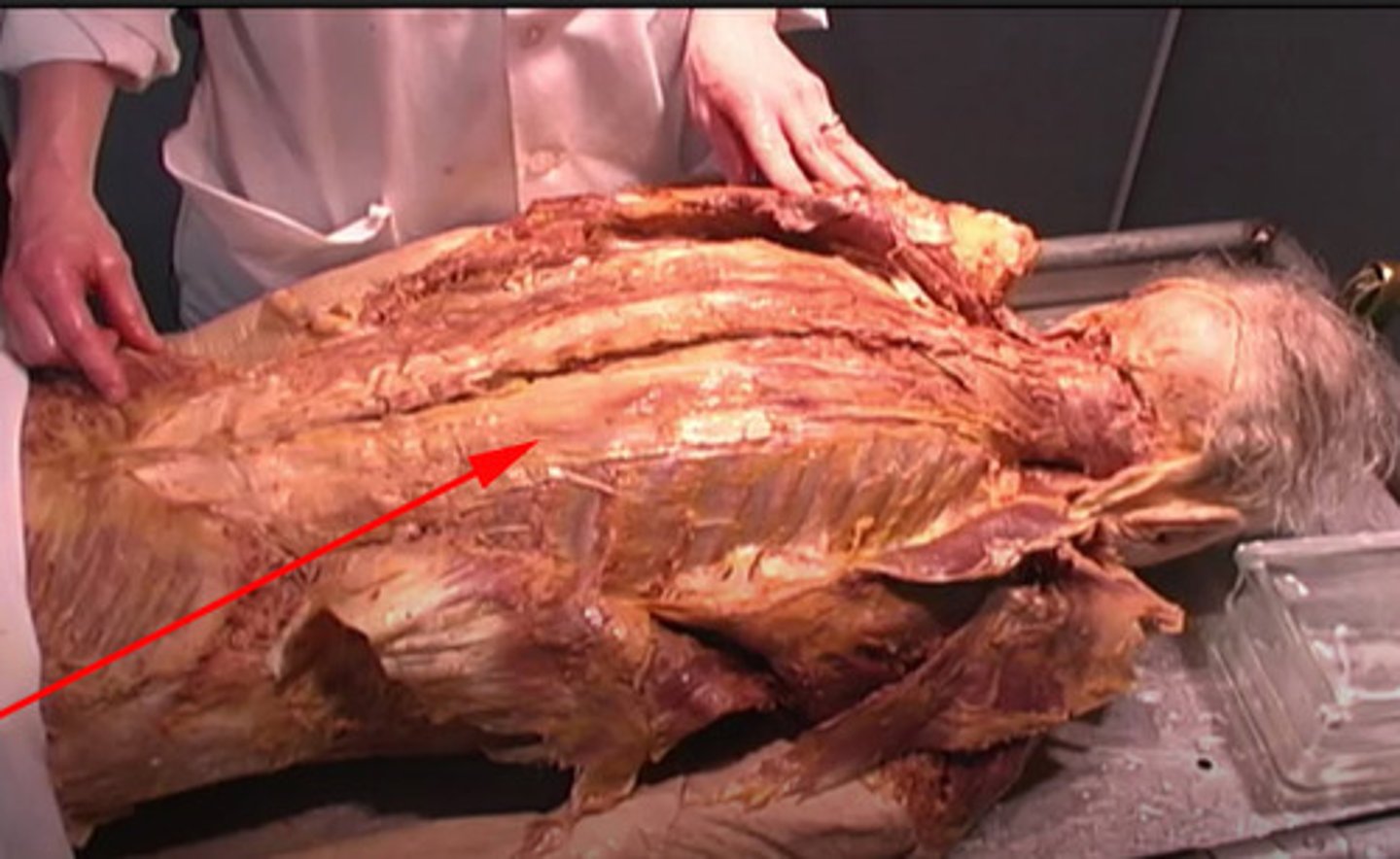
pectoralis major
external oblique
This is the:
pectoralis major
pectoralis minor
external oblique
internal oblique
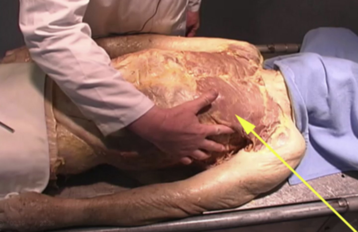
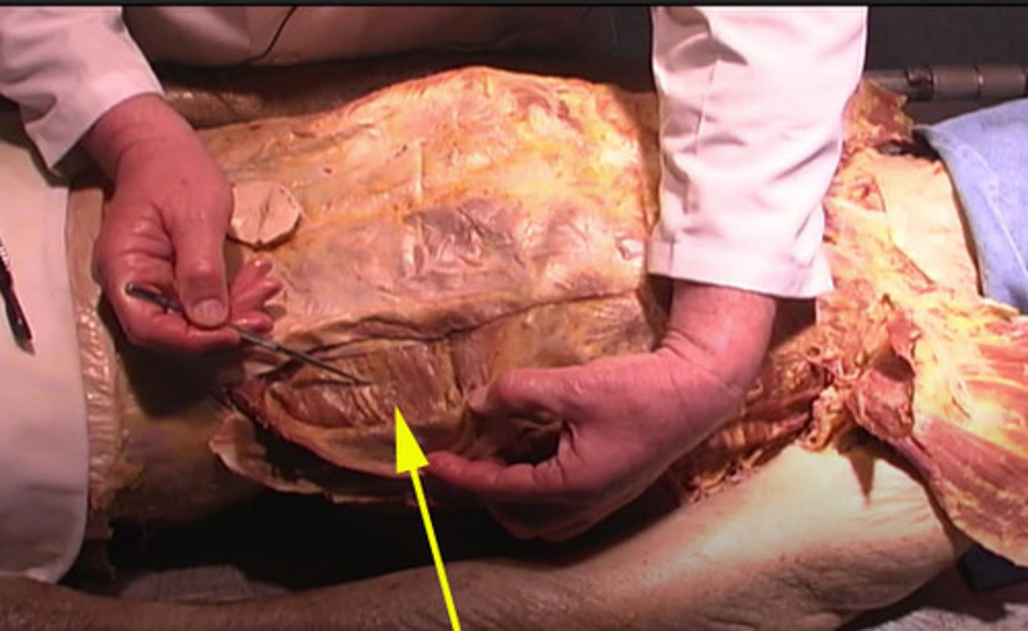
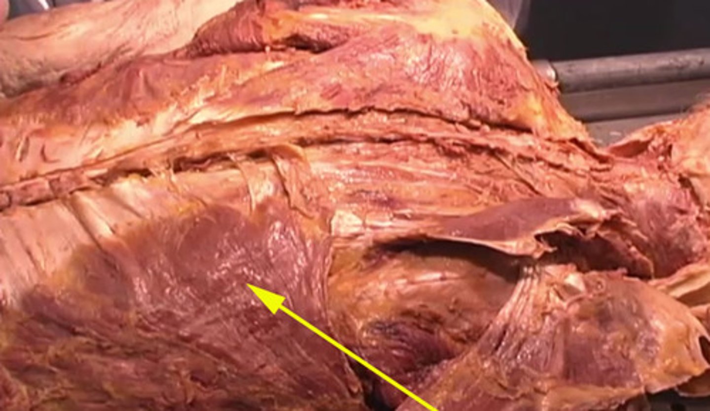
Transversus abdominis
Which structure is this being shown by the arrow?
transversus abdominis
internal abdominis
lateral oblique
oblique
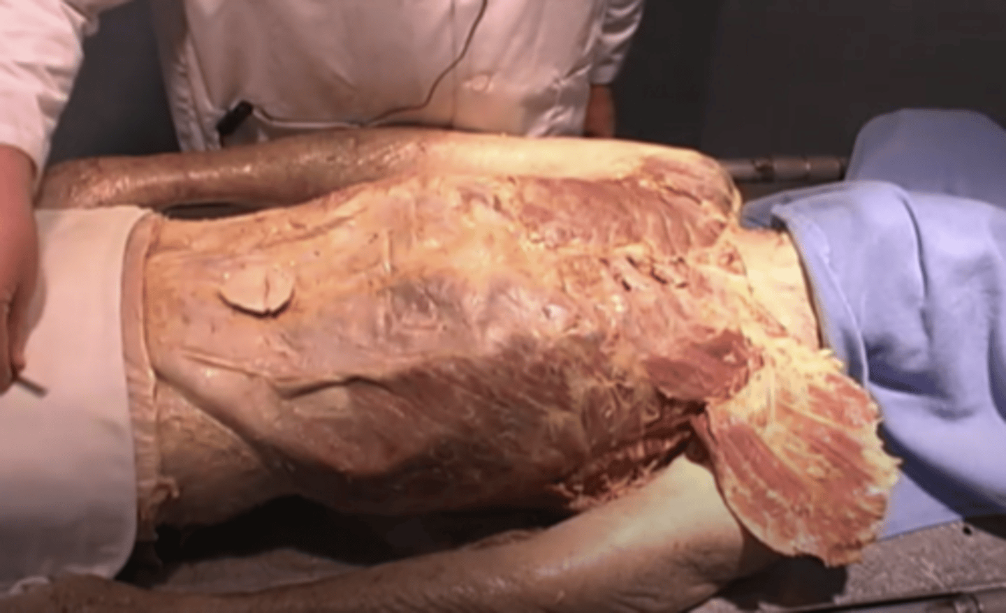
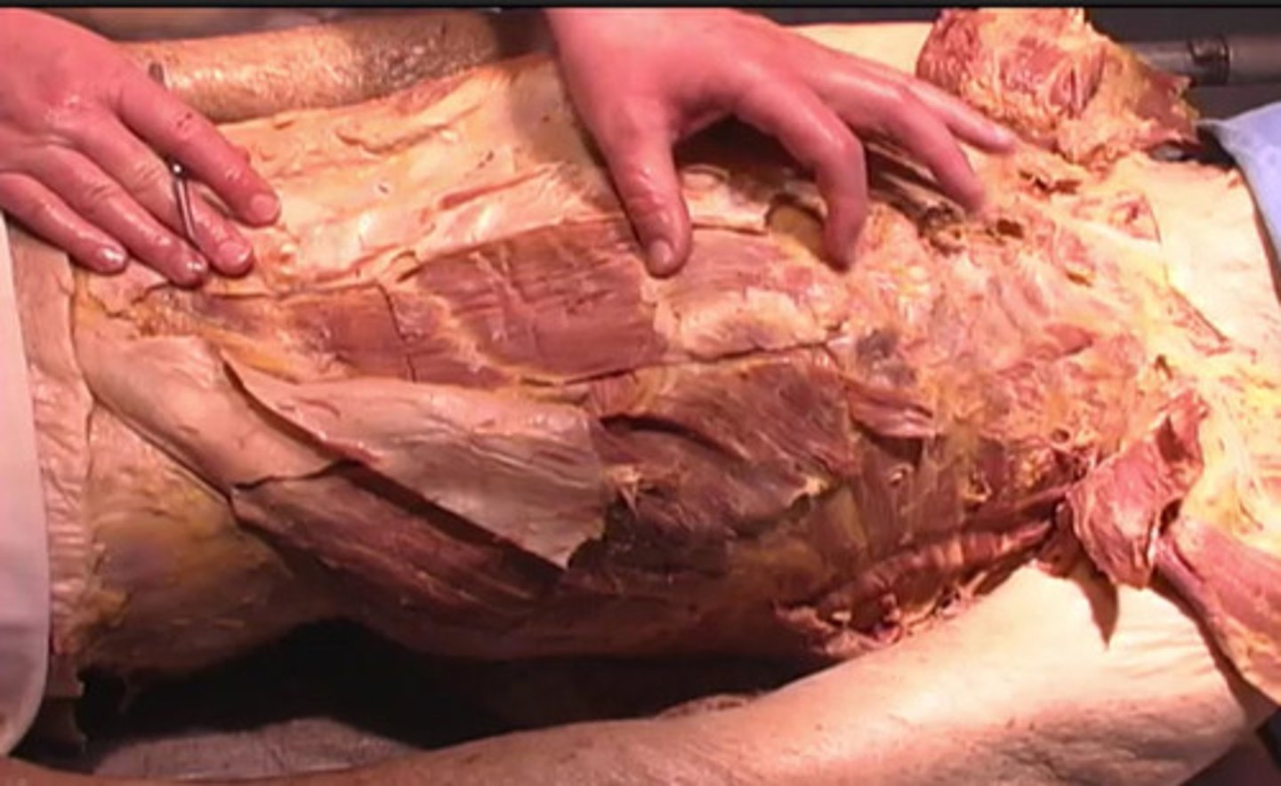
six pack
These portions of the rectus muscle are also known as a(n)?
internal oblique
lateral abdominus
oblique
six pack
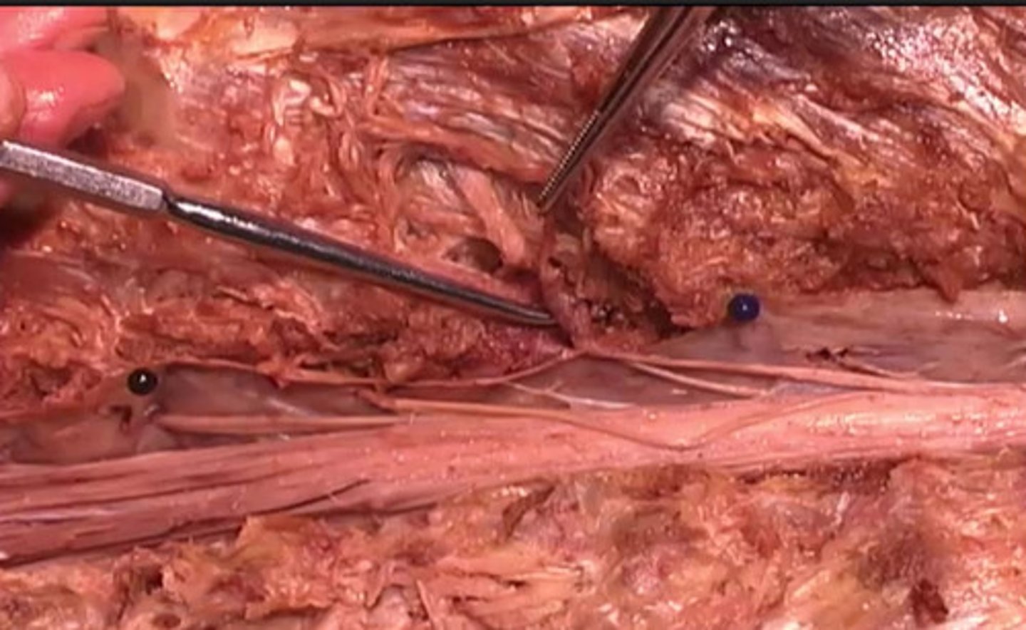
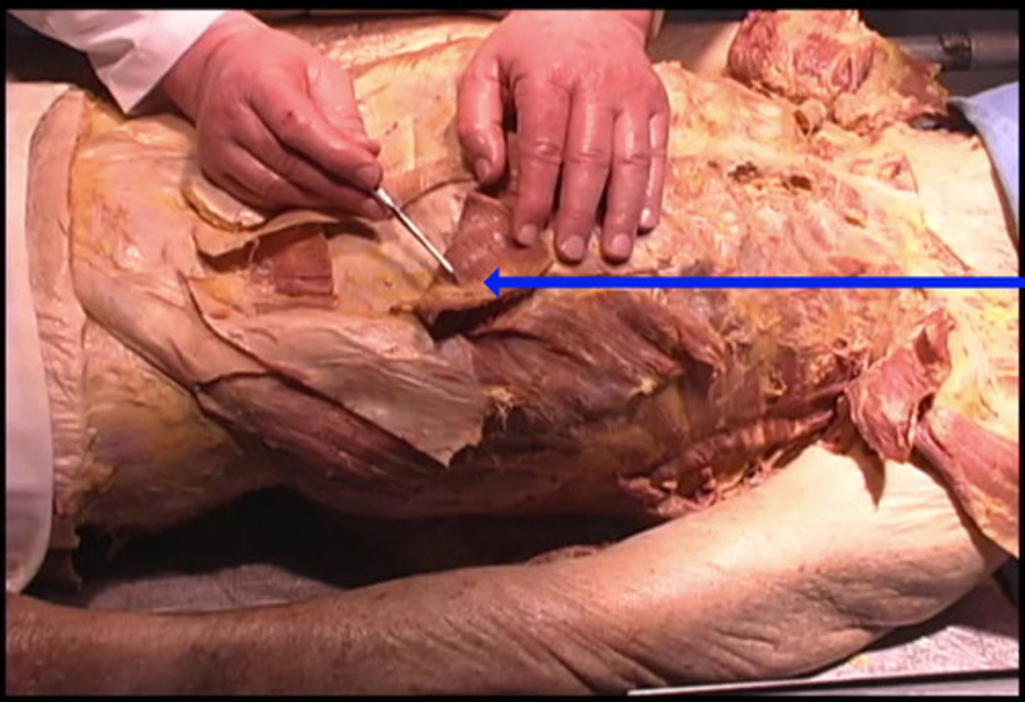
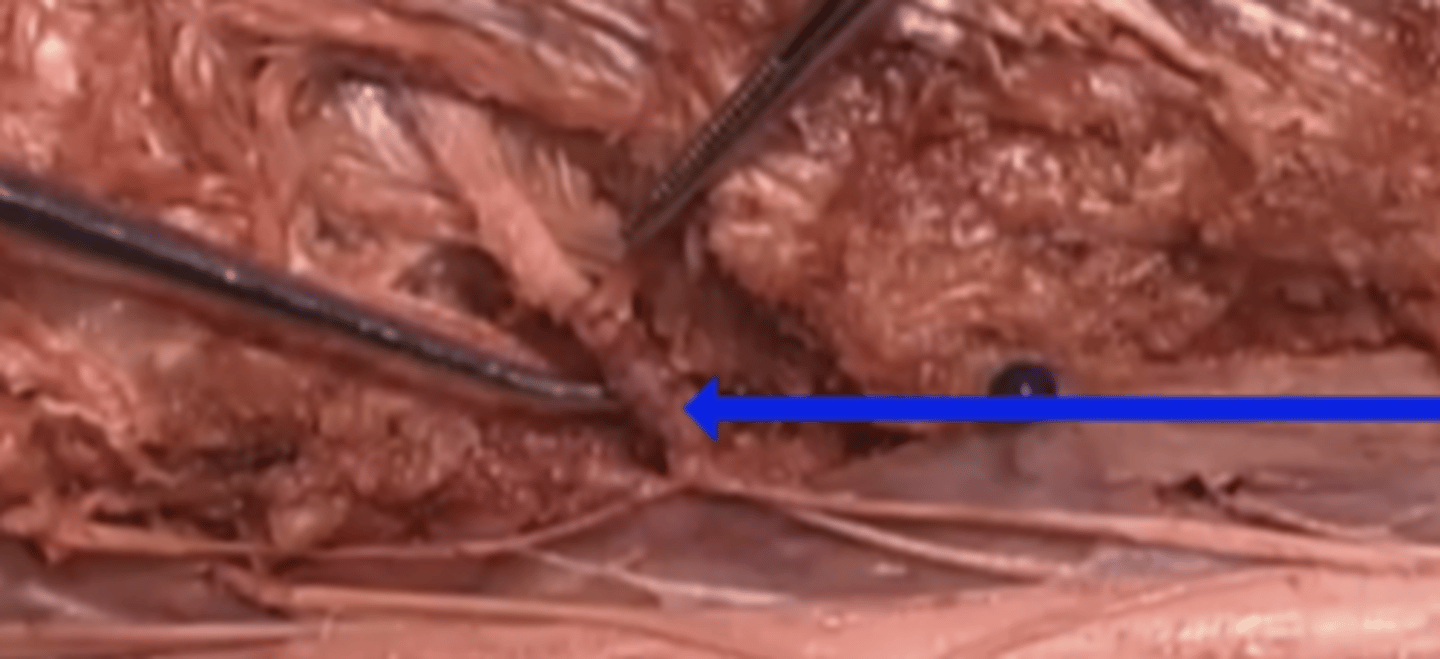
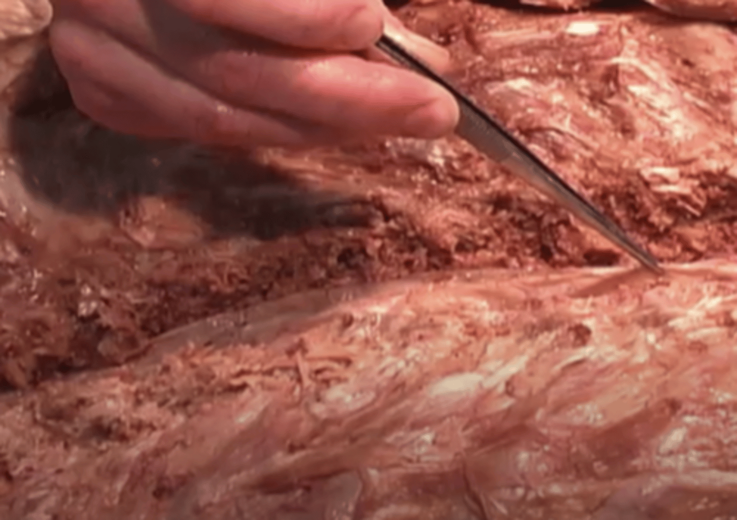
superior epigastric artery
What is the name of the artery being shown here by the blue arrow?
superior intercostal artery
inferior intercostal artery
superior epigastric artery
inferior epigastric artery
rhomboid
What is the name of the muscle being shown here by the blue arrow?
teres major
deltoid
trapezius
rhomboid
arachnoid
What is the name of the muscle being shown here by the blue arrow?
dura mater
pia mater
arachnoid
spinal cord
dorsal root ganglion
What does this region represent?
pia mater
posterior root ganglion
dorsal root ganglion
dura mater
intercostal nerves
What is the name of this structure?
Inferior epigastric artery
Superior epigastric artery
Intercostal nerves
Renal artery
trapezius
What structure is being shown here?
epidermis
pectoralis major
trapezius
lateral spiine
rhomboid
What structure is being shown here?
Rhomboid
Spine
Pectoralis minor
Rib
latissimus dorsi
What structure is being shown here with the pointer?
rib
lateral oblique
latissimus dorsi
spine
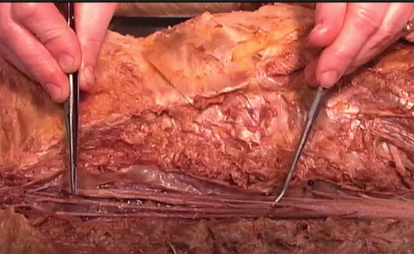
Erector Spinae
To what structure is the red arrow pointing?
Intercostal
Spine
Erector Spinae
Latissimus dorsi
Splenius capitis
What structure is being shown here?
Splenius capitis
Spine
Latissimus dorsi
Erectus spinus
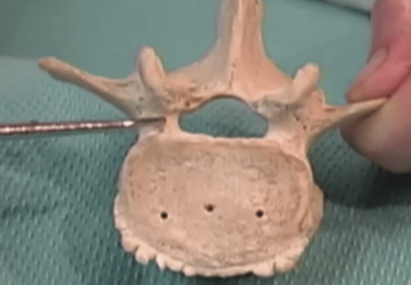
pedicle
What structure is being shown here?
dura mater
What structure is being shown here?
lateral vein
spinus
dura mater
abdominus lateralis
Filum terminale
Which structure is being shown here?
Erectus spinus
Spine
Latissimus dorsi
Filum terminale
dorsal root ganglion
Which swollen structure is being shown here?
anterior ganglion
ventral nerve
dorsal root ganglion
inferior ganglion