Image Planes Flashcards
 Call Kai
 Learn
Learn Practice Test
 Spaced Repetition
 Match
Match Flashcards
 Knowt Play
1/54

There's no tags or description
Looks like no tags are added yet.
Name | Mastery | Learn | Test | Matching | Spaced | Call with Kai | Chat |
|---|
No analytics yet
Send a link to your students to track their progress
55 Terms
Top of ultrasound image
Standard assignment for the scanning surface in any ultrasound image orientation.
Viewing from the feet
The specific perspective used to see transverse images.
Independent image side assignments
The relationship between image sides and the patient's actual position.
Consistent diagram presentation
The characteristic of transverse images remaining the same as the transducer moves.
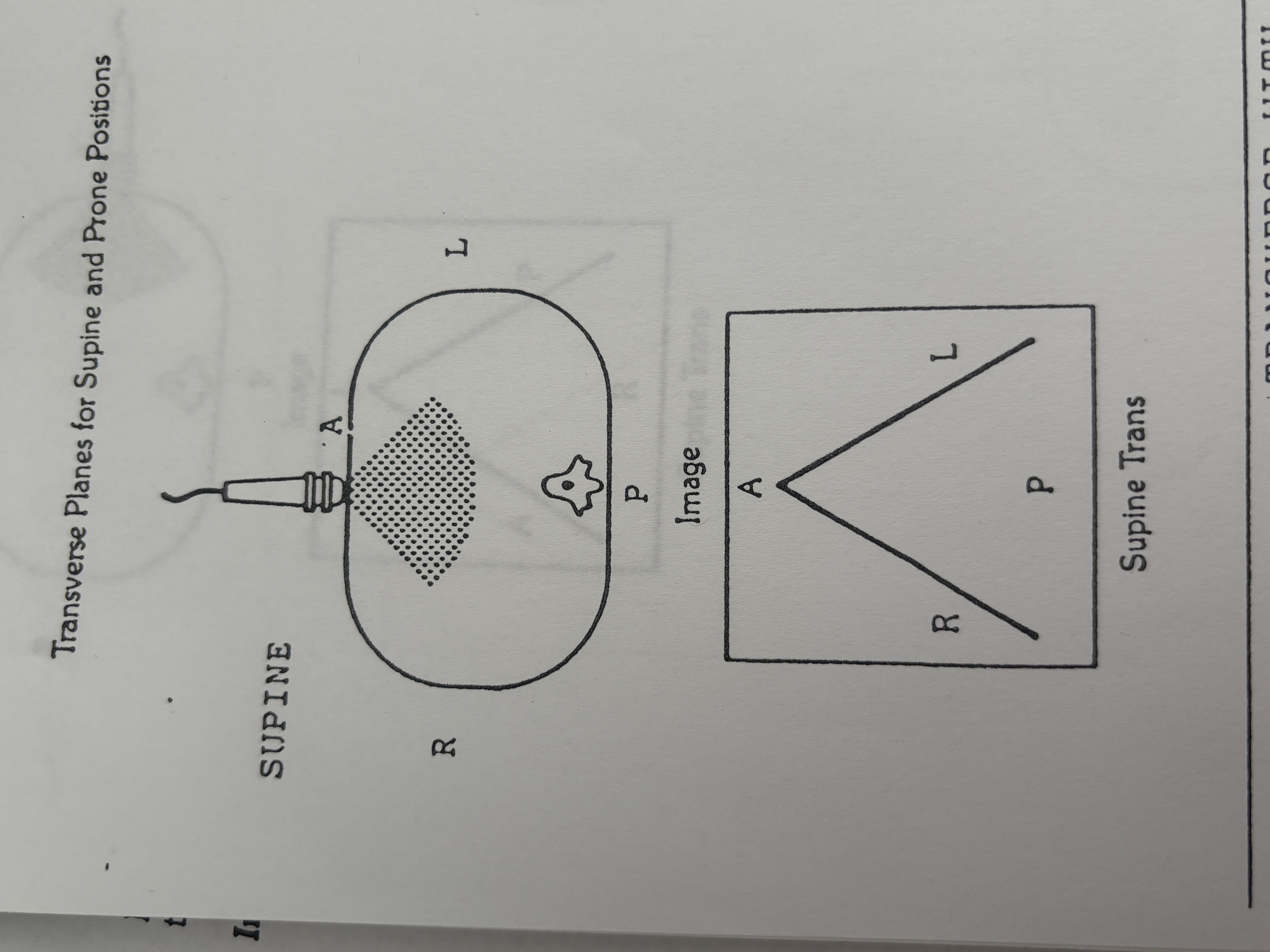
Left side of monitor during anterior transverse scan
Anatomical side shown as Right.
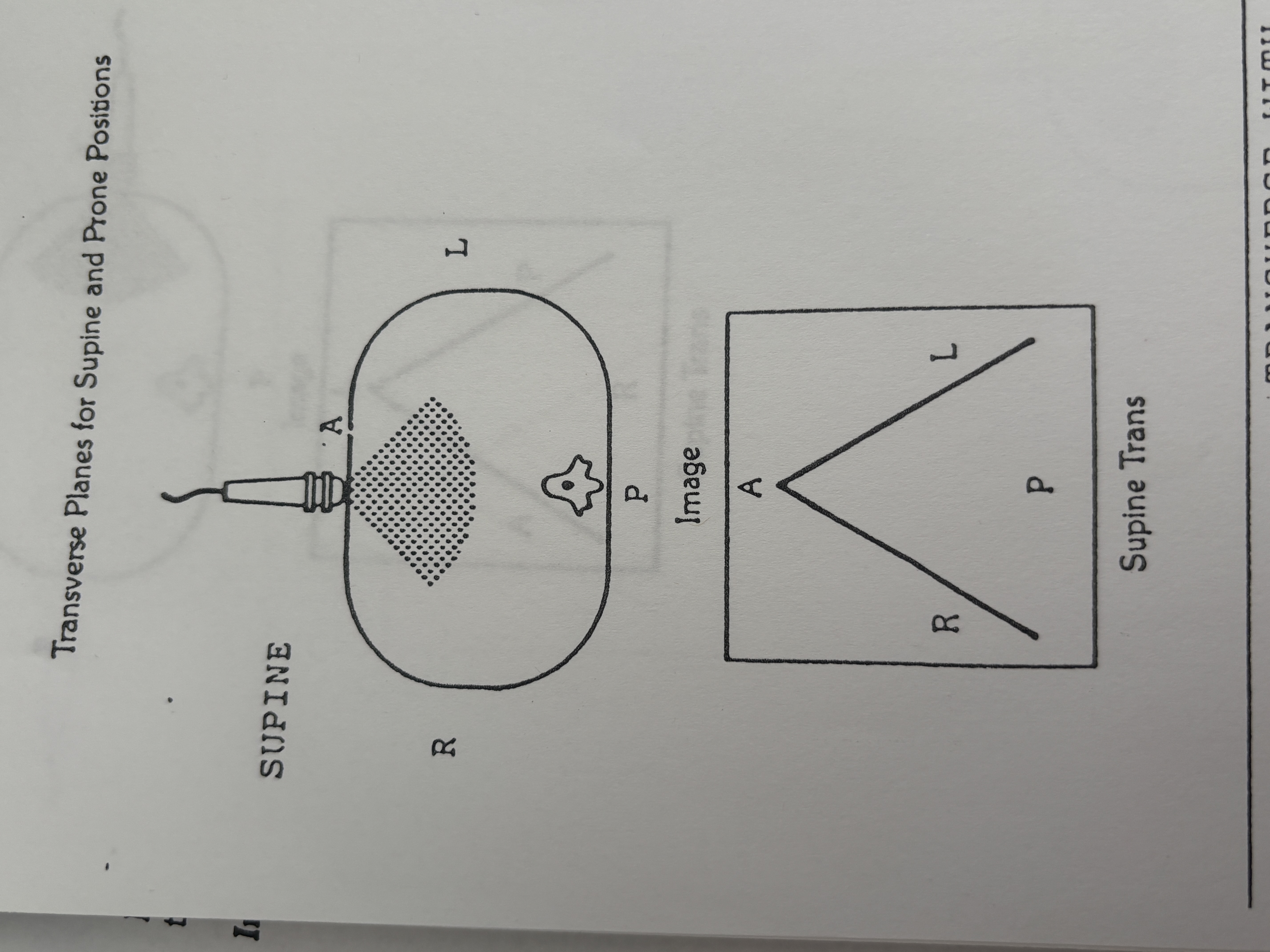
Right side of monitor during anterior transverse scan
Anatomical side shown as Left.

Left side of monitor during posterior transverse scan
Anatomical side shown as Left.

Right side of monitor during posterior transverse scan
Anatomical side shown as Right.

Left side of monitor during right lateral transverse scan
Anatomical side shown as Posterior.

Right side of monitor during right lateral transverse scan
Anatomical side shown as Anterior.

Left side of monitor during left lateral transverse scan
Anatomical side shown as Anterior.
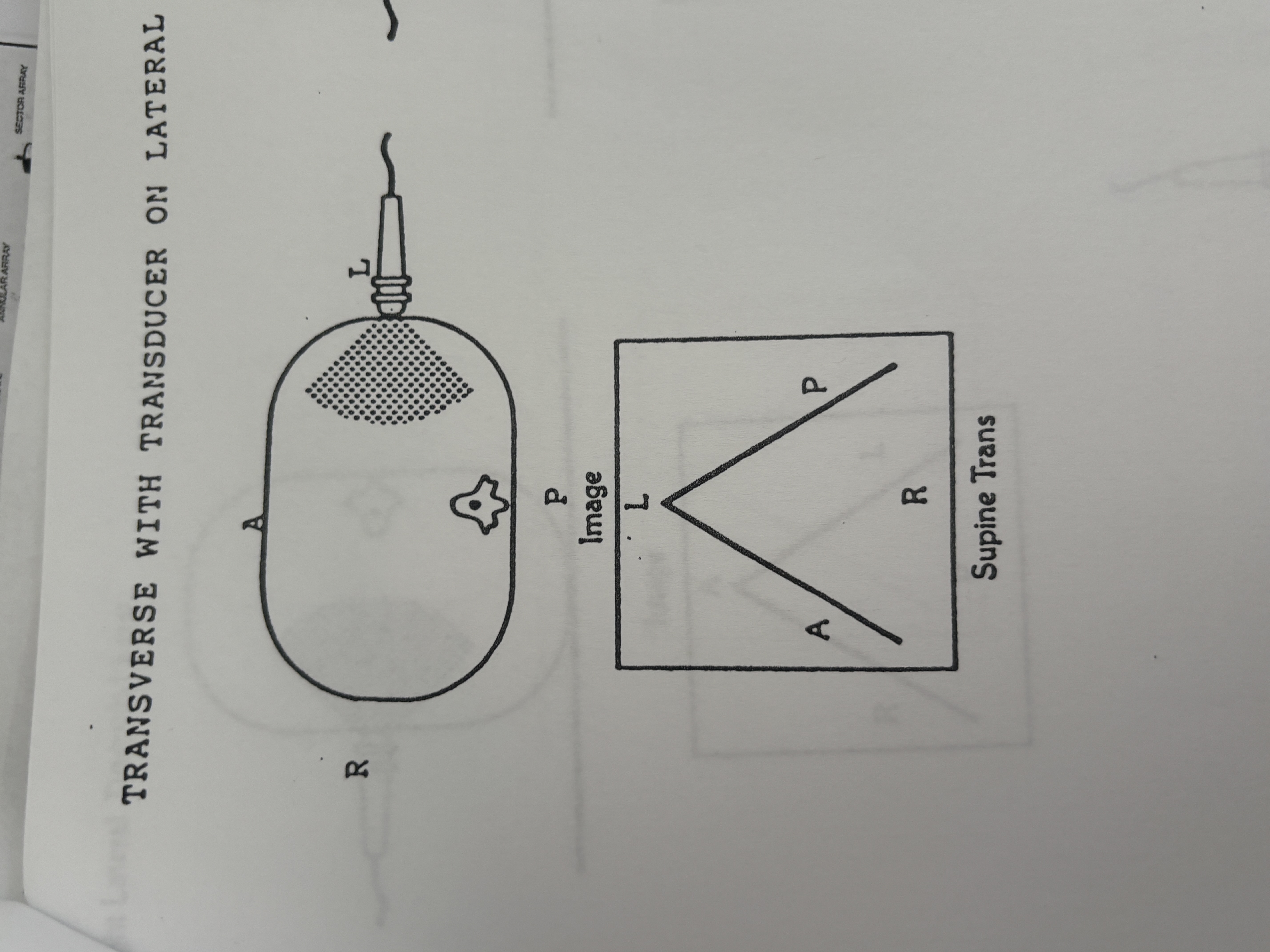
Right side of monitor during left lateral transverse scan
Anatomical side shown as Posterior.
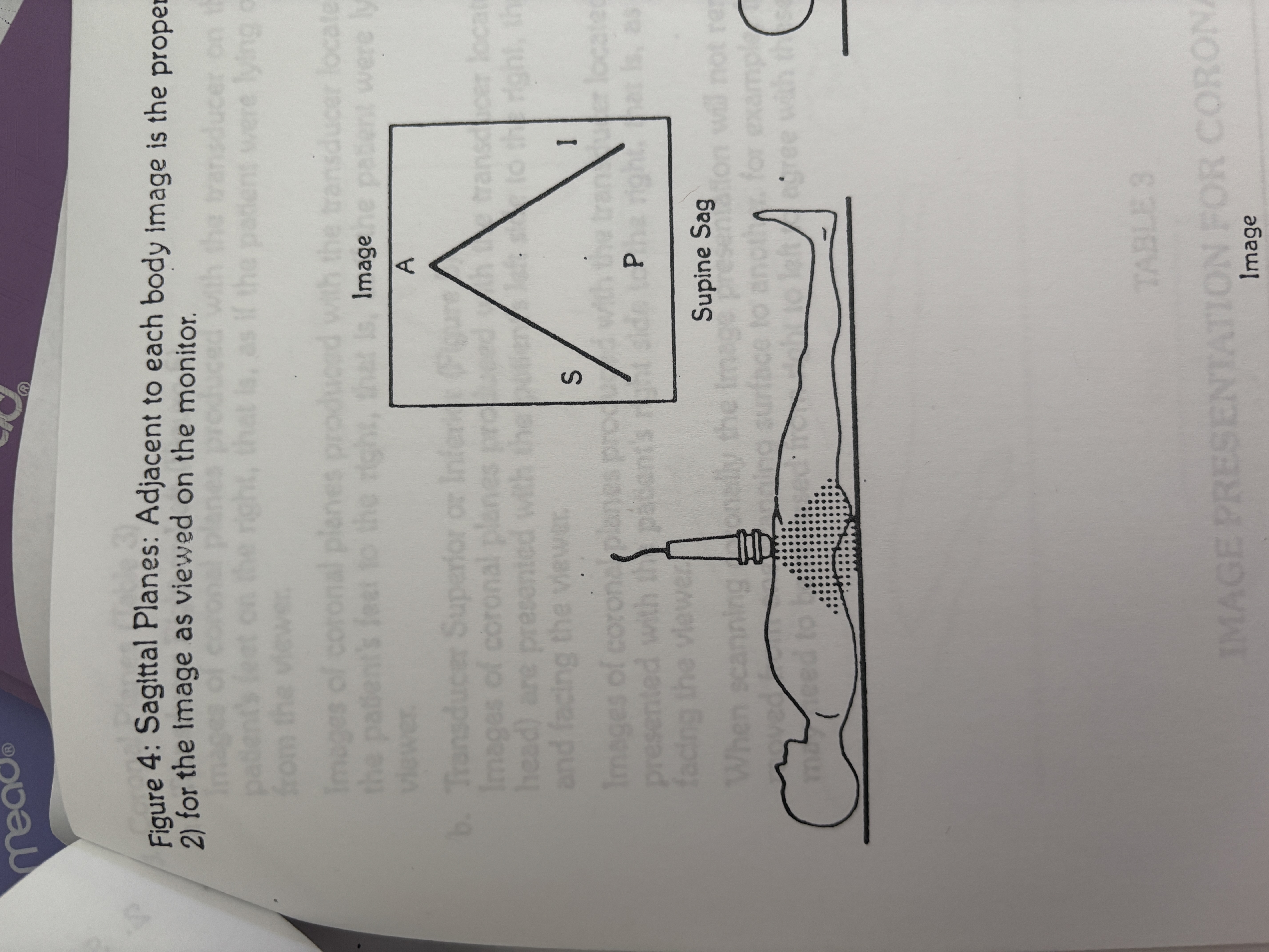
Left side of monitor during anterior sagittal scan
Anatomical part shown as Superior.
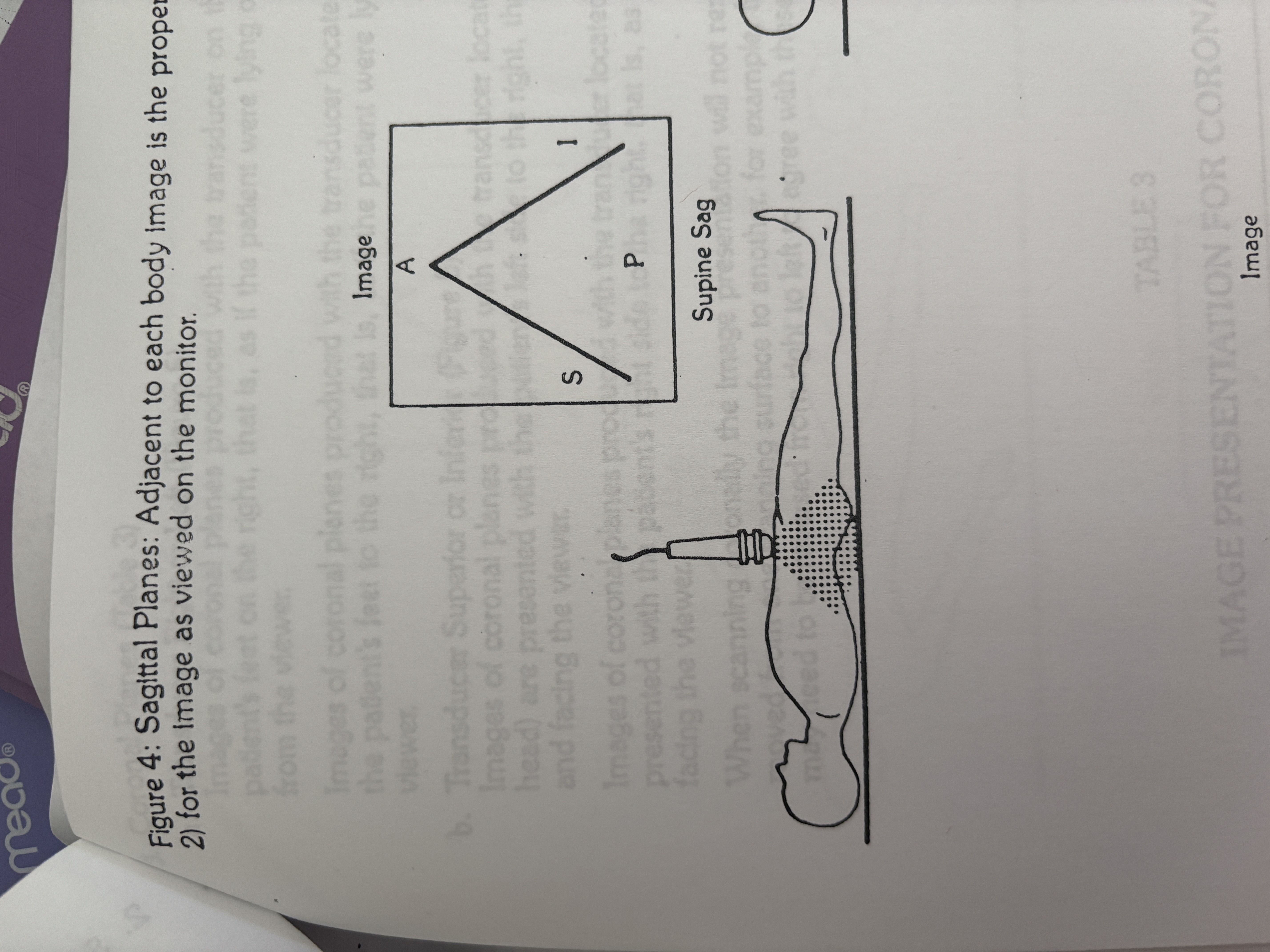
Right side of monitor during anterior sagittal scan
Anatomical part shown as Inferior.
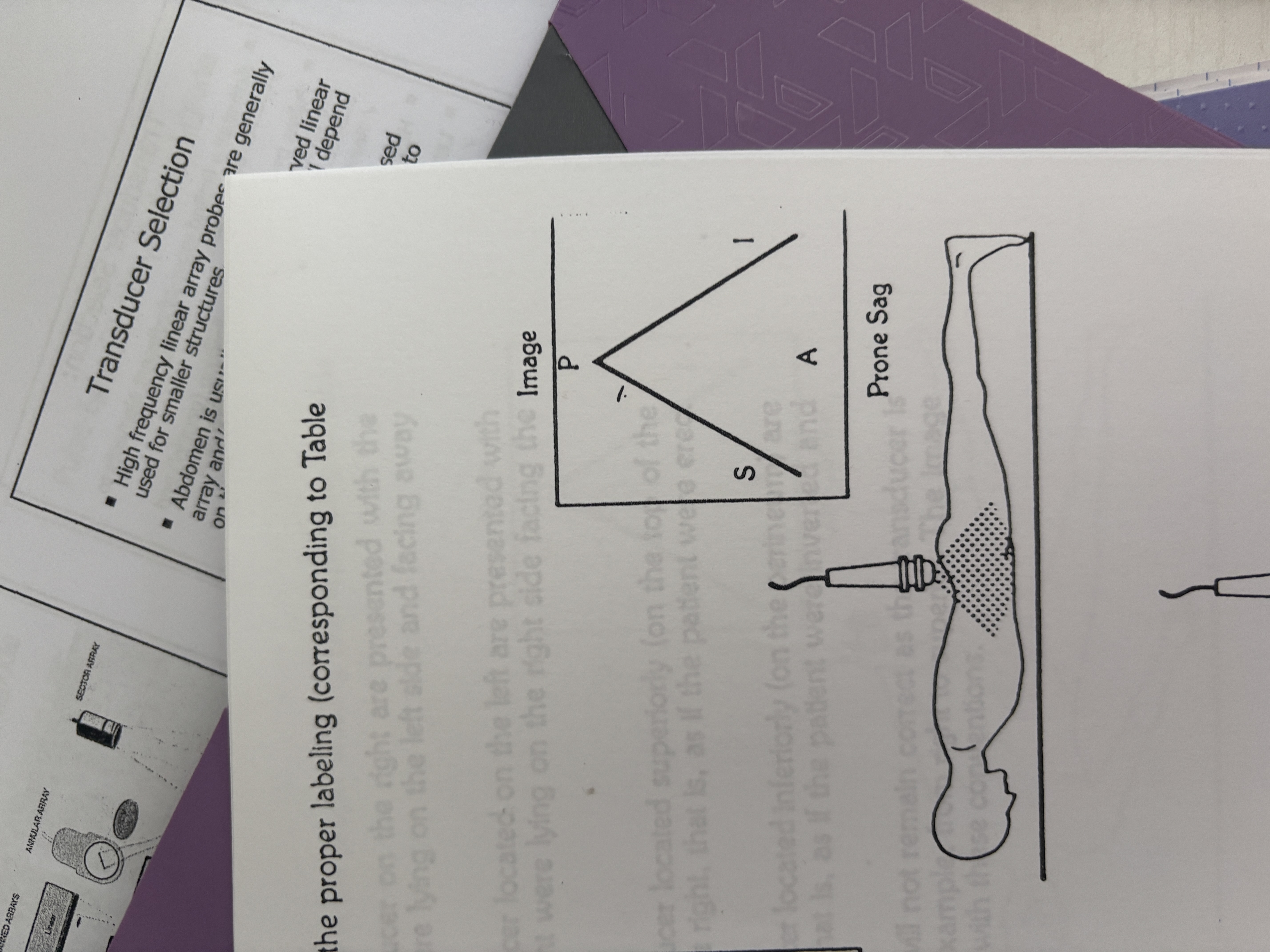
Left side of monitor during posterior sagittal scan
Anatomical part shown as Superior.

Right side of monitor during posterior sagittal scan
Anatomical part shown as Inferior.
Left side of monitor during superior sagittal scan
Anatomical side shown as Anterior.

Right side of monitor during superior sagittal scan
Anatomical side shown as Posterior.

Left side of monitor during inferior sagittal scan
Anatomical side shown as Anterior.
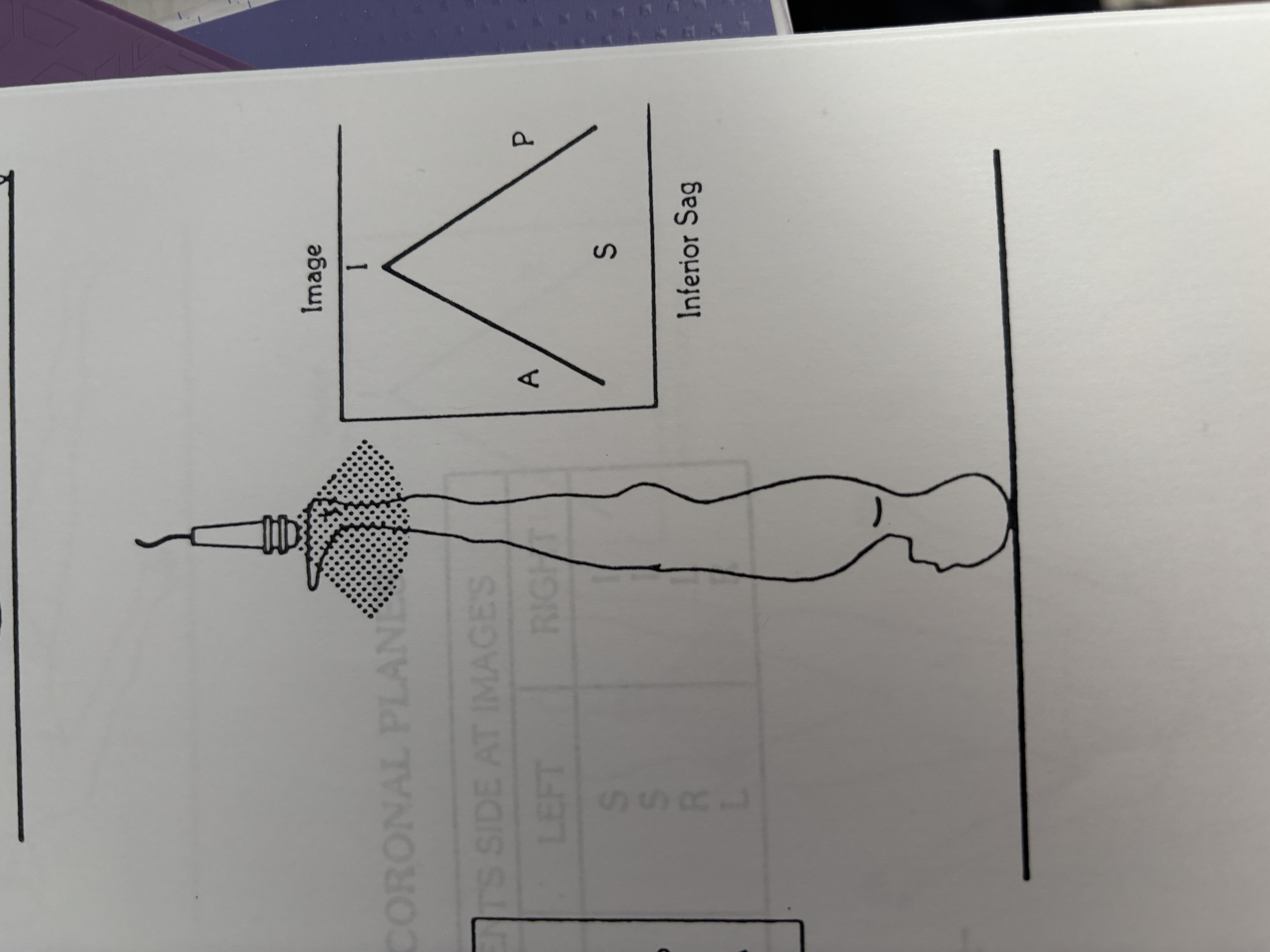
Right side of monitor during inferior sagittal scan
Anatomical side shown as Posterior.
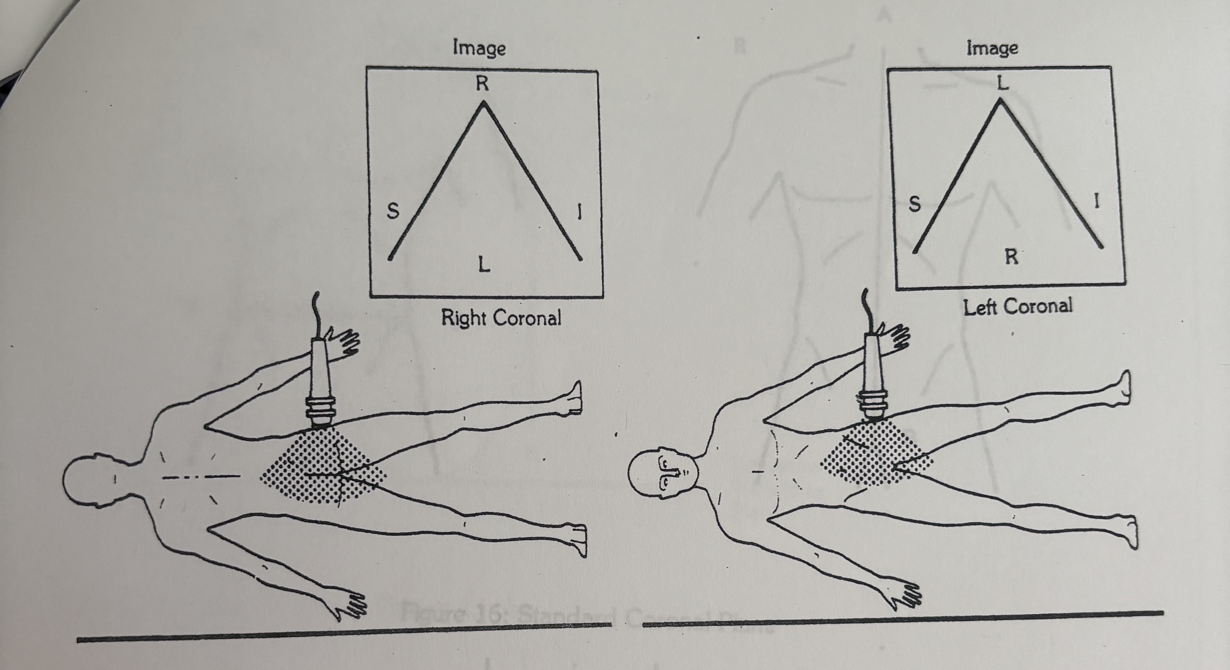
Left side of monitor during lateral coronal scan
Anatomical part shown as Superior.
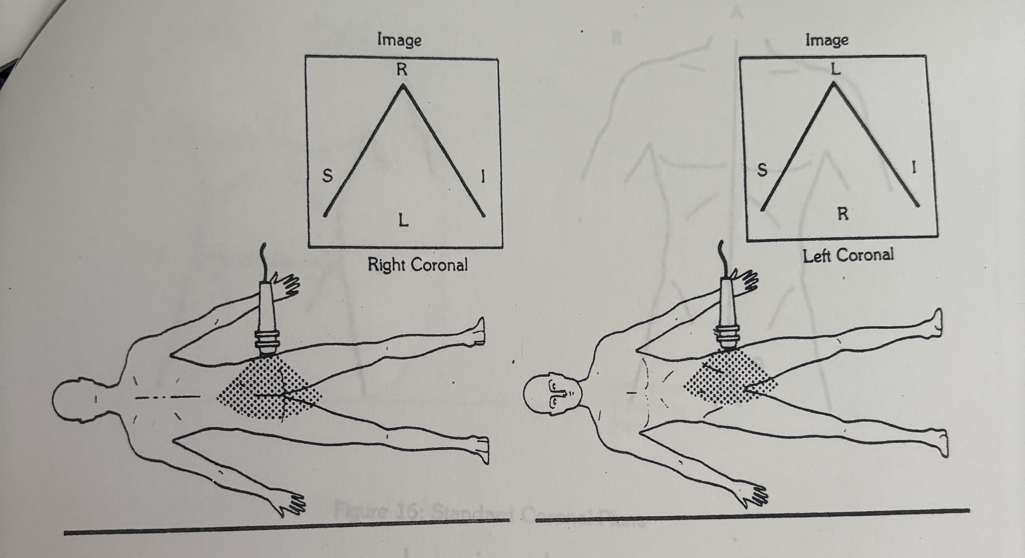
Right side of monitor during lateral coronal scan
Anatomical part shown as Inferior.
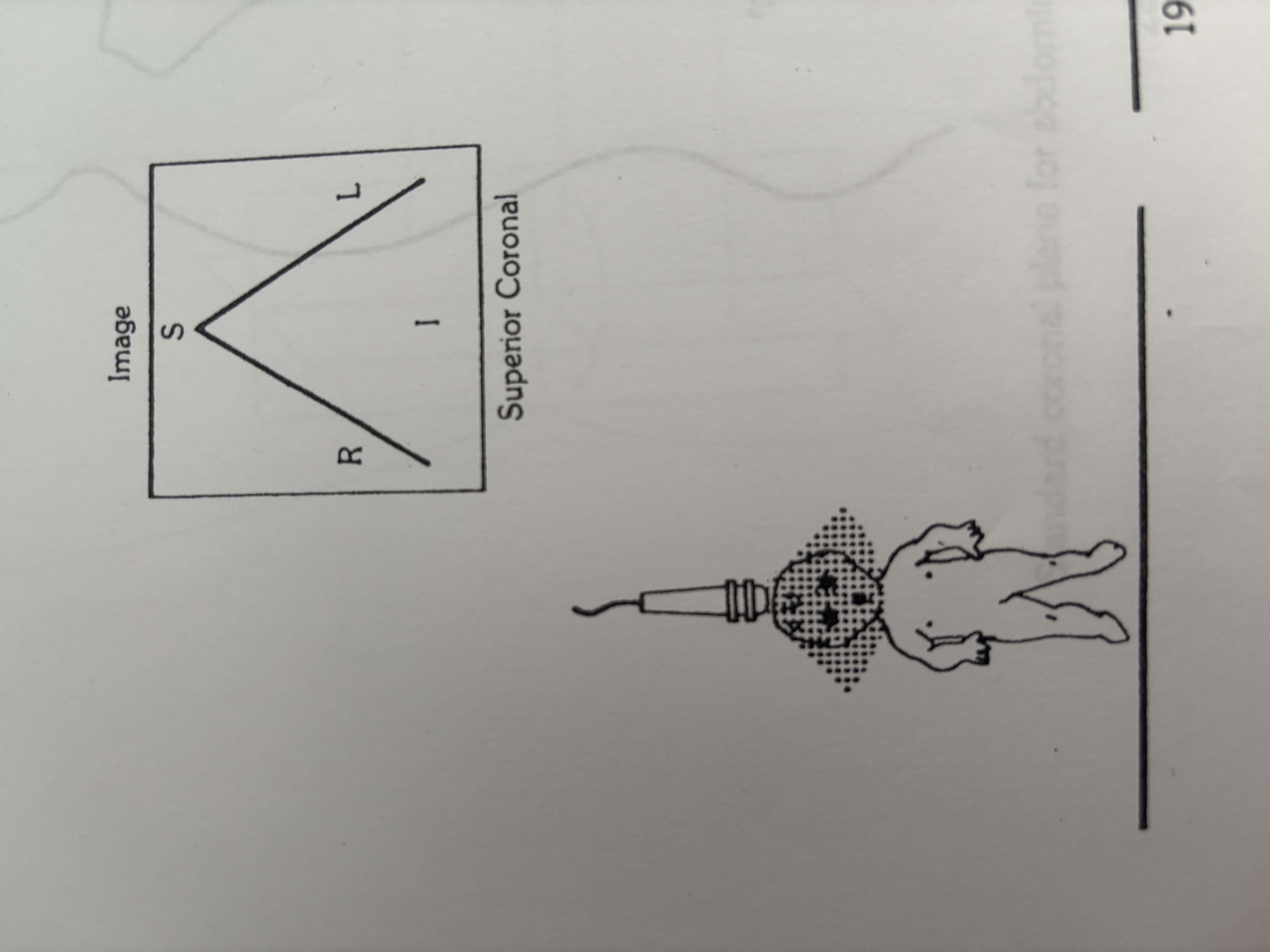
Left side of monitor during superior coronal scan
Anatomical side shown as Right.
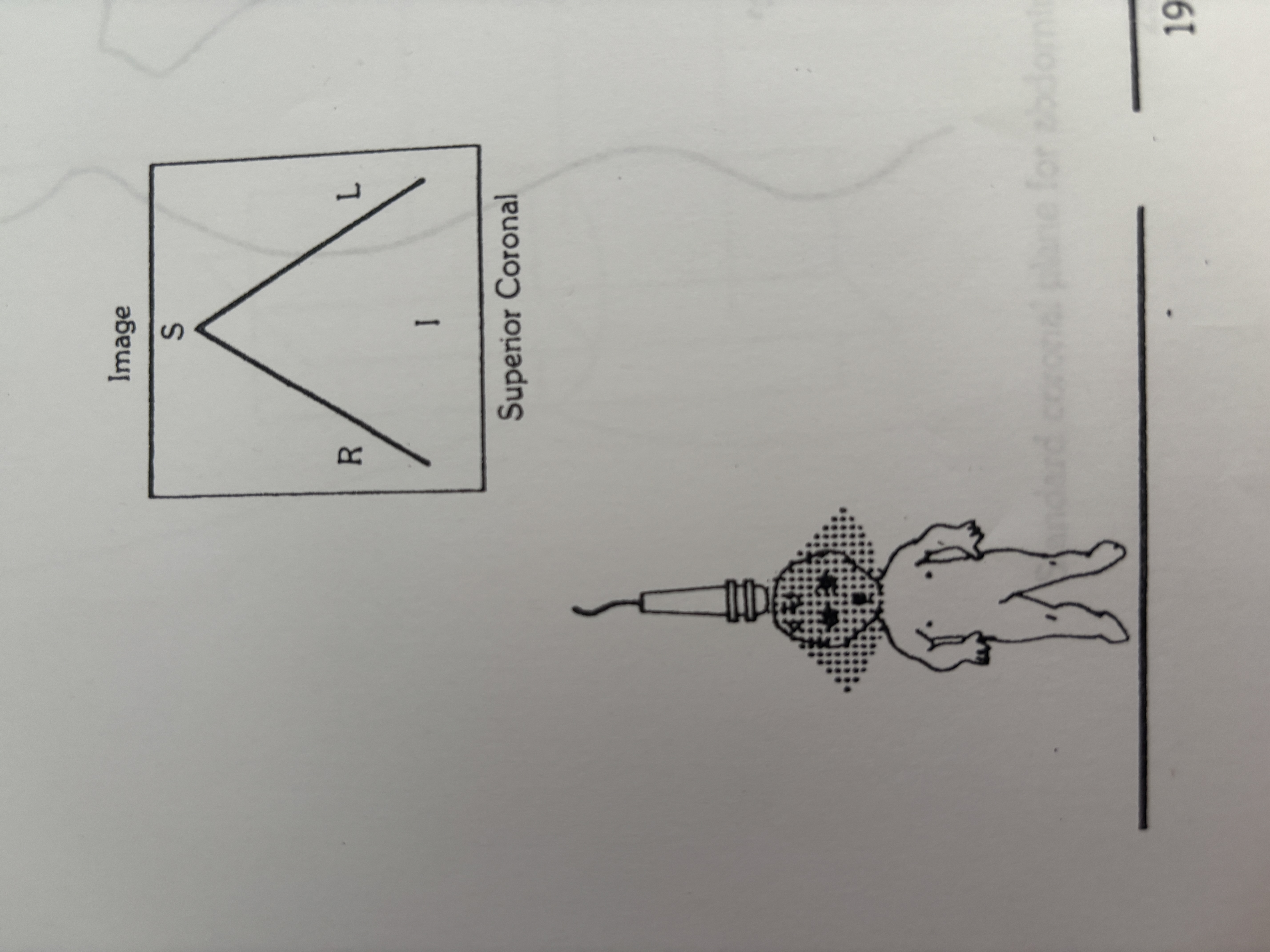
Right side of monitor during superior coronal scan
Anatomical side shown as Left.

Left side of monitor during inferior coronal scan
Anatomical side shown as Left.
Right side of monitor during inferior coronal scan
Anatomical side shown as Right.
'A' in ultrasound labeling
Anterior.
'P' in ultrasound labeling
Posterior.
'R' in ultrasound labeling
Right.
'L' in ultrasound labeling
Left.
'S' in ultrasound labeling
Superior.
'I' in ultrasound labeling
Inferior.
General rule for right side anatomy in sagittal or coronal images
Patient's feet are on the right.
Head position on the monitor during longitudinal scans
Displayed on the left side.
Scanning for images from the perineum
Probe positioned at the inferior surface
Scanning for images from the top of the head
Probe positioned at the superior surface.
Patient position for a right lateral coronal scan
Lying on their left side.
Patient position for a left lateral coronal scan
Lying on their right side.
Action for incorrect sagittal image while moving the transducer
Reverse the image left to right.
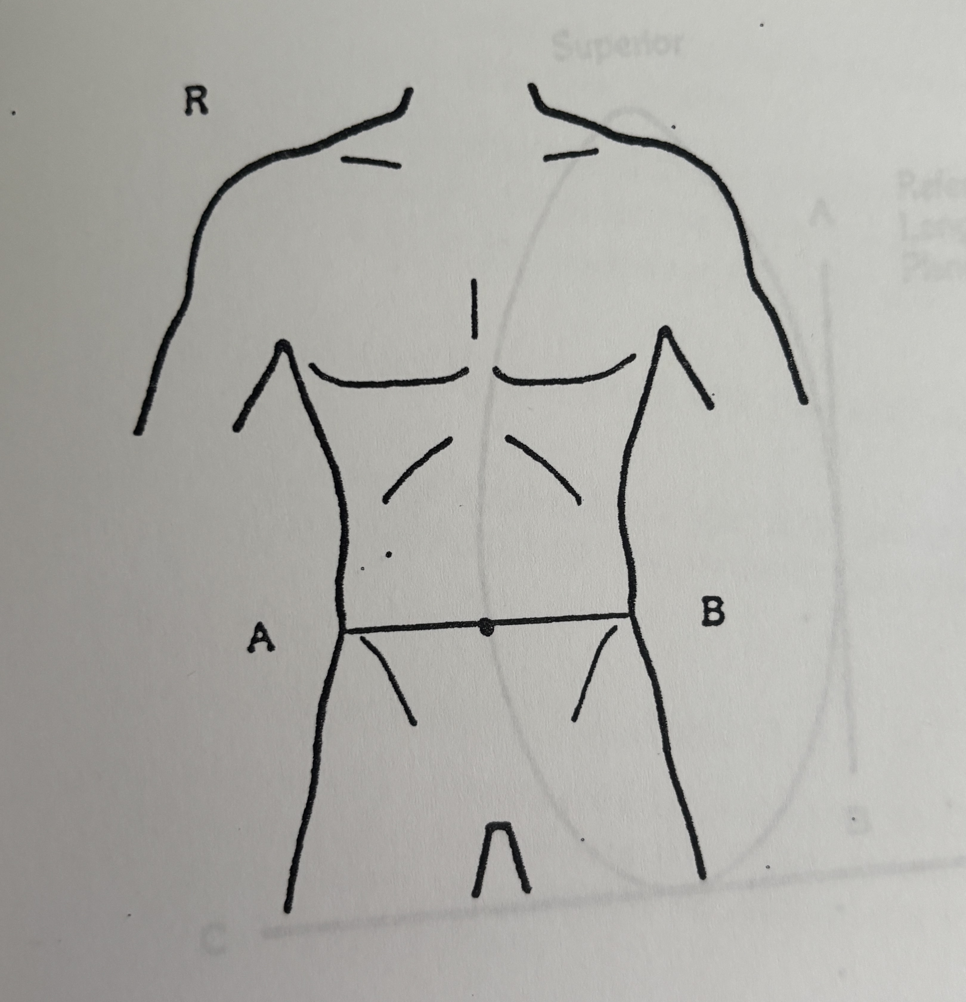
Anatomical plane indicated by line A-B in an image
Standard Transverse Plane.

Anatomical plane indicated by line A-B in an image
Standard Sagittal Plane.

Anatomical plane indicated by line A-B in an image
Standard Coronal Plane.

Plane defined by line A-B along the organ's length
Reference Longitudinal Plane.

Plane defined by line C-D across the organ's short axis
Reference Transverse Plane.

Plane that divides a limb front to back
Reference Anterior-Posterior Plane.

Plane that divides a limb horizontally
Reference Transverse Plane.
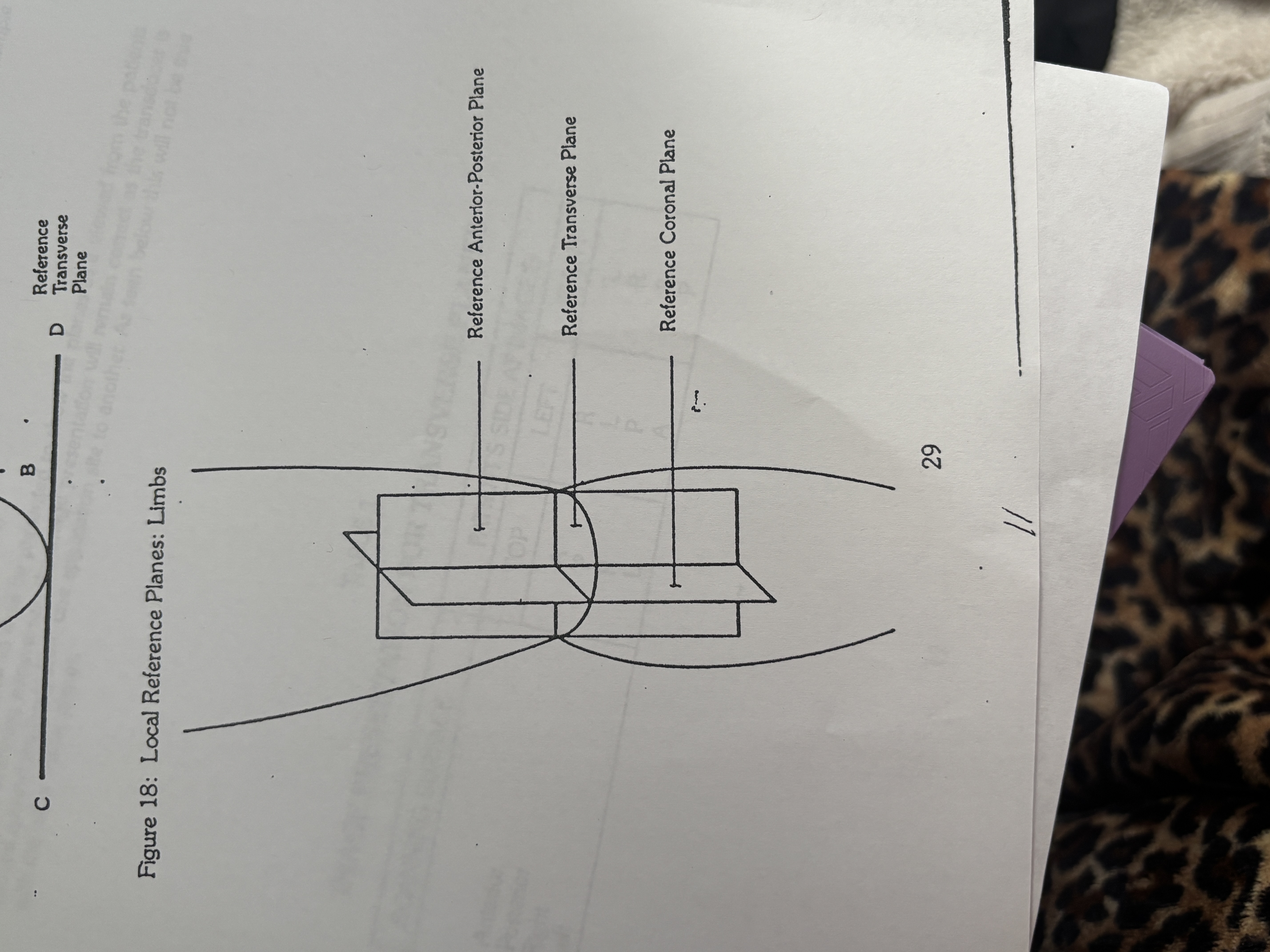
Plane that divides a limb side to side
Reference Coronal Plane.
Total basic scanning surfaces in ultrasound
Six.
Patient lying on their back.
Supine scanning position
Patient lying on their stomach.
Prone scanning position
Patient lying on their side.
Lateral decubitus scanning position
Anatomical part shown right of the image in sagittal superior scan
Posterior (patient's back).
Anatomical part shown left of the image in sagittal superior scan
Anterior.
Patient perspective for coronal scans
Facing the viewer's left side.
Four general scanning surfaces for each image plane
Anterior, posterior, right, and left.