CC1 PART 2
 Call Kai
 Learn
Learn Practice Test
 Spaced Repetition
 Match
Match Flashcards
 Knowt Play
1/245

Earn XP
Description and Tags
*No Tables
Name | Mastery | Learn | Test | Matching | Spaced | Call with Kai | Chat |
|---|
No analytics yet
Send a link to your students to track their progress
246 Terms
Number of sugar units
Number of carbons
Location of the CO group
Stereochemistry
CLASSIFICATION OF CARBOHYDRATES
Number of sugar units
CLASSIFICATION OF CARBOHYDRATES:
polysaccharides, disaccharides, monosaccharide
Number of carbons
CLASSIFICATION OF CARBOHYDRATES:
trioses, tetroses, pentoses, hexoses
Location of the CO group
CLASSIFICATION OF CARBOHYDRATES:
aldoses and ketoses
Stereochemistry
CLASSIFICATION OF CARBOHYDRATES:
D and L enantiomers
D enantiomers
also known as “dextrorotatory”
OH is on the Right
These are examples of:
D-glyceraldehyde
D-Erythrose
D-Glucose
Galactose
L enantiomers
also known as “Levorotatory”
OH is on the Left
These are examples of:
L - Ribose
Form glycosidic bonds with other carbohydrates and with noncarbohydrates
Modify proteins and their function by glycosylation
Some are reducing substances (sugars with hemiacetal group)
GENERAL PROPERTIES OF CARBOHYDRATES
Sucrose
Some are reducing substances (sugars with hemiacetal group)
One of its most notable exception is ______ which has no hemiacetal group (therefore does not interfere)
Glycolysis
Gluconeogenesis
Glycogenesis
Glycogenolysis
Lipogenesis
Lipolysis
TCA Cycle
EXAMPLES OF METABOLIC PATHWAYS
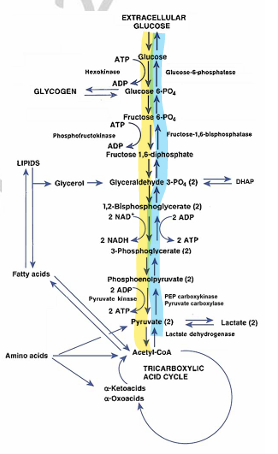
Glycolysis
metabolism of glucose to pyruvate or lactate for production of energy; occurs in the Cytoplasm of Cells
Energy yield - ATPs produced = 2 (net)
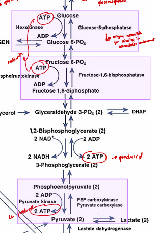
Irreversible steps
All arrow going up (yellow highlight)
Irreversible steps
All violet boxes are ______
Gluconeogenesis
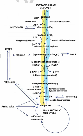
de novo synthesis of glucose from noncarbohydrate sources; occurs in the primarily in the Cytoplasm of cells, primarily in the Liver & Kidneys
All arrow going down (blue highlight)
Glycogenesis
conversion of glucose to glycogen, primarily in the liver and muscle
Glycogenolysis
breakdown of glycogen to glucose
Reverse Gluconeogenesis
Lipogenesis
Happens in the adipose tissue
conversion of carbohydrates to lipids primarily fatty acidsand triglycerides
Lipolysis
breakdown of triglycerides into glycerol and fatty acids
TCA Cycle
oxidation of acetyl CoA derived from the breakdown of carbohydrates (glycolysis), fatty acids, and certain amino acids; occurs in the Mitochondria
also known as “Krebs Cycle”
Hypoglycemic Disorders
Diabetes Mellitus
DISORDERS OF GLUCOSE METABOLISM
Post-absorptive/Fasting
UNDER HYPOGLYCEMIC DISORDERS TYPES:
occurs after 10 hours without food; secondary to hyperinsulinism (insulinoma), hormonal deficiencies, genetic disorders, autoimmunity or drug-induced
Pancreatic tumor that hyper secretes insulin
Post-prandial/Alimentary/Reactive
UNDER HYPOGLYCEMIC DISORDERS TYPES:
occurs usually within 4 hours after eating a meal
Neurogenic
UNDER HYPOGLYCEMIC DISORDERS SYMPTOMS:
tremulousness, palpitations, anxiety, diaphoresis, hunger, and paresthesias
Related to ANS
Predominate in Reactive hypoglycemia or in the autonomic nervous systems
Neuroglycopenic
UNDER HYPOGLYCEMIC DISORDERS SYMPTOMS:
dizziness, tingling, blurred vision, behavioral changes, seizure, and coma
Predominate in fasting hypoglycemia or in decreased glucose supply to the brain.
Panic value: <40 mg/dL
Whipple’s triad of hypoglycemia
UNDER HYPOGLYCEMIC DISORDERS DIAGNOSIS:
symptoms of hypoglycemia, low plasma glucose level, and relief of symptoms with correction of hypoglycemia
≤ 50 mg/dL
Present in all types of hypoglycemia
↑
UNDER HYPOGLYCEMIC DISORDERS DIAGNOSIS:
Change in plasma glucose ≥25 mg/dL (under controlled fasting condition) coincident with:
__ insulin level (≥41.7 pmol/L)
__ proinsulin level (≥5 pmol/L)
__ C-peptide level (≥0.2 nmol/L)
↓
UNDER HYPOGLYCEMIC DISORDERS DIAGNOSIS:
Change in plasma glucose ≥25 mg/dL (under controlled fasting condition) coincident with:
__ βHA (β-Hydroxybutyric acid) levels (≤2.7 mmol/L) - ketone
Insulinoma
Also endogenous hyperinsulinism
endogenous − increased proinsulin and C-peptide level
exogenous − overdose in insulin
Most common type of hypoglycemia (#1 cause of fasting)
“-oma” meaning pancreatic tumor cells that hypersecrete insulin
Uncontrollable, does not respond to feedback mechanism
Insulinoma is the exact opposite of Type 1 DM
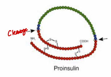
Proinsulin
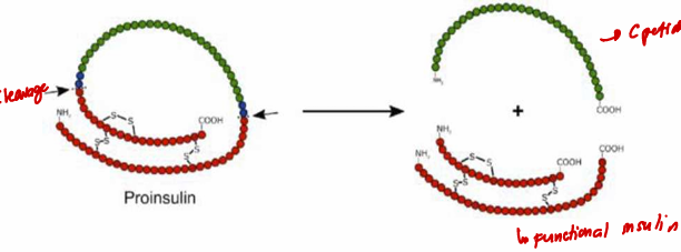
a precursor of C-peptide and insulin
Insulin is from proinsulin
C peptide needs to be cleaved so that proinsulin becomes insulin
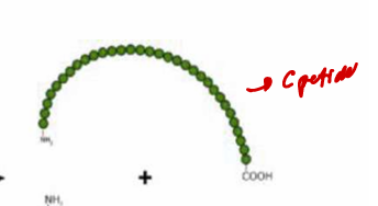
C - peptide
Marker and indicator of endogenous hyperinsulinism
Fragment released from proinsulin molecule
β-Hydroxybutyric acid
major ketone body
most abundant ketone (78% of ketone bodies)
primary marker of DM and insulinoma
High in Type 1 DM
Low in Insulinoma
Type 1 DM
UNDER DIABETES MELLITUS ADA CLASSIFICATION:
B Cell destruction leading to absolute insulin deficiency
Immune mediated - (+) auto antibodies; about 99% cases
idiopathic
DEFICIENT
Type 2 DM
UNDER DIABETES MELLITUS ADA CLASSIFICATION:
Insulin resistance with progressive insulin deficiency
No problem in insulin production; receptor; intracellular defects (no cell response)
RESISTANT
Genetic defects of B cell function
Genetic defects in insulin action
diseases of the exocrine pancreas
Endocrinopathies
Drug-or chemical-induced
Infections
Uncommon forms of immune-mediated diabetes
Other genetic syndromes
UNDER DIABETES MELLITUS ADA CLASSIFICATION:
OTHER TYPES OF DM:
diseases of the exocrine pancreas
These are examples of What Type of DM:
pancreatitis, pancreatectomy, pancreatic cancer/neoplasia, cystic fibrosis, hemochromatosis, fibrocalculous pancreatopathy
Gestational
UNDER DIABETES MELLITUS ADA CLASSIFICATION:
glucose intolerance during pregnancy that disappears post-partum but may convert to type 2 DM in 30-40% of cases within 10 years;
due to metabolic and hormonal changes
Every 3 Years
TESTS AND DIAGNOSTIC CRITERIAFOR DM:
Screening Tests for Adults ≥ 45 years old
Every Year
TESTS AND DIAGNOSTIC CRITERIAFOR DM:
Screening Tests for Individuals ≥ 1 risk factor
HbA1C and FPG
TESTS AND DIAGNOSTIC CRITERIAFOR DM:
*In the absence of unequivocal hyperglycemia, diagnosis requires two abnormal test results obtained at the same time (e.g., ________________) or same test at two different time points.
24-28
TESTS AND DIAGNOSTIC CRITERIA FOR GDM:
All nondiabetic pregnant women should be screened for GDM at _______ weeks of gestation.
Whole Blood Glucose
TESTS FOR MONITORING GLYCEMIC CONTROL:
Daily Monitoring
most commonly analyzed using POCT devices at home or at the patient’s bedside; 3-4 times daily for type 1 DM patients according to ADA guidelines
about 10-15% lower than plasma glucose
ADA glycemic goals:
Pre-prandial (before meal): 70-130 mg/dL
Post-prandial (after meal): <180 mg/dL
HBA1C (Glycated Hemoglobin)
TESTS FOR MONITORING GLYCEMIC CONTROL:
Long Term Monitoring
Hb A with glucose irreversibly attached to one or both N-terminal valines of the β-chains
Provides an index of average blood glucose levels over the past 2-3 months
Unreliable in patients with hemolytic disorders (e.g. hemoglobinopathies, autoimmune hemolytic anemia)
ADA glycemic goal: < 7%
Poor control: >12%
eAG = (28.7*A1c) 46.7
120 Days
Life span of RBCs
glycosylation occurs also while exposed to hyperglycemic plasma
Fructosamine
TESTS FOR MONITORING GLYCEMIC CONTROL:
Represents glycosylated proteins, especially albumin.
Used to assess glycemic control over the past 2-3 weeks in cases when HbA1c is unreliable.
Half life of Alb = 17 days
Has the advantage of using serum samples but is unreliable when serum albumin level is < 3 g/dL or if patient has hypoalbuminemia/hypoproteinemia.
Reference values: 205-285 umol/L
Microalbuminuria
TESTS FOR MONITORING GLYCEMIC CONTROL:
Early indicator of diabetic nephropathy
Persistent albuminuria in two out of three urine collections 20-200 ug/min (AER), 30-300 mg/24 h, or an albumin-creatine ratio of 30-300 ug/mg (ACR) or 3.5-30 mg/mmol within a 3 to 6 month period
30-300 mg/24h - Spx = Timed specimen
3 - 6 month period - Spx random specimen
7mg/dl @ RT; 2 mg/dl/h (ref)
METHODS OF GLUCOSE MEASUREMENT:
SPECIMEN CONSIDERATIONS
Use of NaF or SST to prevent glycolysis. Otherwise, glycolysis will take place at a rate of _______________________
For NaF use iodoacetate (if BUN or CREA; does not inhibit urease)
Effect of dextrose contamination ↑PG (500 mg/dl)
Copper Reduction
Ferric Reduction
Condensation
What are example the Chemical Methods?
Copper Reduction
CHEMICAL METHODS:
Benedicts Reaction
Common initial reaction of the _______
Non - specific because glucose is not the only one that can catalyze the initial reaction
Folin Wu
UNDER COPPER REDUCTION:
Cu + PMA → phosphomolybdenum
PMA = OH
Disadvantage: non specific
Increases due to other reducing agents
Nelson-Somogyi
UNDER COPPER REDUCTION:
Cu + AMA → arsenomolybdenum
Neocuproine
UNDER COPPER REDUCTION:
Cu + neocuproine → Cu − neocuproine
is a simple complex formation between Cuprous ion and neocuproine
this is in yellow orange color
blue
Phosphomolybdenum and Arsenomolybdenum is _____ in color.
Intensity of color is monitored spectrophotometrically
Color is directly proportional to the level of Cuprous ions.
Ferric Reduction
CHEMICAL METHODS:
_______ (yellow orange) is reduced to ferrocyanide (colorless)
_________ is also called as Inverse colorimetry or Hagedorn Jensen method
Monitors the disappearance of color

Condensation
CHEMICAL METHODS:
Dubowski Method - Most specific, popular, and best amongst all chemical methods
Not affected by oxidizing and reducing agents
The intensity of the color is monitored spectrophotometrically.
Color is proportional to the level of glucose

Glucose oxidase
ENZYMATIC METHODS:
Most popular and most commonly used enzymatic method
Glucose is oxidized to gluconic acid and hydrogen peroxide (H2O2)
Catalyzed by the enzyme glucose oxidase
Hydrogen peroxide is used as an oxidizing agent
Peroxidase-coupled/Trinder reaction
2 Detection Methods for Glucose Oxidase:
subject to many interferences
Colorimetric / Spectrophotometric
More routinely used
Serum glucose determination chromogen:
4-aminophenozone oxidized to quinonimine (red purple)
Absorbance: 500nm

Polarographic
2 Detection Methods for Glucose Oxidase:
measurement of the degree of O2 consumption using a pO2 (Clark) electrode (internal glucose)
requires addition of molybdate and iodide or catalase and ethanol to prevent the re-formation of oxygen (source of error)
Better because it is not affected by reducing and oxidizing agents
Measures the amount of oxygen consumed in the reaction by detecting the amount of oxygen left
Oxygen consumed is proportional to the amount of glucose in the sample
Hexokinase
ENZYMATIC METHODS:
reference method for glucose; coupling reaction is highly specific
effect of hemolyzed/icteric sample F↓ (source of error)
Hemoglobin interferes of glucose
UV Method ; Involves UV measurement at 340nm

Glucose Dehydrogenase
ENZYMATIC METHODS:
Highly specific for glucose, not subject to interference from substances normally found in serum and provides results in close agreement with hexokinase method.
Not commonly used except in glucose POCT (glucometer)
Formula of Non-Colorimetric Method
This is the Formula of Non-Colorimetric or Colorimetric Method of Glucose Dehydrogenase?
Measure using NADPH at A 340 nm
Tetrazolium dye may be used to enable colorimetric spectrophotometric measurement

Colorimetric Method
This is the Formula of Non-Colorimetric or Colorimetric Method of Glucose Dehydrogenase?
Formazan = BLUE

Galactosemia
INBORN ERRORS OF CARBOHYDRATE METABOLISM:
due to a deficiency of one of three enzymes involved in galactose metabolism: galactose-1-phosphate uridyl transferase (GALT), galactokinase (GALK), or uridine diphosphate galactose-4-epimerase (GALE)
affected children present with mental retardation, failure to thrive, and galactosuria
Hepatic glycogenoses
INBORN ERRORS OF CARBOHYDRATE METABOLISM:
UNDER Glycogen Storage Diseases/Glycogenoses:
Usually manifest with hepatomegaly, hypoglycemia, and growth retardation
von Gierke disease (Type 1a)
Most common hepatic glycogenosis
Deficiency of the enzyme glucose-6-phosphatase
Present with hypoglycemia, lactic acidosis, and hyperuricemia.
von Gierke disease
▪ Most common hepatic glycogenosis
▪ Deficiency of the enzyme glucose-6-phosphatase
▪ Present with hypoglycemia, lactic acidosis, and hyperuricemia.
Muscle glycogenosis
Manifests with exercise intolerance, muscle cramps, fatigue, and weakness (Type V & VIII)
↑ skeletal muscle enzymes: CK, AST LD, aldolase
Surface
Core
Components of Lipoproteins are:
Surface
These are under what component of lipoproteins?
Apolipoproteins - protein moietics
Phospholipids
Non-esterified cholesterol
Phosphilipid (PL) and Non-esterified Cholesterol (NEC)
These Two Surface proteins are Amphipathic Lipids (w Polar/Non-Polar grps)
Core
These are under what component of lipoproteins?
Triglycerides
Cholesterol esters
Free fatty acids
Triglycerides
Cholesterol esters
Free fatty acids
These Three Core proteins are Neutral Lipids
Lecithin cholesterol acyltransferase (LCAT)
catalyzes the esterification of cholesterol
LDL Receptor (LDL-R)
mediates the endocytosis of lipoproteins especially LDL
Lipoprotein lipase (LPL)
catalyzes the hydrolysis of TG in lipoproteins releasing free fatty acids and glycerol to tissues
Exogenous pathway
This is the process of _____?
Dietary Lipids → Intestine → Chylomicrons → needs LPL & CII → to hydrolyze to Chylomicrons Remnants → liver will use the chylomicrons remnants in the synthesis of the VLDL
to be continued
huge size
Note that chylomicrons are not taken up by the liver because of ______
Endogenous pathway
This is the process of _____?
VLDL → needs LPL → hydrolyze into smaller IDL → needs LPL → to much smaller LDL
LDL
_____ is considered as a metabolite of VLDL as it comes with the metabolism of VLDL that can easily be taken up by the liver and the peripheral cells.
Atherogenic
if the amount of cholesterol delivered to peripheral tissues is in excess.
De Novo Synthesis
Liver Synthesizes using ____________
Phospholipids
What is the major lipid component (excluding the protein) of the HDL?
IDL
MINOR AND ABNORMAL LIPOPROTEINS:
lipolytic product of VLDL catabolism taken up by the liver or converted to LDL;
density, electrophoretic mobility, protein and lipid contents are intermediate between those of VLDL and LDL
It is minor because it is only intermediate in the metabolism of VLDL
β-VLDL
MINOR AND ABNORMAL LIPOPROTEINS:
“floating β lipoprotein”
richer in cholesterol than VLDL (VLDL-C/TG ratio>0.3); associated with Apo E2 isoform which has low affinity for the LDL receptor leading to dysbetalipoproteinemia
Structure is similar to VLDL
It has a much lower density than the typical VLDL
The higher the lipid content, the lower the density, the more that the particle floats
Lp(a)
MINOR AND ABNORMAL LIPOPROTEINS:
“sinking pre-β lipoprotein”
Structure is similar to LDL
Contains the prothrombotic Apo(a) which is homologous with plasminogen
Associated with stroke, MI, CHD (increased protein content)
inhibits fibrinolysis.
Plasmin
________ is an important fibrinolytic factor
LpX
abnormal lipoprotein associated with obstructive biliary disease and LCAT Deficiency (catalyzes the esterification of cholesterol)
Consists mostly of phospholipids and non-esterified cholesterol
Xanthomas
cutaneous lesions due to accumulation of lipids in the eyelids (xanthelasma), Achilles and extensor tendons (tendinous xanthoma), extensor surfaces (eruptive xanthoma), and palm and wrist creases (palmar xanthoma)
↑ Chylo and/or VLDL
FREDRICKSON CLASSIFICATIONOF HYPERLIPOPROTEINEMIAS additional info:
↑TG = acute pancreatitis (xanthomas) = ↑risk pancreatitis
↑ LDL
FREDRICKSON CLASSIFICATIONOF HYPERLIPOPROTEINEMIAS additional info:
↑ Total Cholesterol (xanthelasmas) = ↑ risk Coronary heart disease
mutation or genetic mutation
Main cause of Hypolipoproteinemia is _____________
Abetalipoproteinemia
HYPOLIPOPROTEINEMIAS:
AKA Bassen-Kornzweig Syndrome
An autosomal recessive disorder involving mutations in the MTTP gene with absolute nonexistent levels of apoB48 and apoB100
Absence of Apo B-containing liproproteins (VLDL, LDL, CM)
Total Cholesterol = Very low
Triglyceride = Nearly undetectable
Defect in APOB containing lipoprotein
Hypobetalipoproteinemia
HYPOLIPOPROTEINEMIAS:
autosomal dominant disorder caused by nonsense or missense mutations in the apo B gene;
low total cholesterol and triglyceride levels
Decreased level of TC and TG
Defect in APOB containing lipoprotein
Tangier disease
HYPOLIPOPROTEINEMIAS:
Rare
Autosomal recessive disorder characterized by low to undetectable HDL due to a mutation in the ABCA1 gene
Defect in HDL
Hypoalphalipoproteinemia
HYPOLIPOPROTEINEMIAS:
Defect in HDL
Common autosomal dominant disorder characterized by HDL-C Levels → <30 mg/dL in men → <40 mg/dL in women
High risk for CHD because of low HDL level
HDL is inversely related to cardiac risk.
Abetalipoproteinemia & Hypobetalipoproteinemia
These Hypolipoproteinemias are Associated with Defects in Apo B
Tangier disease & Hypoalphalipoproteinemia
These Hypolipoproteinemias are Associated with Defects in HDL
Chemical Methods for Cholesterol
PLASMA LIPID ANALYSIS (ANALYTICAL METHOD)
This formula is for?
Saponification using alcoholic potassium hydroxide which catalyzes the hydrolysis of cholesterol or cholesteryl ester to separate or release free cholesterol from fatty acid

Saponification
What Step for Chemical Method?
Reagent:
Alcoholic potassium hydroxide (KOH)
Purpose:
Hydrolysis of cholesterol esters
Extraction
What Step for Chemical Method?
Reagent:
Bloor’s reagent (Ethanol-ether)
Purpose:
Removal of protein interference
Purification
What Step for Chemical Method?
Reagent:
Digitonin
Purpose:
Precipitation of free cholesterol
Colorimetry
What Step for Chemical Method?
Reagent:
H2SO4, acetic anhydride or ferric ions
Purpose:
Formation of colored compound
Liebermann-Burchardt
Chemical Methods for Cholesterol:
Colorimetry
Acetic anhydride is the reagent used
Cholestadienly MSA is green in color
