Lab Heart/Blood Vessel
 Call Kai
 Learn
Learn Practice Test
 Spaced Repetition
 Match
Match Flashcards
 Knowt Play
1/52

There's no tags or description
Looks like no tags are added yet.
Name | Mastery | Learn | Test | Matching | Spaced | Call with Kai | Chat |
|---|
No analytics yet
Send a link to your students to track their progress
53 Terms

What is the line encircled?
anterior interventricular sulcus

Name the string structures?
chordae tendineae
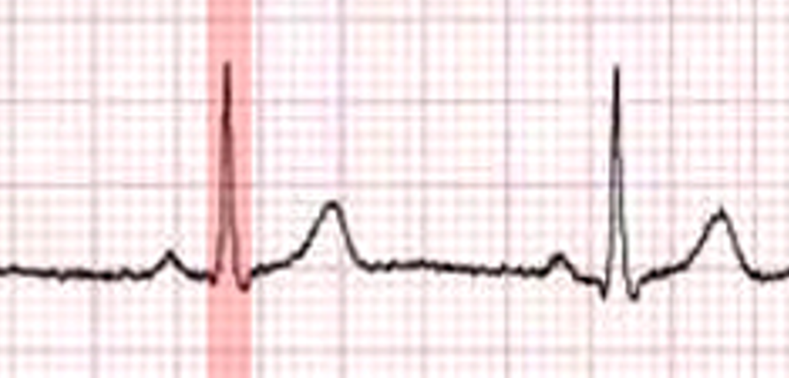
Name the highlighted deflection on EKG?
QRS complex

Name the valve ( be specific )
aortic semilunar valve
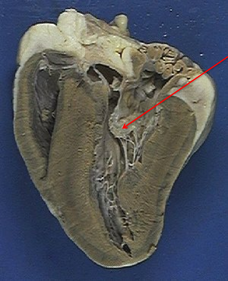
Name the valve?
bicuspid valve — mitral valve
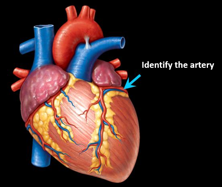
Identify the artery?
circumflex artery

Identify the artery?
brachiocephalic artery

Identify the artery?
femoral artery

Identify the artery?
left common carotid artery

Identify the artery?
internal carotid artery

Identify the artery?
celiac trunk

Identify the artery?
right common carotid artery.
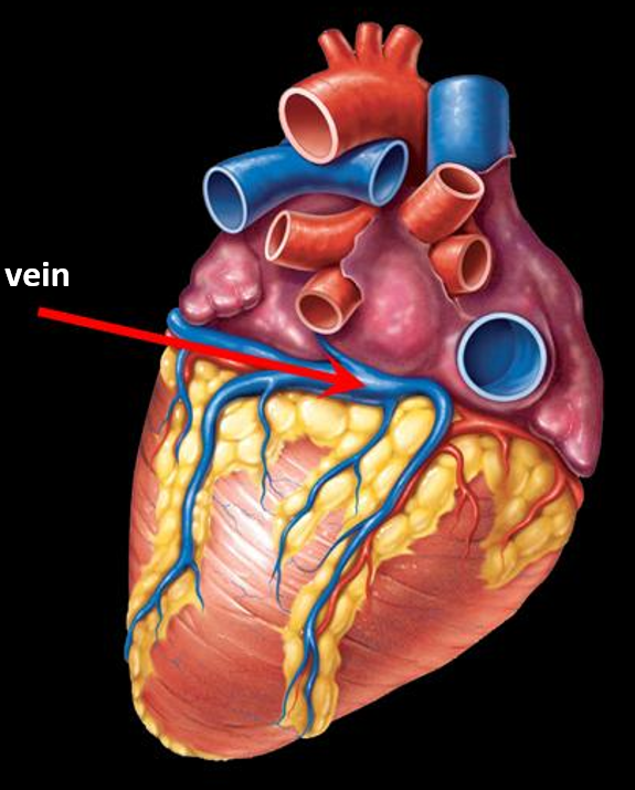
Identify the vein?
coronary sinus

Identify the vein?
superior vena cava

Identify the vein?
Right renal vein

Identify the artery?
External iliac artery

Identify the blood vessel?
Left pulmonary veins

Identify the artery
Right subclavian artery
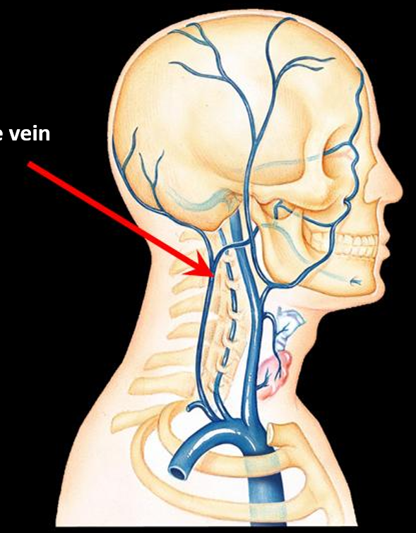
Identify the vein?
External jugular vein

Identify the artery?
Radial artery
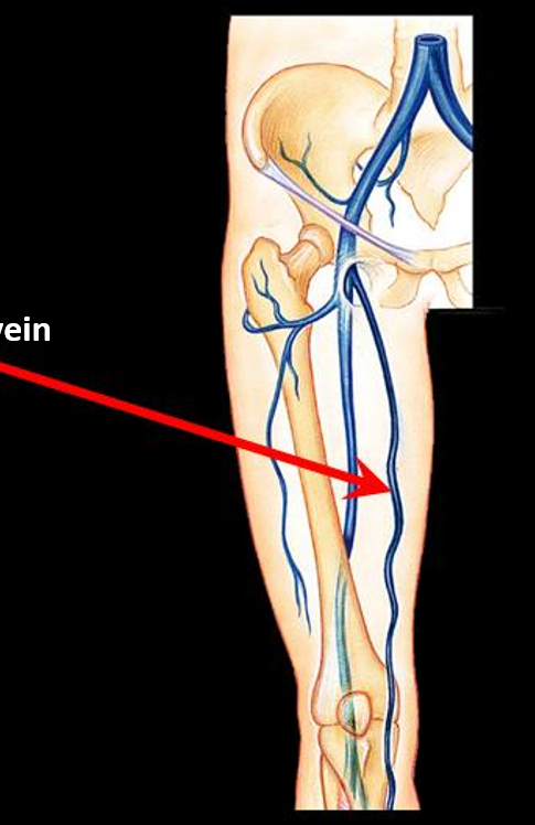
Identify the vein?
Great saphenous vein

Identify the vein?
Cephalic vein
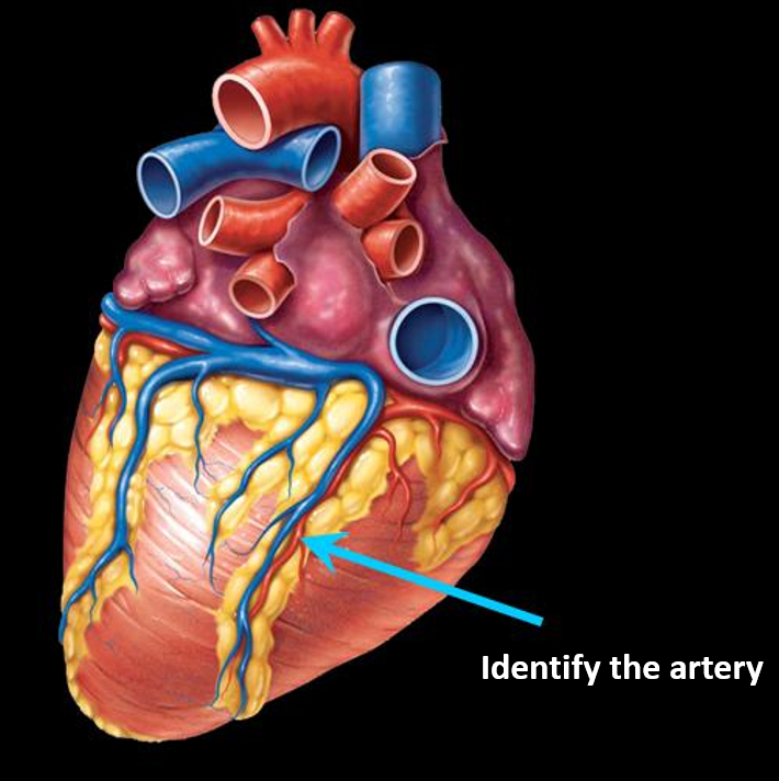
Identify the artery?
Anterior interventricular artery

Identify the artery?
Internal jugular vein

Identify the vein?
Femoral vein

Identify the vein?
Great saphenous vein

Identify the artery?
Basilar artery
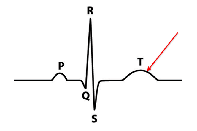
What is the condition of the heart reflected at this peak?
Ventricular systole

Identify the blood group?
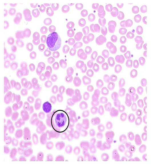
What disease is this individual suffering from?
Sickle Cell Anemia

What is this cell? How can you identify this?
Neutrophil — segmented, multi-lobed nucleus
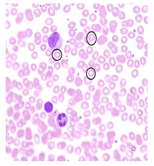
What is the major protein found in this cell?
Hemoglobin

A deficiency of this cell type is called what?
Thrombocytopenia
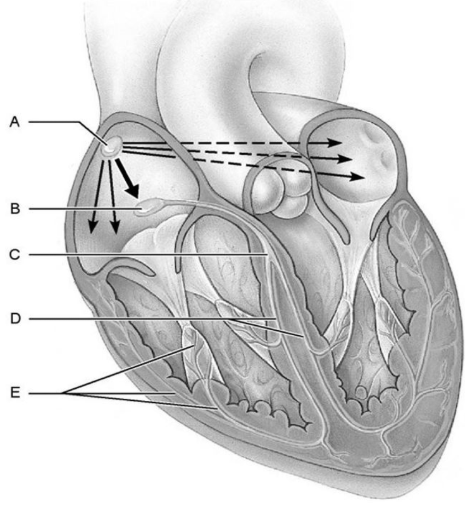
A?
Sinoatrial (SA) node

B?
Atrioventricular (AV) node

C?
Atrioventricular (AV) bundle
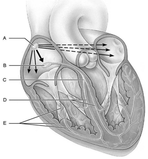
D?
Bundle branches

E?
Subendocardial conducting network (Purkinje fibers)

In the ECG diagram, what does label A represent and what is its physiological meaning?
P wave — Atrial depolarization.

What structure do labels B, C, and D collectively form, and what electrical event does it represent?
QRS Complex — Ventricular depolarization.

What does label E stand for on the ECG trace, and what is its significance?
T wave — Ventricular repolarization

Point after which pressure begins to rise in the aorta?
D
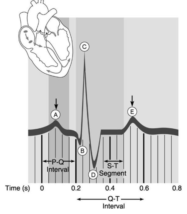
Point that represents the "dup" sound made by the heart.
E

Based on the diagram's timeline, how long does a normal cardiac cycle last, and what resting heart rate does this correspond to?
It lasts approximately 0.8 seconds and resting heart rate of about 75 beats per minute (BPM).

Normal sinus rhythm

Junctional rhythm

Second-degree heart block

Ventricular fibrillation.
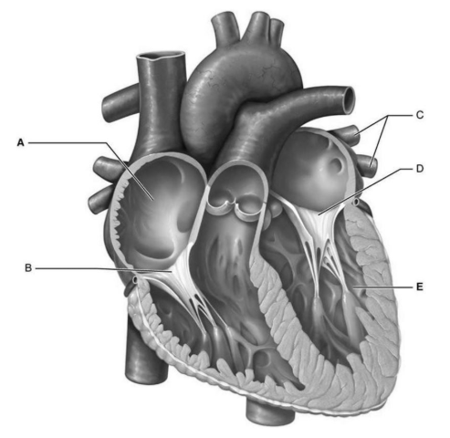
A
Right atrium
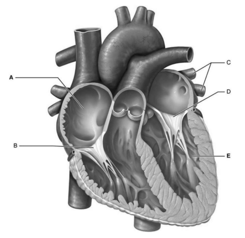
B
Tricuspid valve

C
Pulmonary veins

D
Mitral (bicuspid) valve
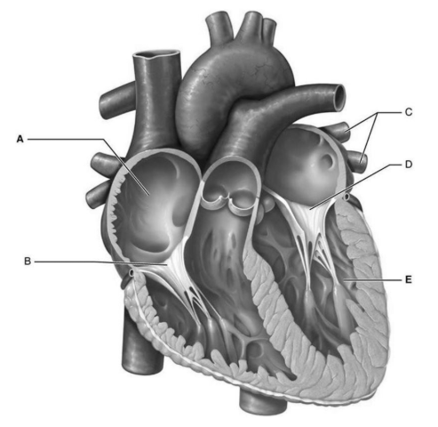
E
Left ventricle