Histo block 4
1/24
There's no tags or description
Looks like no tags are added yet.
Name | Mastery | Learn | Test | Matching | Spaced |
|---|
No study sessions yet.
25 Terms
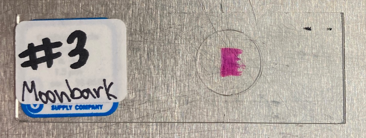
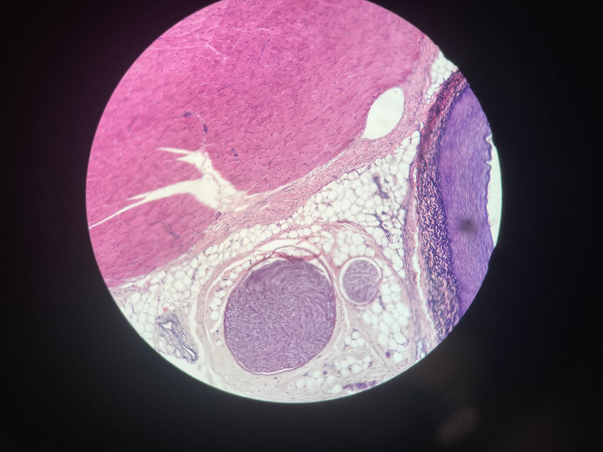
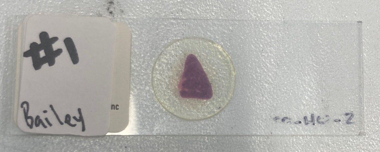
Cardiac muscle
Pink Rectangle
Intercalated discs, centrally located nuclei
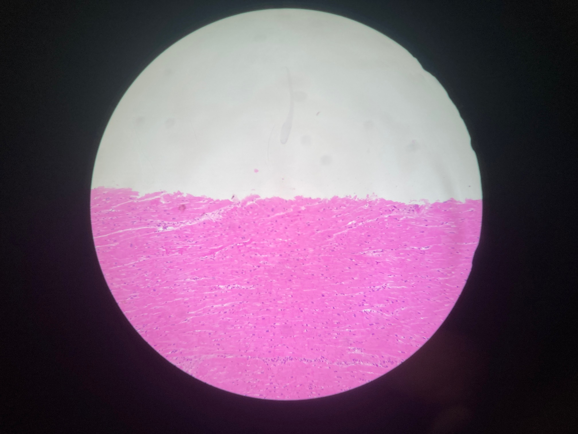
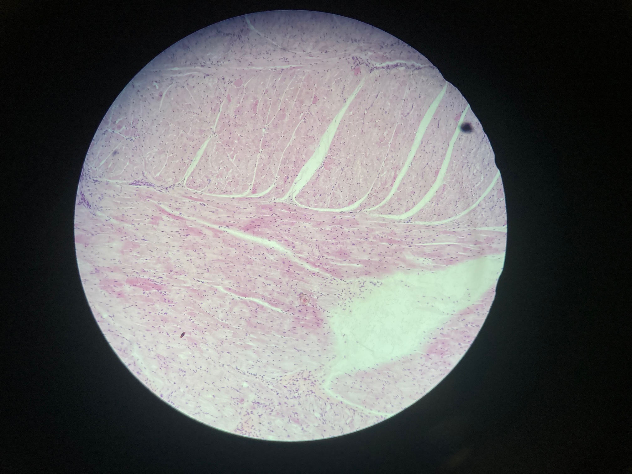
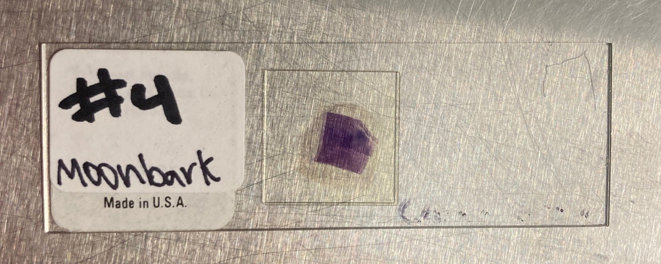
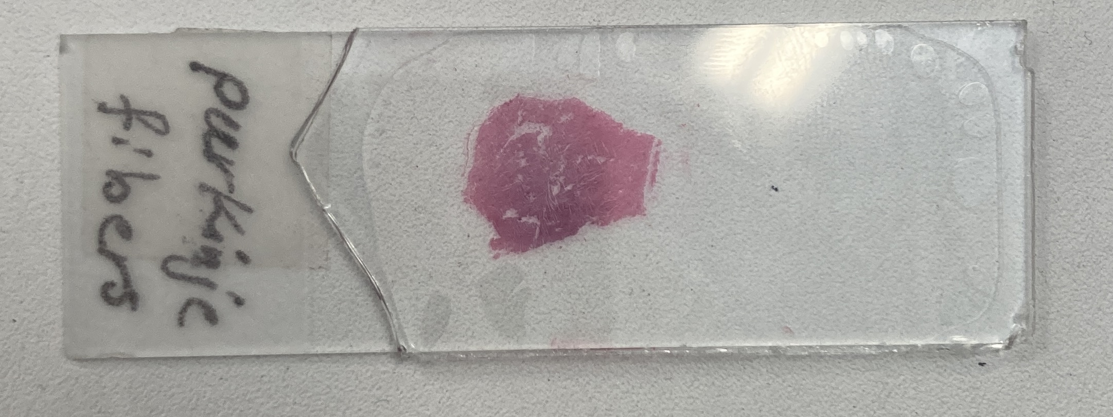
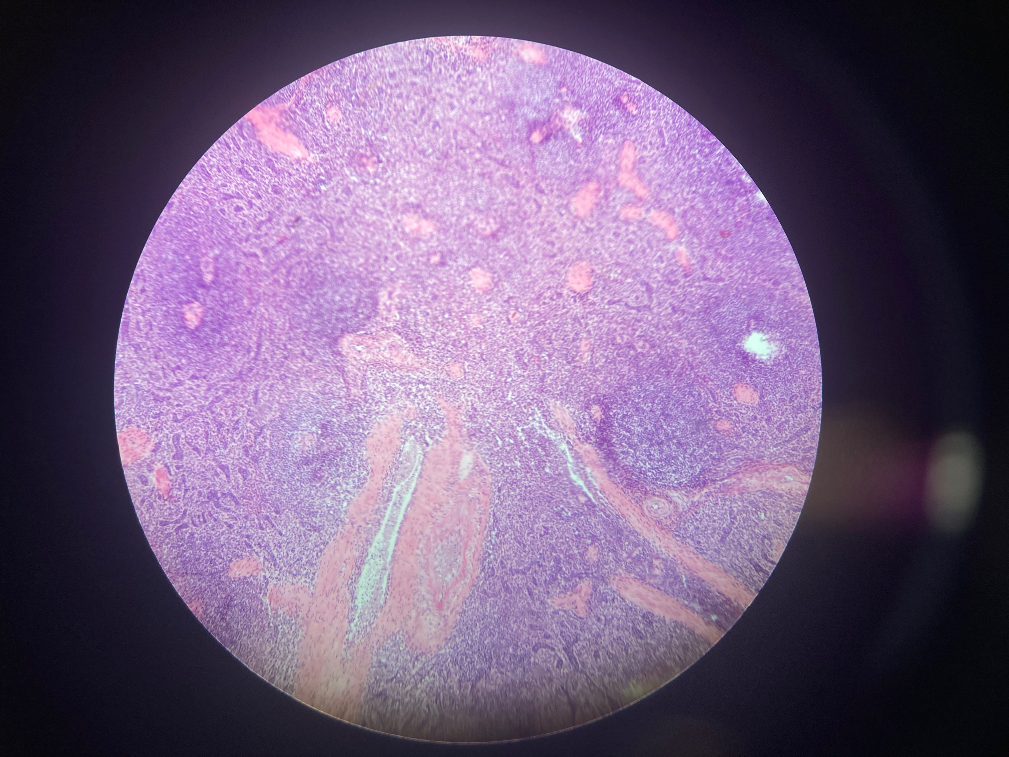
Cardiac muscle (Purkinje fibers)
Purple rectangle
Elongated cells, fewer myofibrils than regular cardiac cells
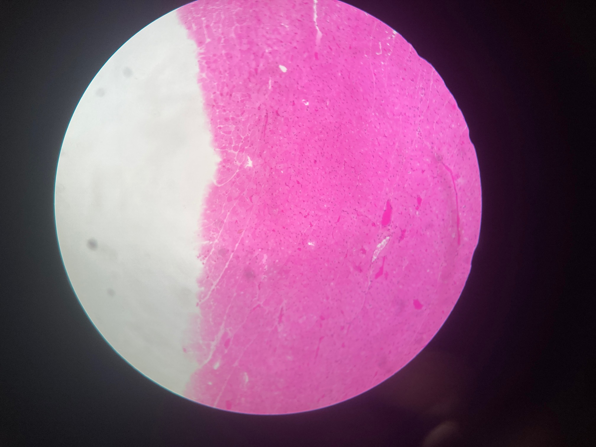
Cardiac muscle
Pink rectangle
Intercalated discs, centrally located nuclei
Heart (intercalated disk)
Purple square
Intercalated discs, centrally located nuclei
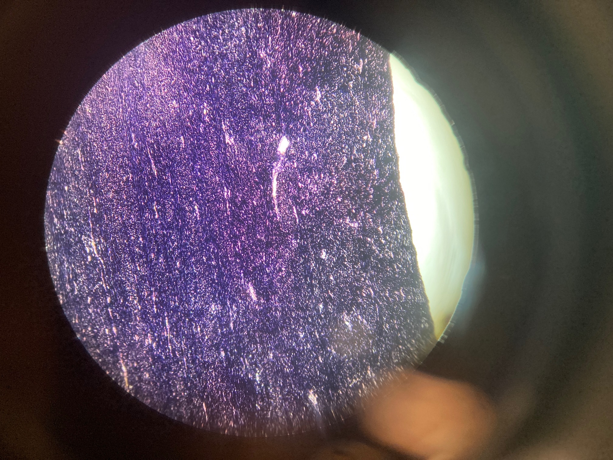
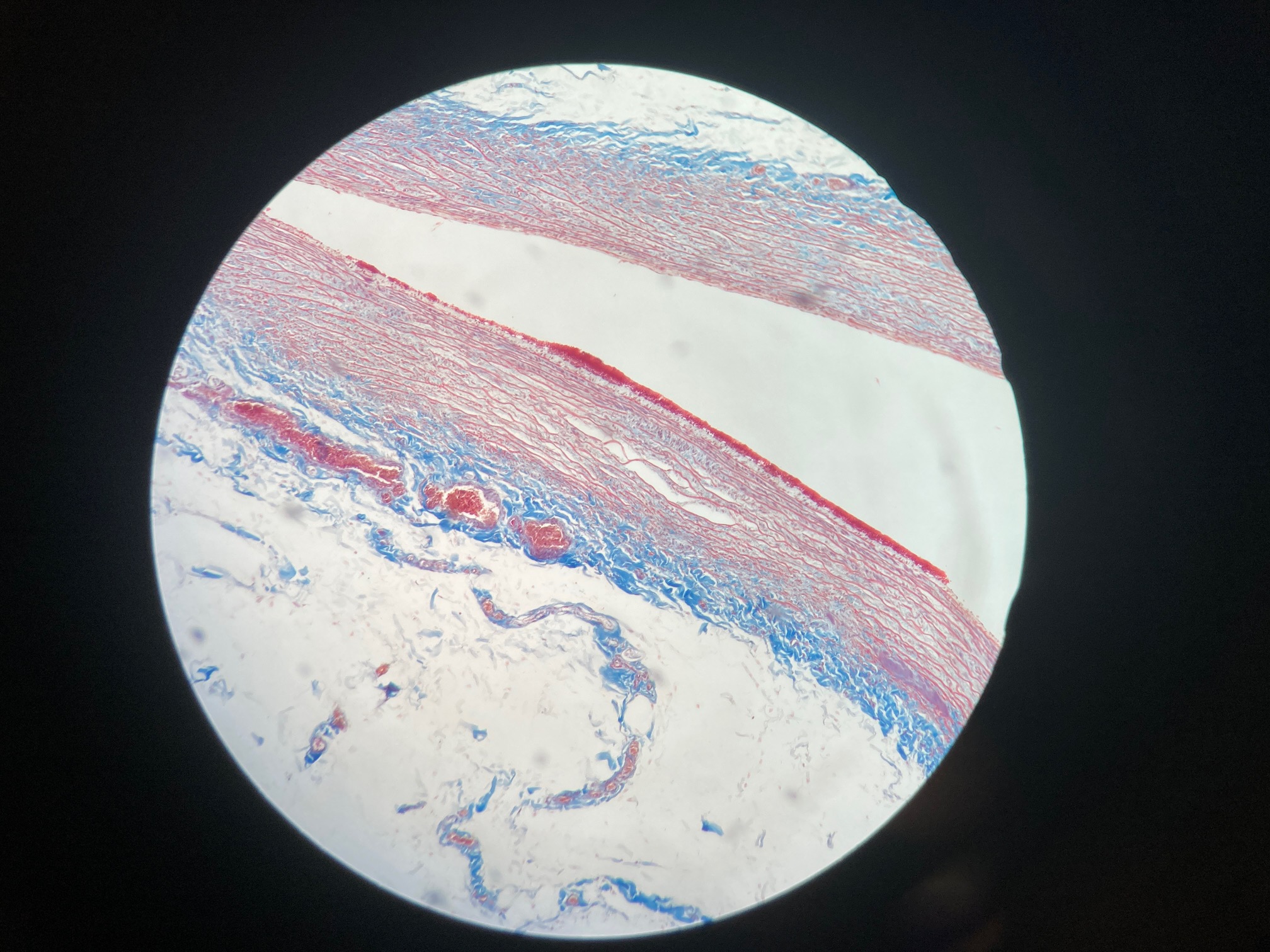
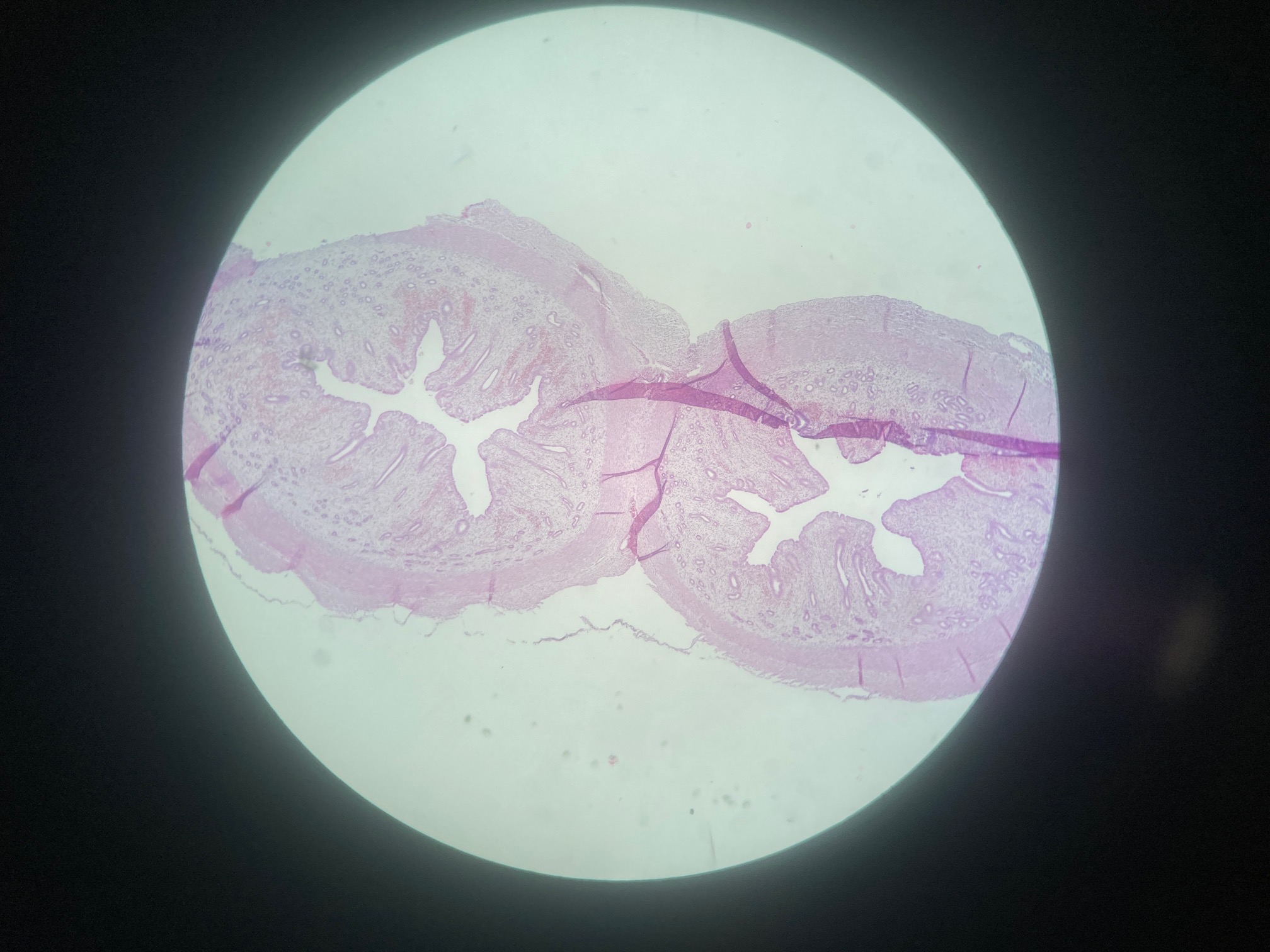
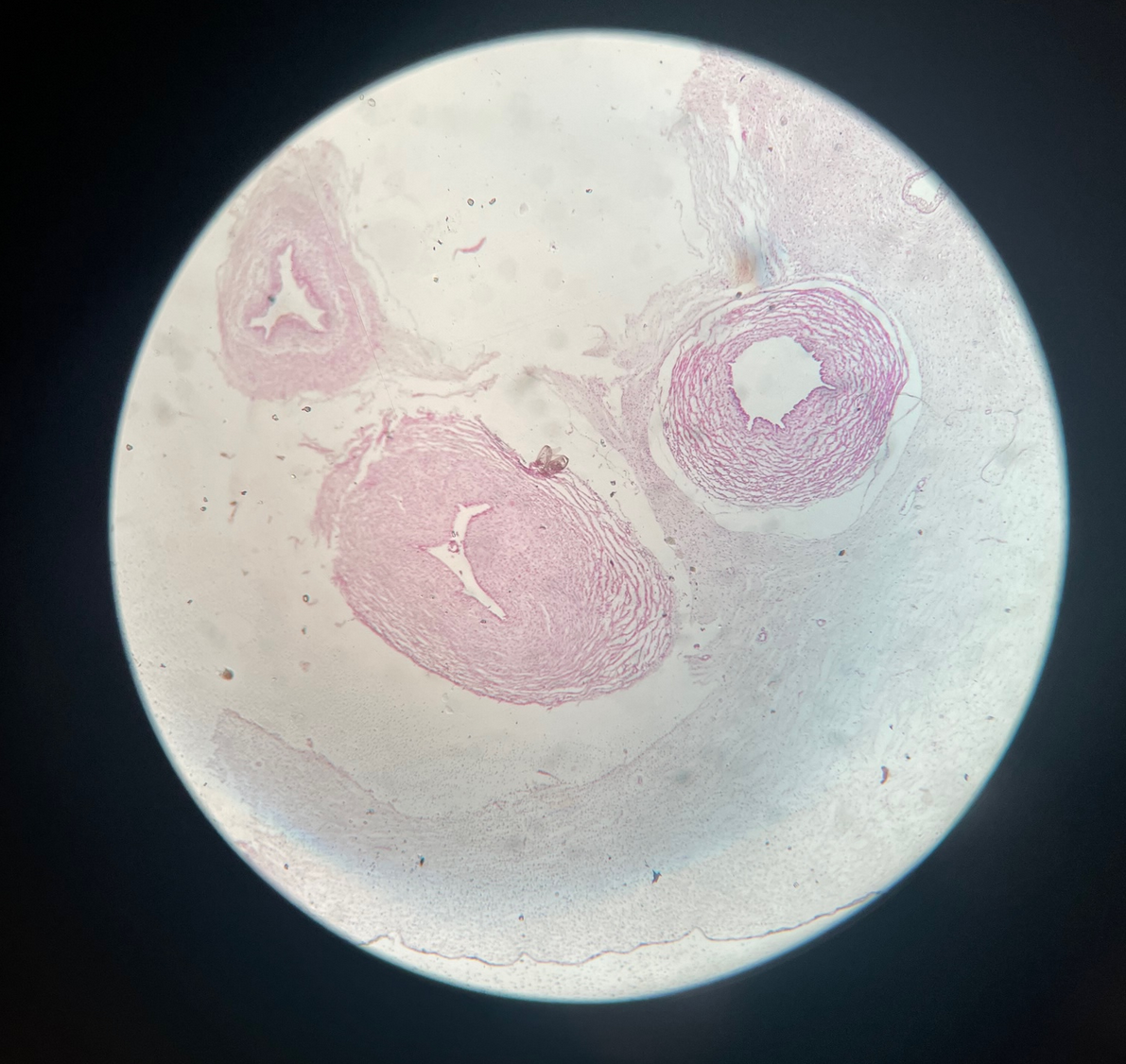
Aorta
4 dark rectangles
tunica intima, tunica adventitia
CCA common carotid artery
3 question marks
tunica intima, tunica adventitia
Artery, Vein, Nerve
Shy guy/Among us
tunica intima, tunica adventitia
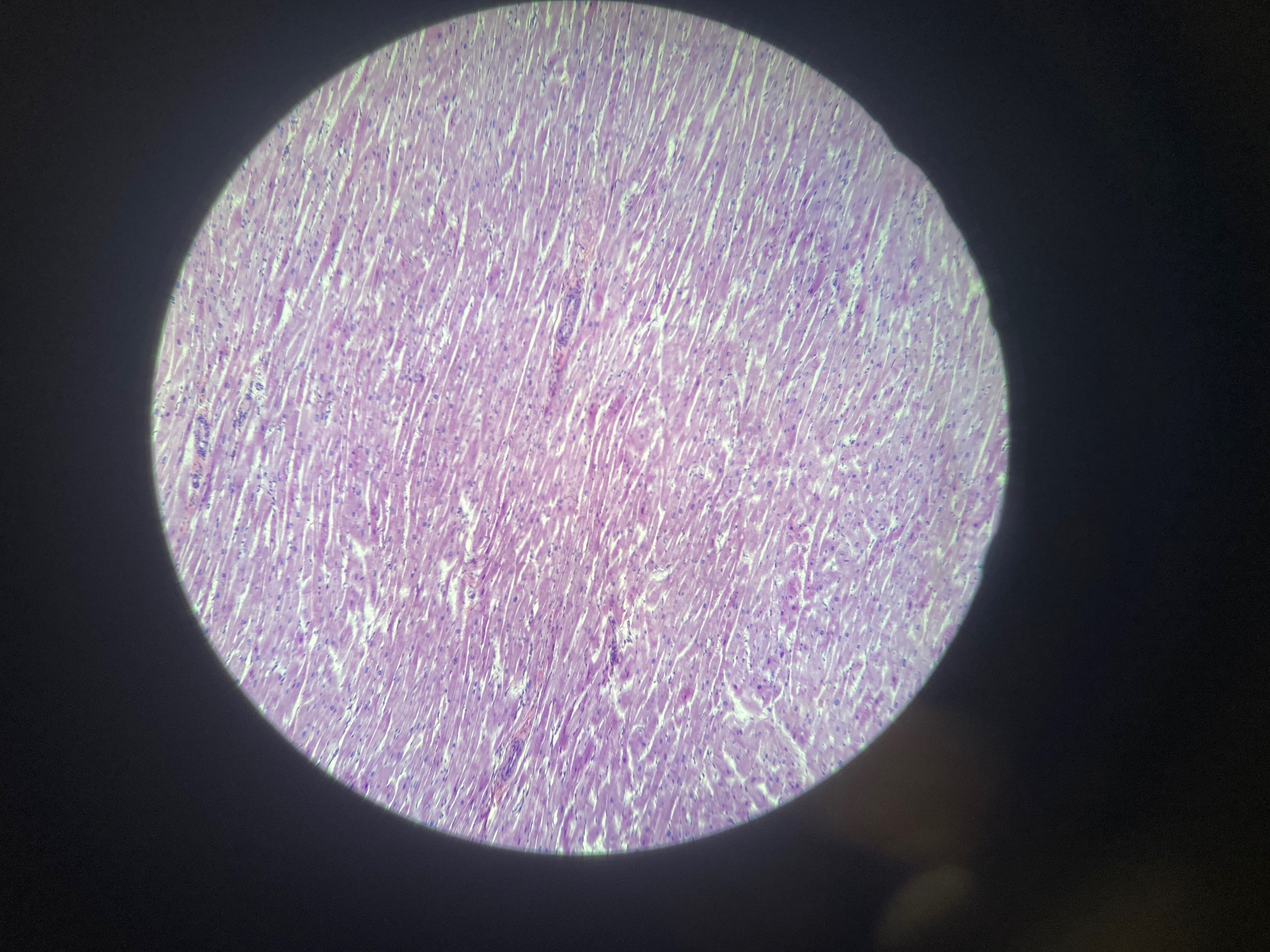
Purkinje Fibers
Elongated cells
Fewer myofibrils compared to regular cardiac muscle cells
Larger than typical cardiac muscle cells
Large number of mitochondria
Prominent glycogen stores
Well-developed sarcoplasmic reticulum network
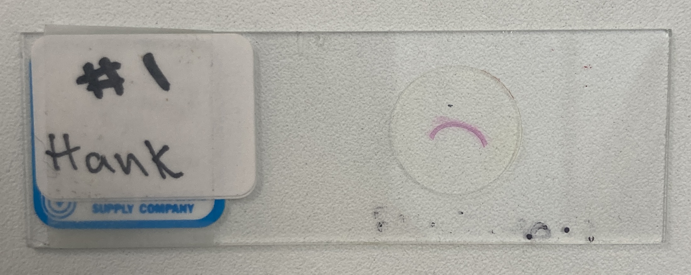
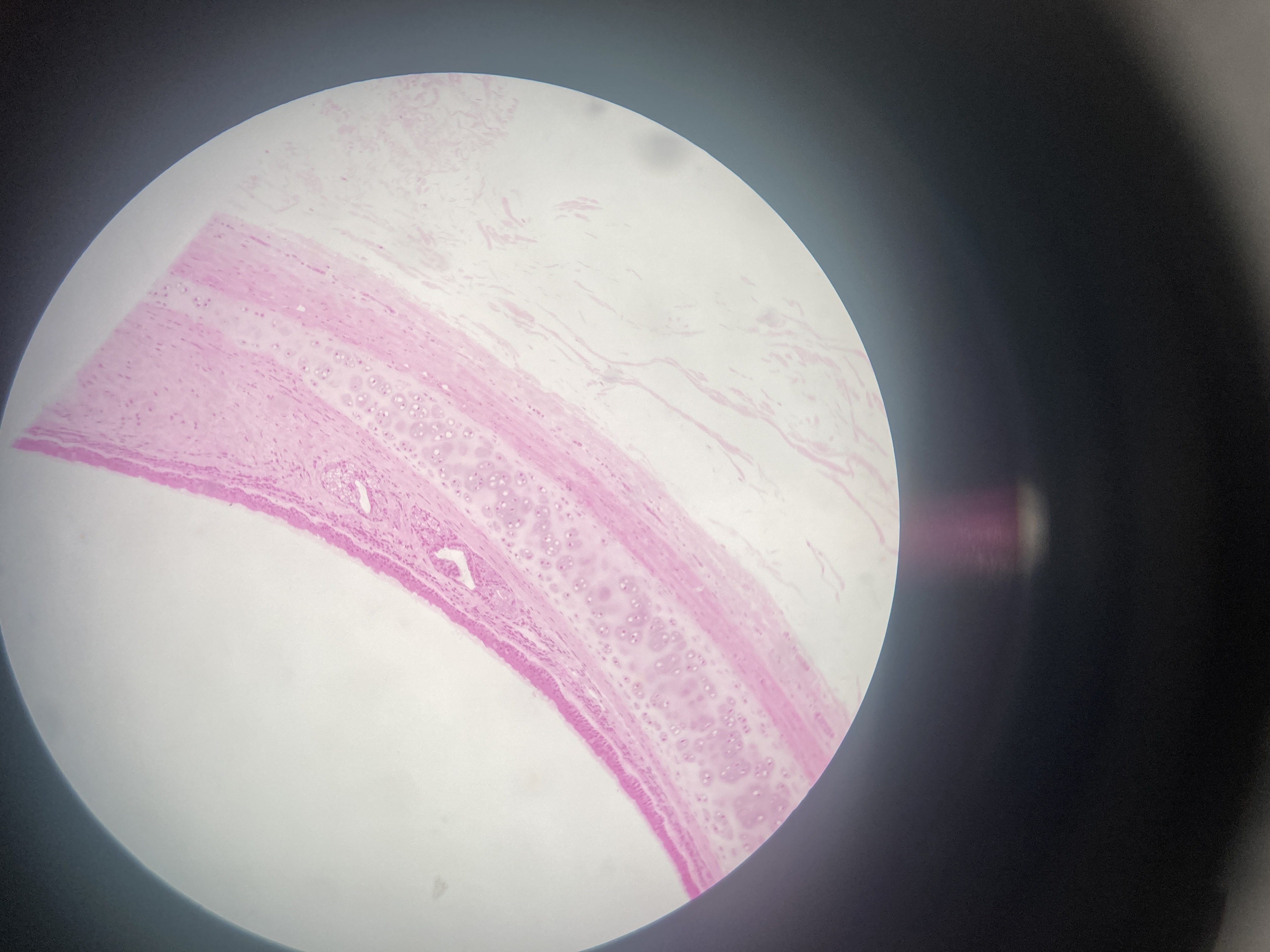
Trachea (monkey)
Pseudostratified ciliated columnar epithelial
Goblet cells
Basal cells
Brush cells
Submucosal glands
Smooth muscle cells
Cartilage
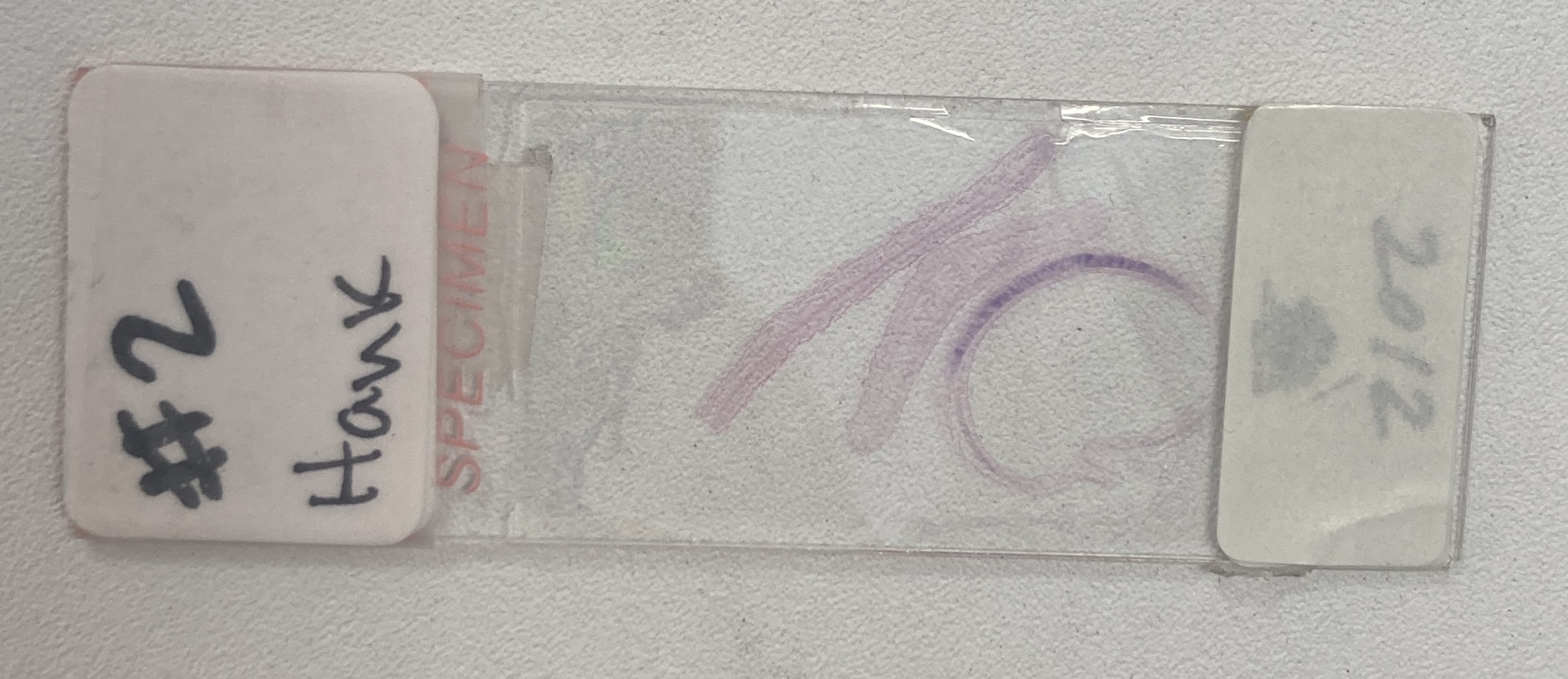
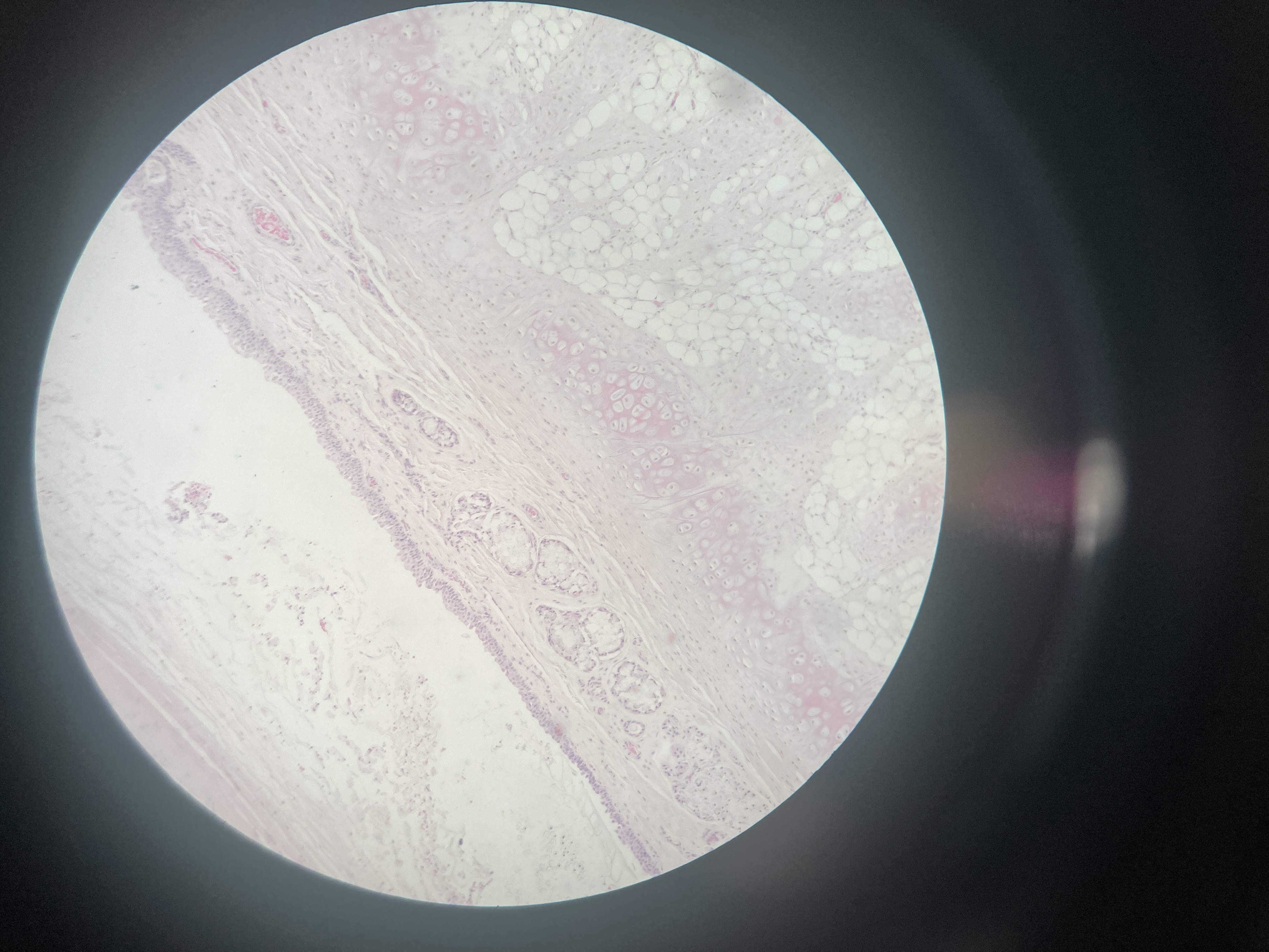
Trachea (cat)
Pseudostratified ciliated columnar epithelial
Goblet cells
Basal cells
Brush cells
Submucosal glands
Smooth muscle cells
Cartilage
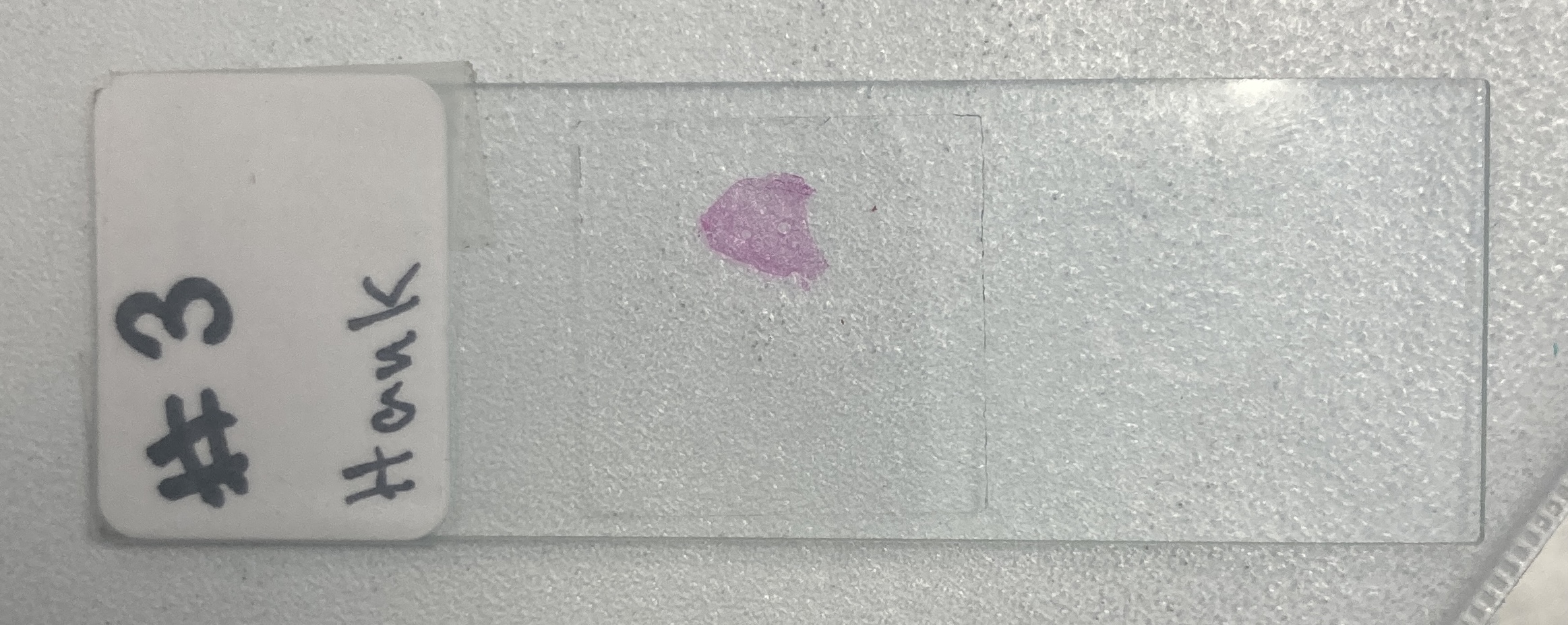
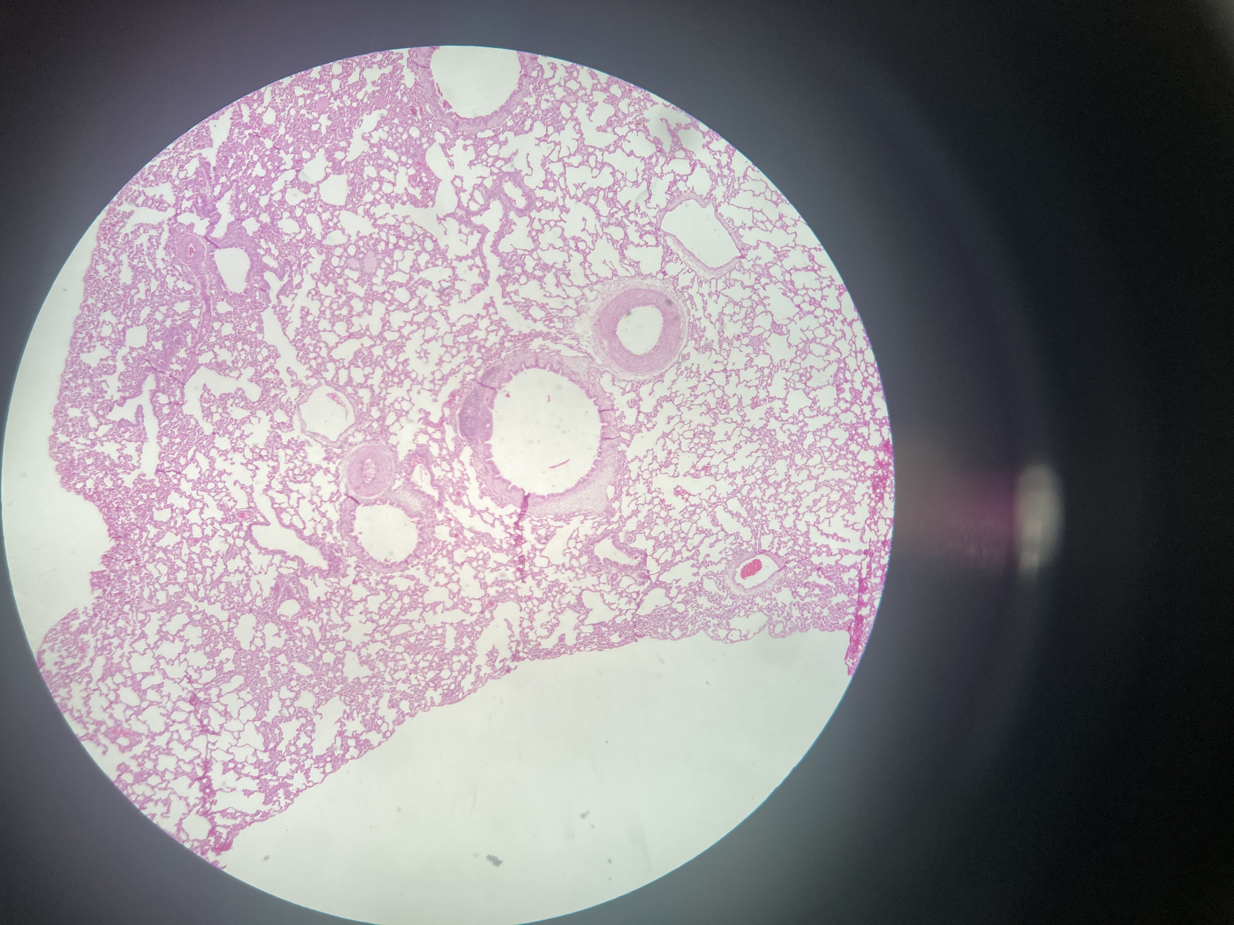
Lung (mouse)
Type I pneumocytes
Type II pneumocytes (or septal cells)
Alveolar macrophages (dust cells)
Capillary endothelial cells
Fibroblasts
Smooth muscle cells
Goblet cells
Ciliated epithelial cells
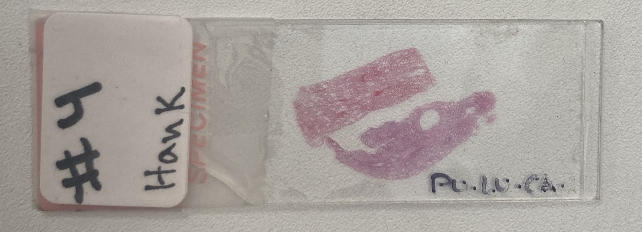
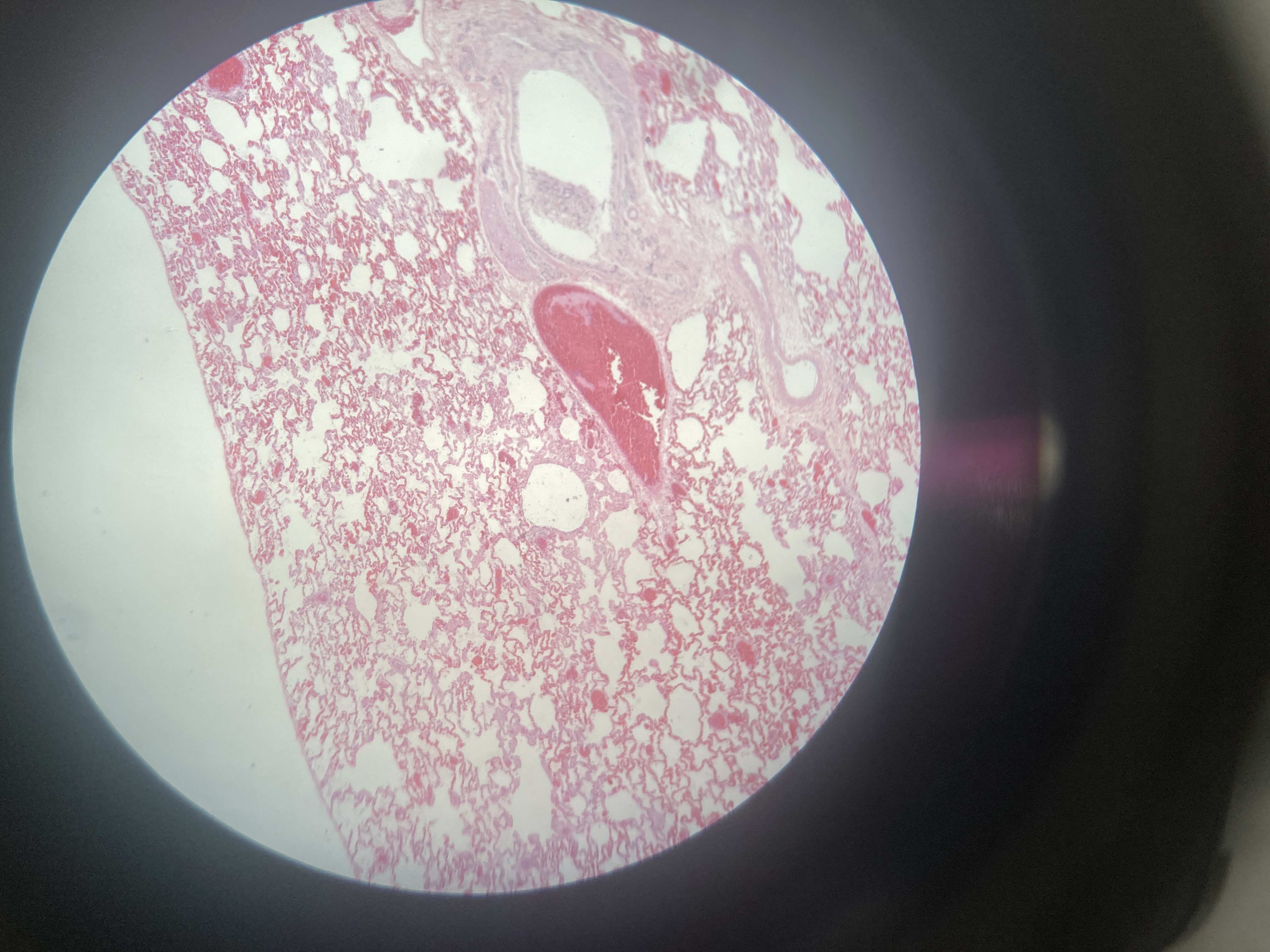
Lung (cat)
Type I pneumocytes
Type II pneumocytes (or septal cells)
Alveolar macrophages (dust cells)
Capillary endothelial cells
Fibroblasts
Smooth muscle cells
Goblet cells
Ciliated epithelial cells
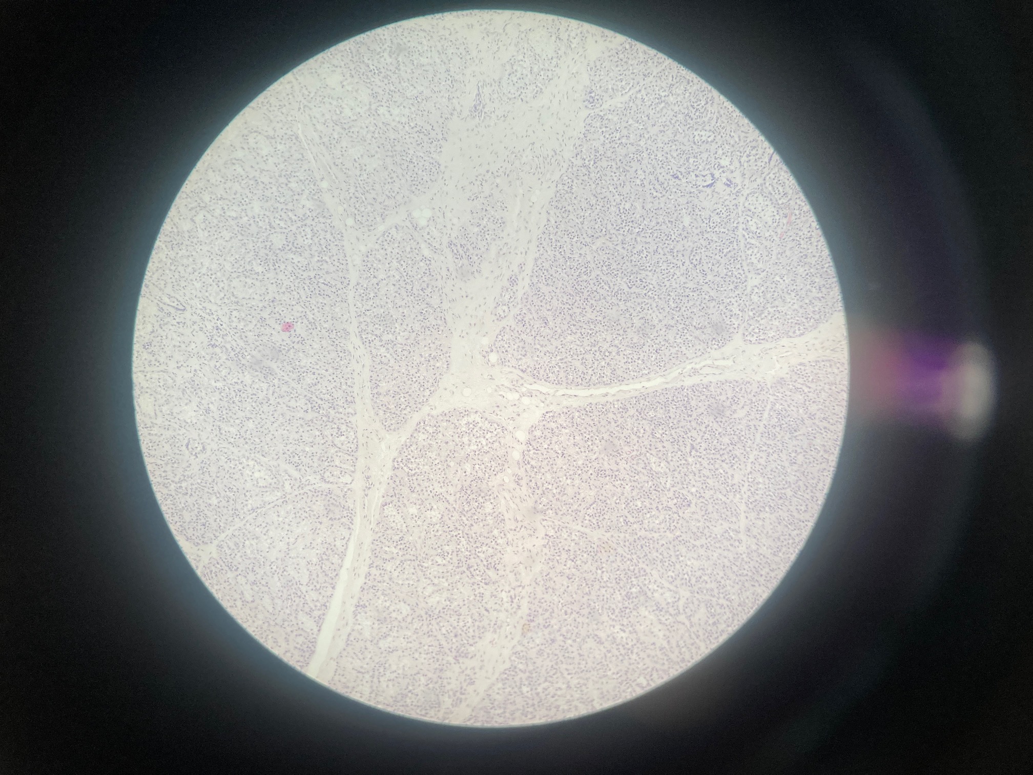
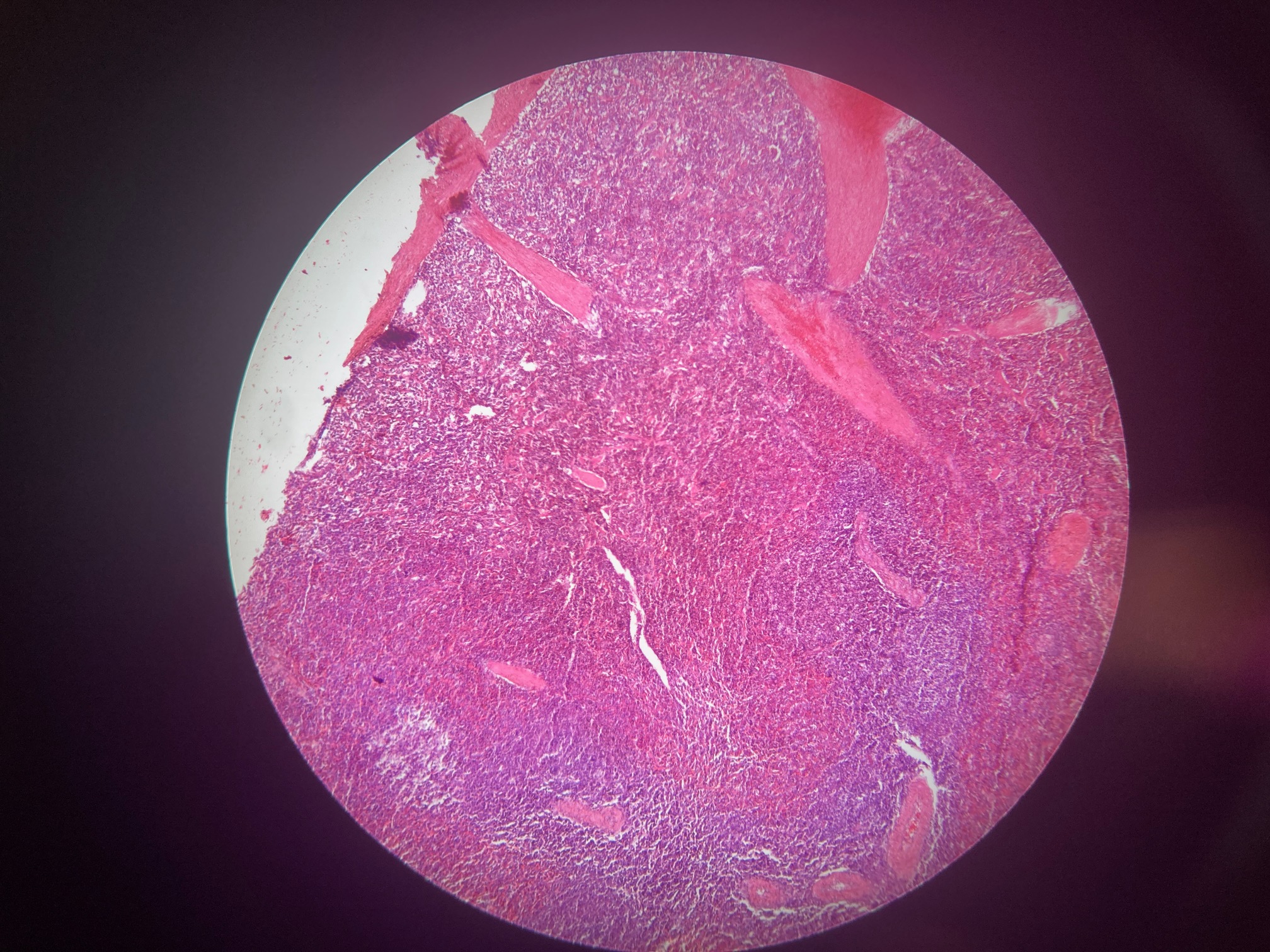
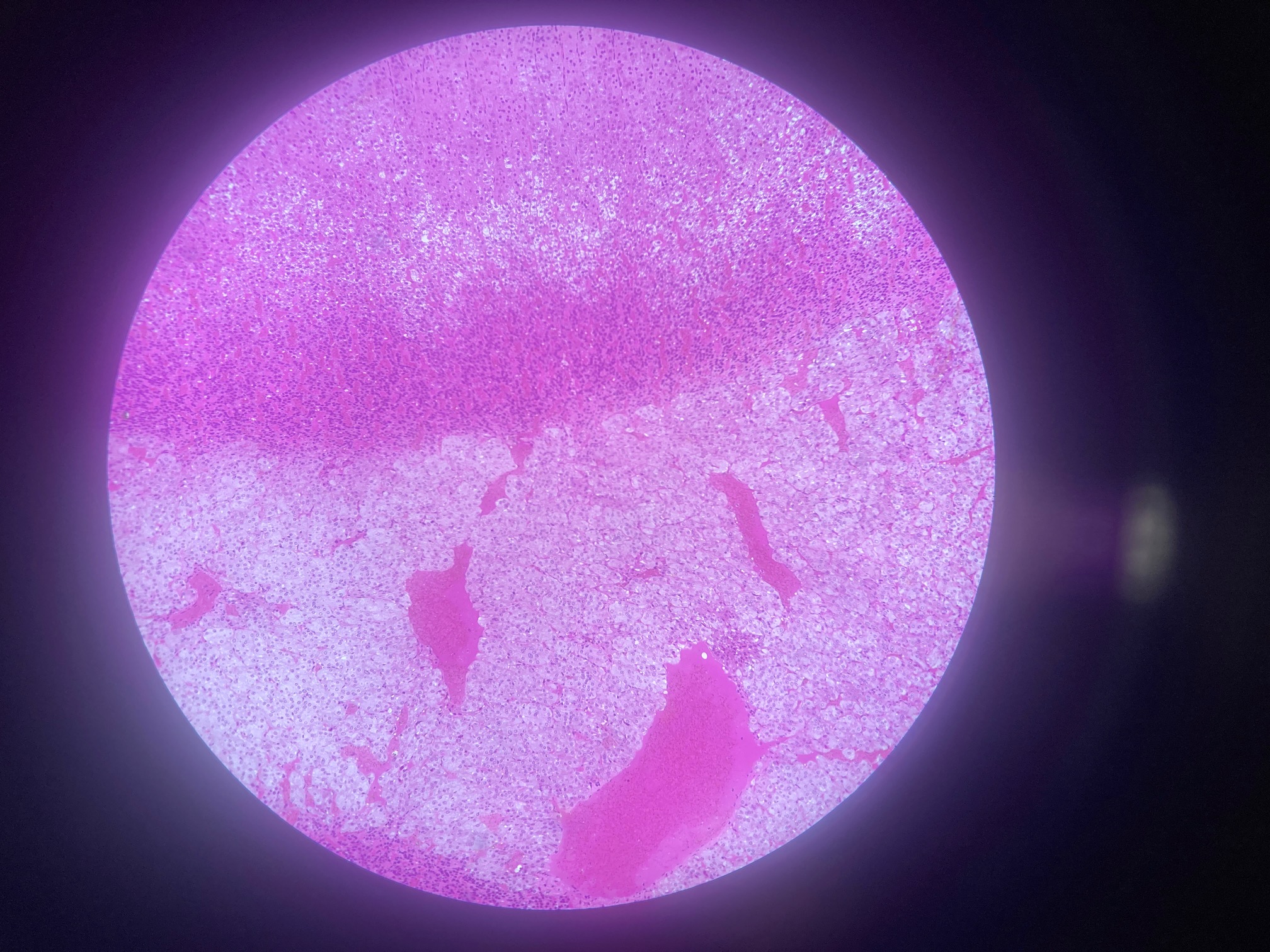
Spleen
Red pulp macrophages
White pulp cells:
Lymphocytes (B / T cells)
Dendritic cells
Macrophages
Periarteriolar lymphoid sheath (PALS) cells
Marginal zone macrophages
B cells in lymphoid follicles
Spleen
Red pulp macrophages
White pulp cells:
Lymphocytes (B / T cells)
Dendritic cells
Macrophages
Periarteriolar lymphoid sheath (PALS) cells
Marginal zone macrophages
B cells in lymphoid follicles

Spleen
Red pulp macrophages
White pulp cells:
Lymphocytes (B / T cells)
Dendritic cells
Macrophages
Periarteriolar lymphoid sheath (PALS) cells
Marginal zone macrophages
B cells in lymphoid follicles
Lymph Node
Lymphocytes (T / B cells)
Macrophages
Dendritic cells
Reticular cells
Plasma cells
Histiocytes
Sinusoidal cells
Mast cells
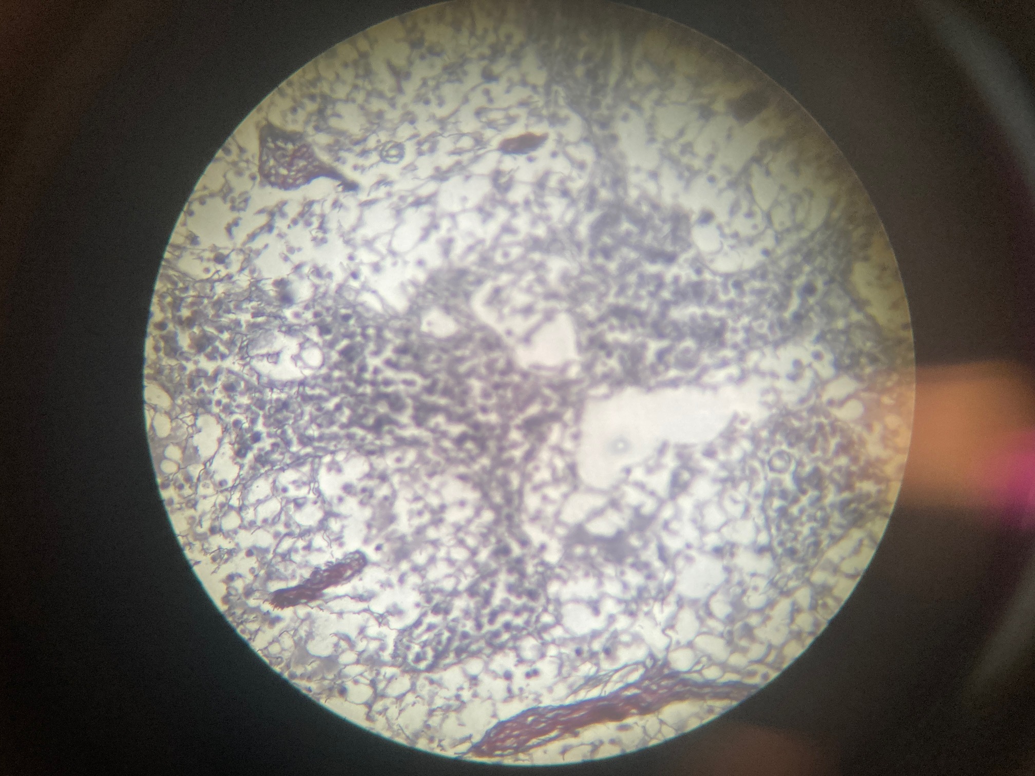
Adrenal gland (monkey)
Adrenal cortex:
Chromaffinrulosa
Zona fasciculata
Zona reticularis
Capsule cells
Adrenal medulla:
Chromaffin cells
Ovary (dog)
Oocyte
Granulosa cells
Theca cells
Luteal cells (corpus luteum)
Surface epithelial cells
Stromal cells
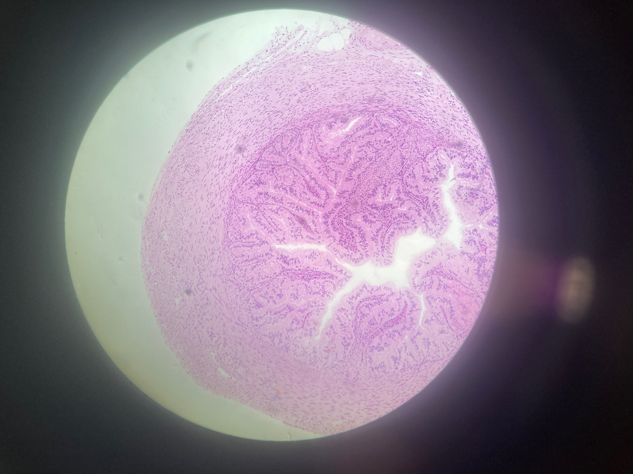
Uterine (monkey)
Endometrial epithelial cells
Endometrial stromal cells
Myometrial smooth muscle cells
Perimetrial cells
Uterine glandular cells
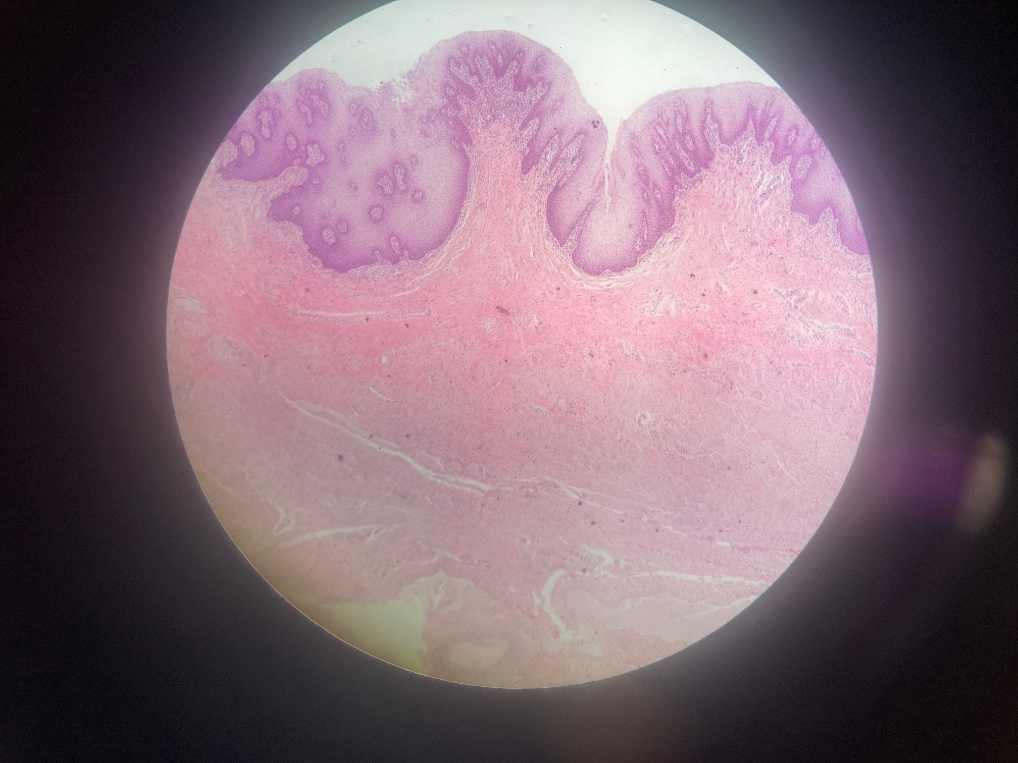
Vagina (human)
Squamous epithelial cells
Intermediate cells
Basal cells
Langerhans cells
Goblet cells
Uterus (dog)
Endometrial epithelial cells
Endometrial stromal cells
Myometrial smooth muscle cells
Perimetrial cells
Uterine glandular cells
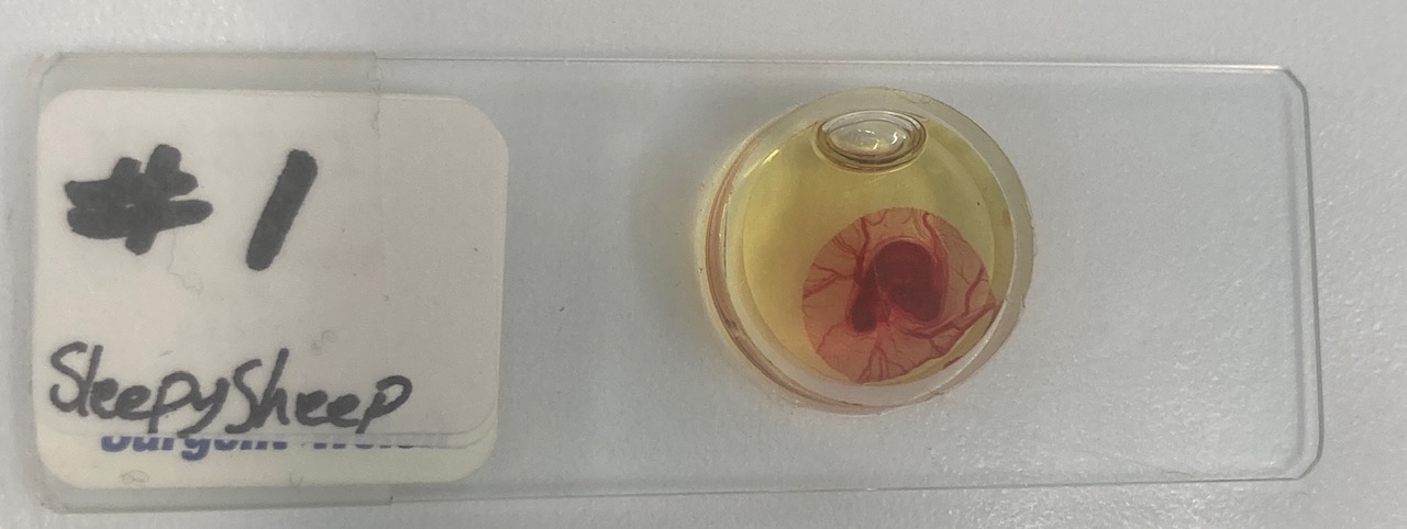
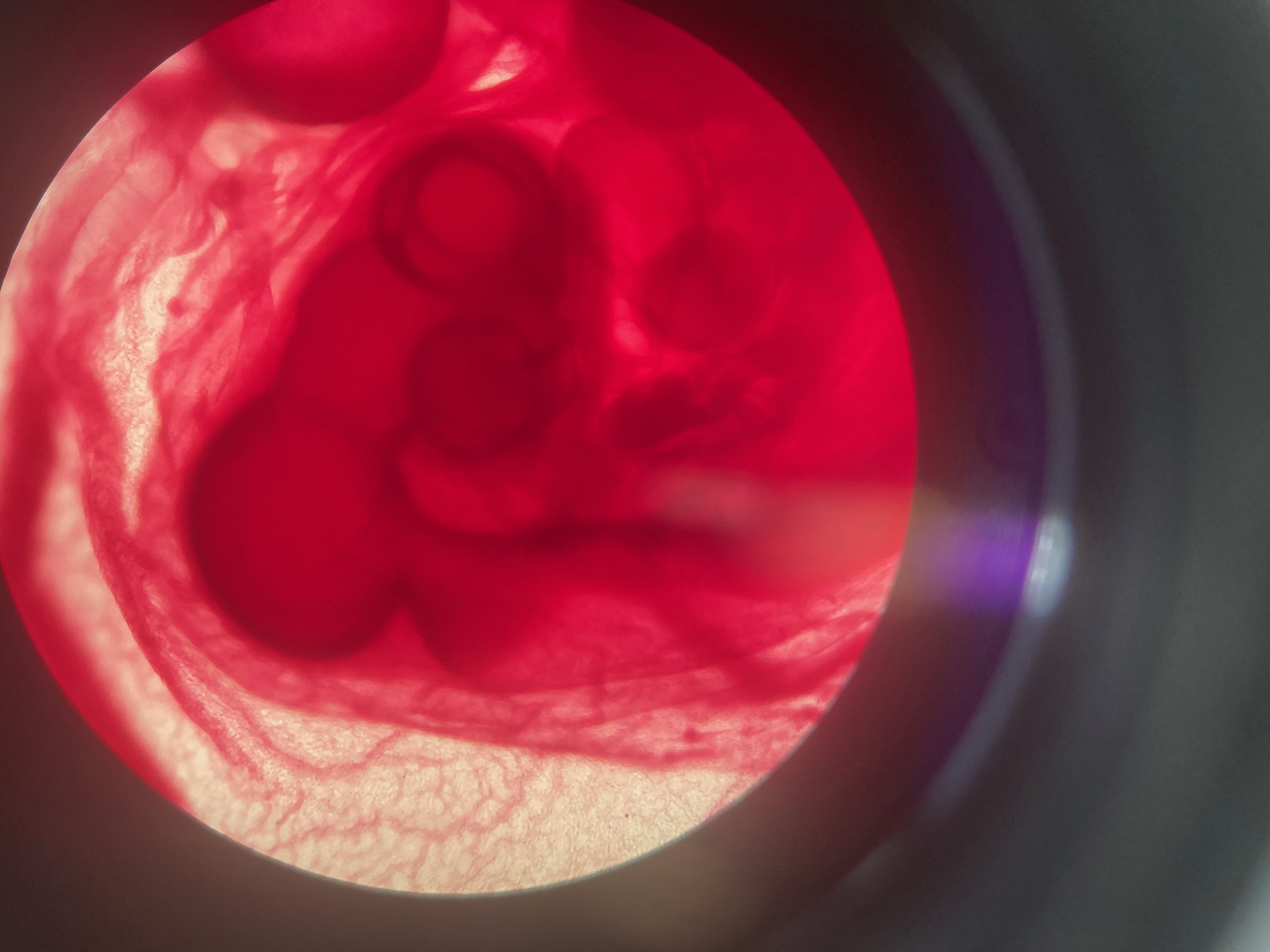
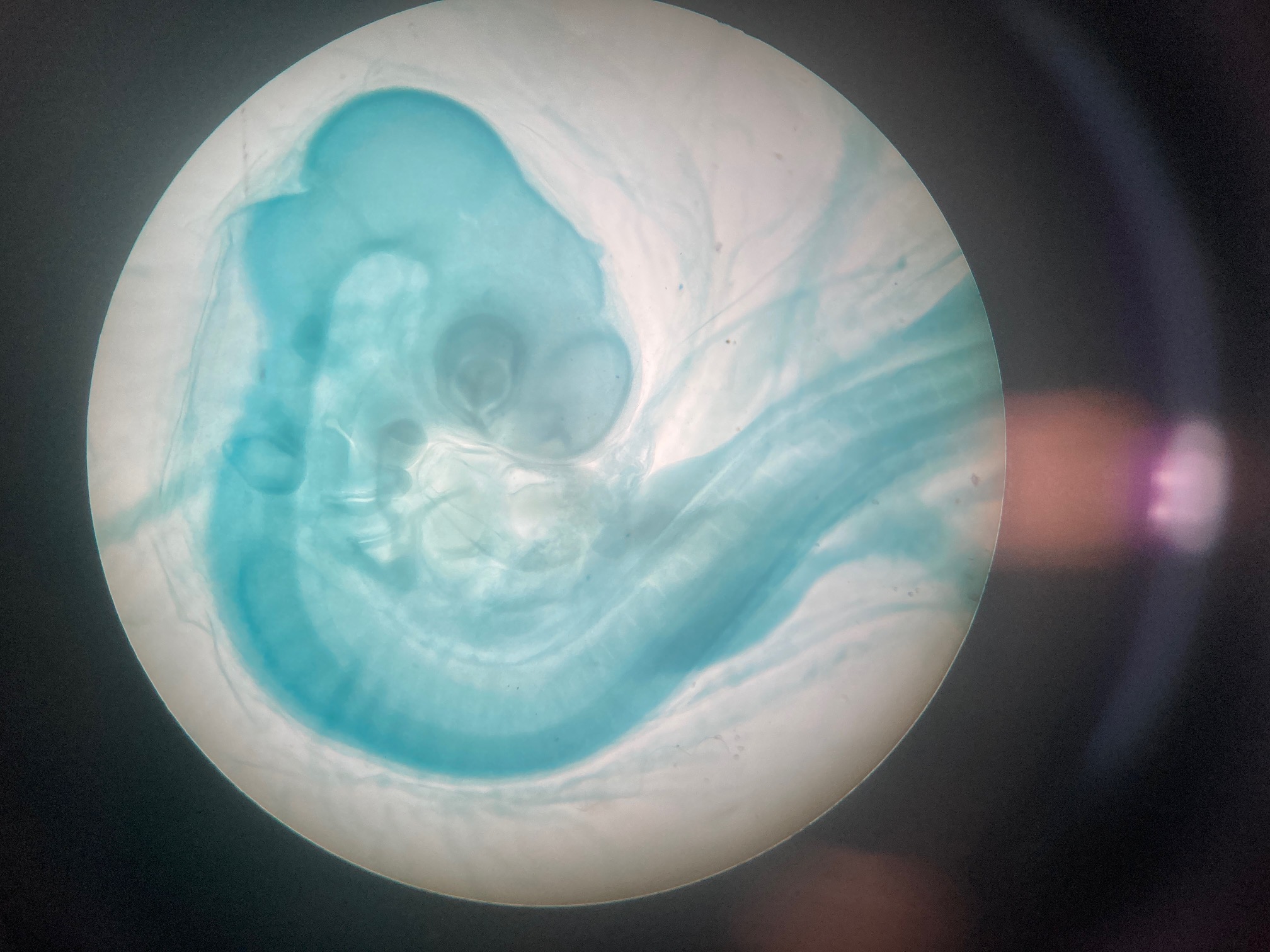
Chicken embryo
Epiblast cells
Hypoblast cells
Mesoderm cells
Endoderm cells
Ectoderm cells
Neural crest cells
Extraembryonic cells
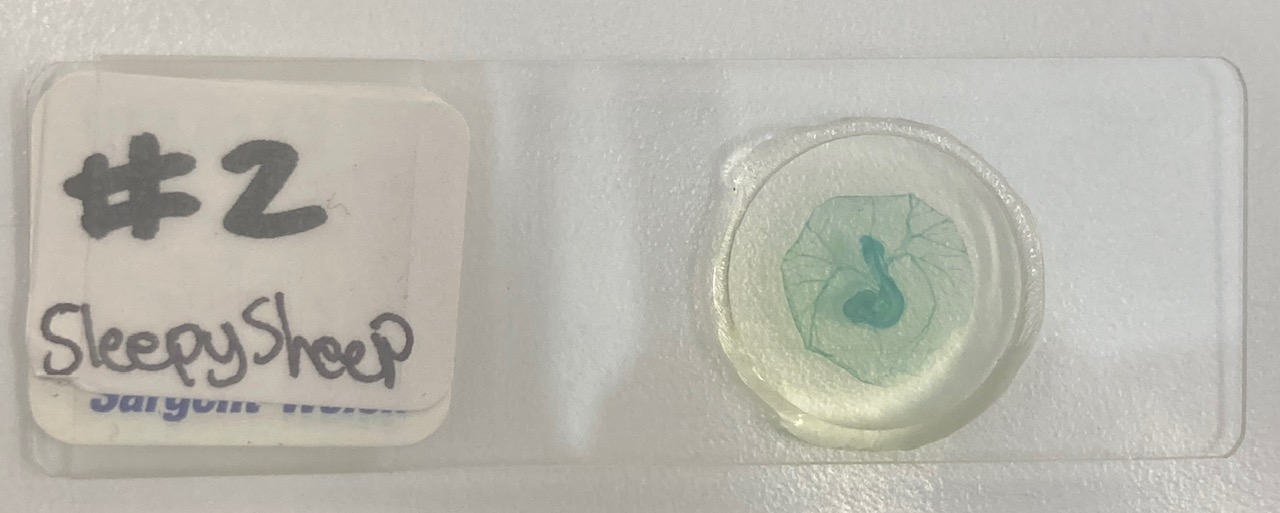
Chicken embryo
Epiblast cells
Hypoblast cells
Mesoderm cells
Endoderm cells
Ectoderm cells
Neural crest cells
Extraembryonic cells
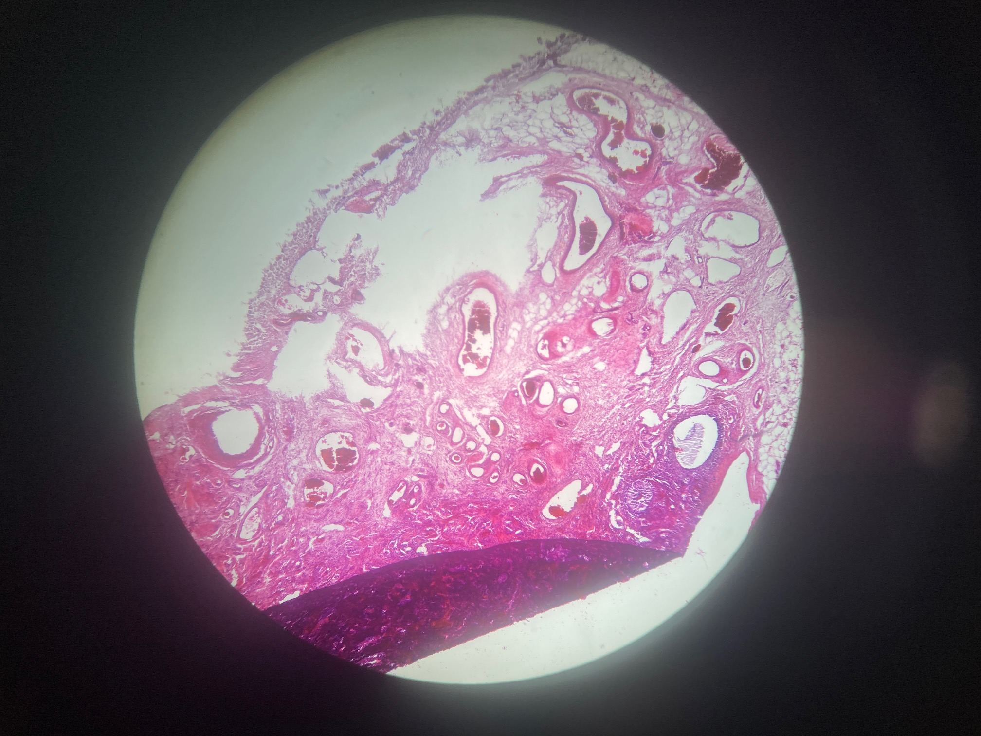
Umbilical cord
Umbilical arteries, vein, urachus
Lactating mammary gland
Myoepithelial cells