Nervous System - Lecture Notes
 Call Kai
 Learn
Learn Practice Test
 Spaced Repetition
 Match
Match Flashcards
 Knowt Play
1/40

There's no tags or description
Looks like no tags are added yet.
Name | Mastery | Learn | Test | Matching | Spaced | Call with Kai | Chat |
|---|
No analytics yet
Send a link to your students to track their progress
41 Terms
Nervous System: Function
Detects impulses from the senses; control center
Primary Functions: Sensory input
Detects stimuli from the environment and internal conditions
Primary Functions: Integration
processes and interprets sensory information
Primary Functions: Motor output
activates muscles and glands in response to stimuli
Nervous System: Major Organs
Brain
Spinal Cord
Senses
Nerves
Major Components: The Brain
Primary control center for processing information
Major Components: Spinal Cord
a conduit for signals between the brain and the body
Major Components: Peripheral nerves
link the CNS to sensory receptors, muscles, and glands
Major Components: Sensory Organs
Include eyes, ears, taste buds, and olfactory receptors
How is the nervous system organized?
Central Nervous System (CNS)
Peripheral Nervous System (PNS)
Cranial Nerves
Spinal Nerves
The peripheral nervous system is then further divded
What are the types of nerve cells?
Neurons - conduct impulses around the body (make up only 10% of the nerve cells)
Neuroglia (nerve glue) - support, insulate & protect neurons (90% of nerve cells)
What is the structure of a neuron
Neurons have different structures, but have a main cell body with processes extending outward
The cell body contains the nucleus, cytoplasm & organelles
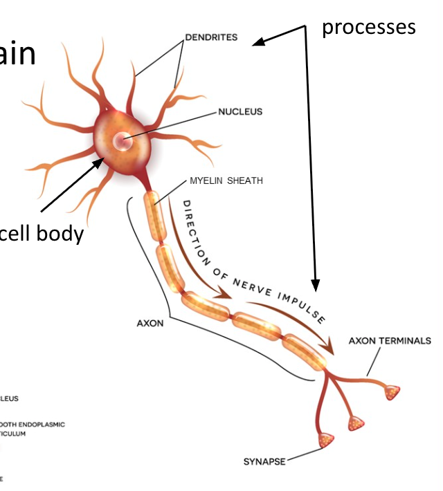
Processes are divided into:
Dendrites
Axons
At the end of axons are axon terminals, which release neurotransmitters to pass impulses to the next neuron
How is a nerve impulse passed along a neuron?
A nerve impulse is an electrical signal that travels along the neuron
It arises from the movement of ions, causing a change in electrical charges
Key principle: Nerve impulses are all-or-none; the neuron either fires completely or not at all
Electrical Signaling: Resting Potential
The neuron’s membrane is polarized
Intracellular space is negatively charged
Extracellular space is positively charged
Electrical Signaling: Depolarization (action potential)
When the neuron is stimulated, either by the environment or another neuron:
Sodium (Na+) rushes into the neuron and reverses the charges
Electrical Signaling: Repolarization
As impulses passes
Potassium (K+) flows out, restoring resting potential
Electrical Signaling: Sodium-potassium pump
Actively restores the ion concentrations to normal, and the resting potential returns
Synaptic Transmission
Neurons communicate at synapses, where the axon terminal of one neuron meets the dendrite or soma of another
Neurotransmitters transmit signals across the synaptic cleft, enabling communication between neurons
Synaptic Transmission Steps:
Action potential reaches the presynaptic terminal
Voltage-gated Ca2+ channels open
Neurotransmitters are released from synaptic vesicles
Neurotransmitters bind to postsynaptic receptors
Sodium channels open, triggering an action potential in the next neuron
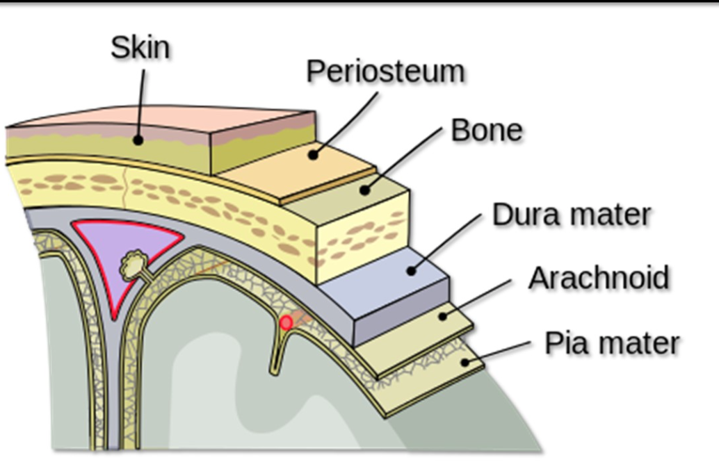
Protection of the Central Nervous System
Three connective tissue layers surround the brain and spinal cord:
Dura mater: Tough, outermost layer
Arachnoid mater: Thin, web-like middle layer
Pia mater: Delicate inner layer, highly vascularized
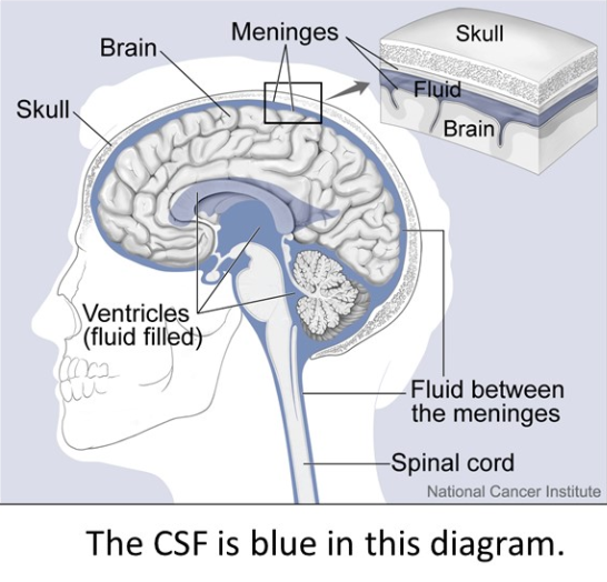
Cerebrospinal Fluid (CSF)
Found in the subarachnoid space (between the arachnoid layer and the pia mater)
Cushions the CNS, maintains homeostasis, and prevents infection
Produced by choroid plexuses in the ventricles
Lateral ventricles (right and left) → Third ventricle → Fourth ventricle.
Reabsorbed into the bloodstream via arachnoid granulations.
How is the Cerebrospinal Fluid produced
The CSF is produced in spaces within the brain called ventricles.
There are 4 ventricles: 2 lateral ventricles (right & left), third ventricle & fourth ventricle.
The lateral ventricles are connected to the third ventricle by the thin interventricular foramen (foramen of Monro).
Structure of the Brain: Cerebrum
Largest brain region, divided into right and left hemispheres connected by the corpus callosum
The surface is covered by
Gyri: ridges
Sulci: Shallow grooves
Fissures: Deep grooves separating lobes
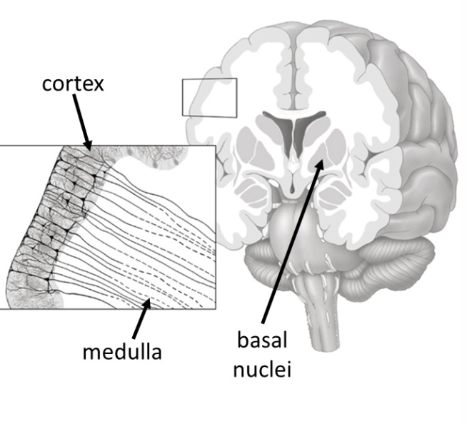
3 Major Layers of the Brain
From Superficial to Deep
Cerebral Cortex: gray matter; cell bodies, dendrites; processes information
Cerebral Medulla: white matter; myelinated axons; transmits signals
Basal Nuclei: Control voluntary movements and motor planning
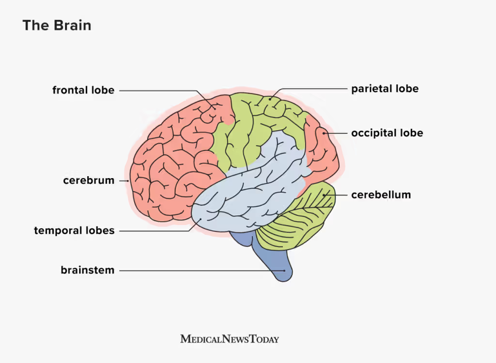
Lobes of the cerebrum: Frontal Lobe
Controls:
Voluntary movements (walking)
Reasoning & decision-making
Memory
Ability to predict consequences of actions
Planning
Verbal communication, specifically in Broca’s area
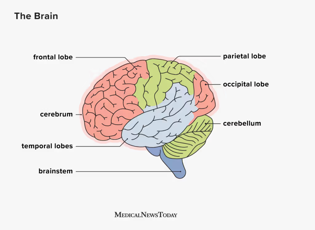
Lobes of the cerebrum: Parietal Lobe
Separated from the frontal lobe by the central sulcus
Controls:
Sensations (pain, temperature, touch)
Visual-spatial processing
Body position
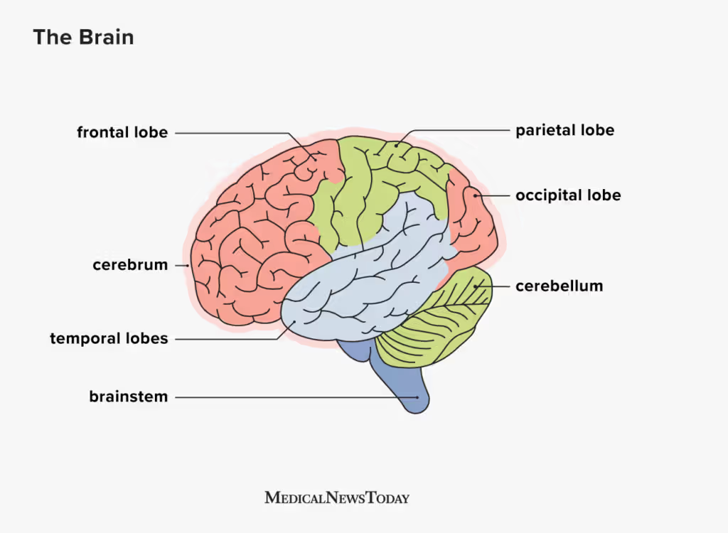
Lobes of the cerebrum: Occipital Lobe
Controls:
Visual processing
Memory of objects
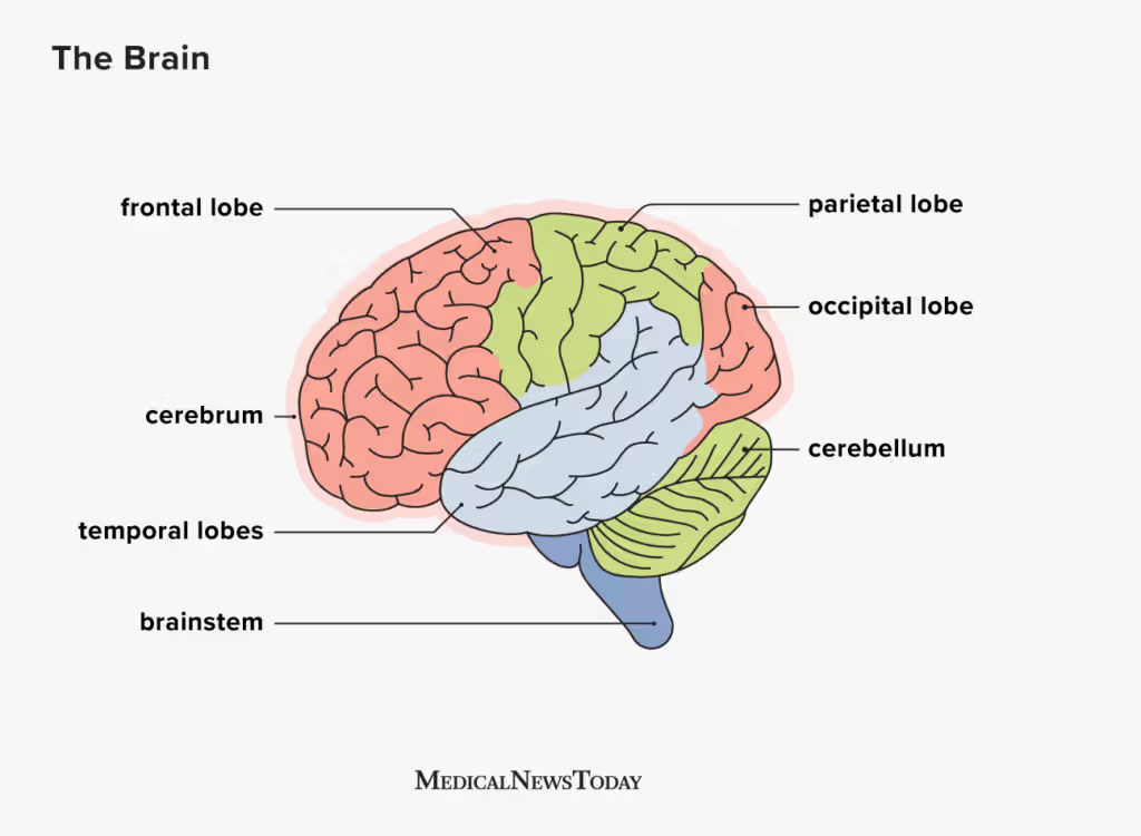
Lobes of the cerebrum: Temporal Lobe
Separated from the frontal lobe by the lateral sulcus
Controls:
Memory
Comprehension & pronunciation of works
Sensations of smell and sound
Emotional association of memories
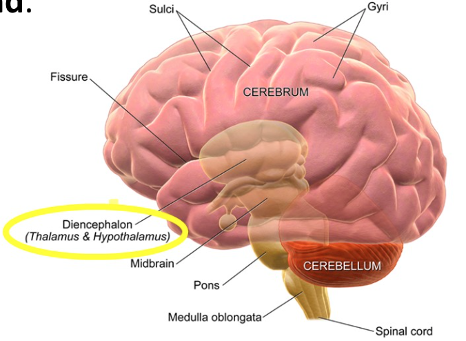
Structure of Diencephalon
Located beneath the cerebrum, small nondescript region
It contains the thalamus, the hypothalamus, and the pineal gland
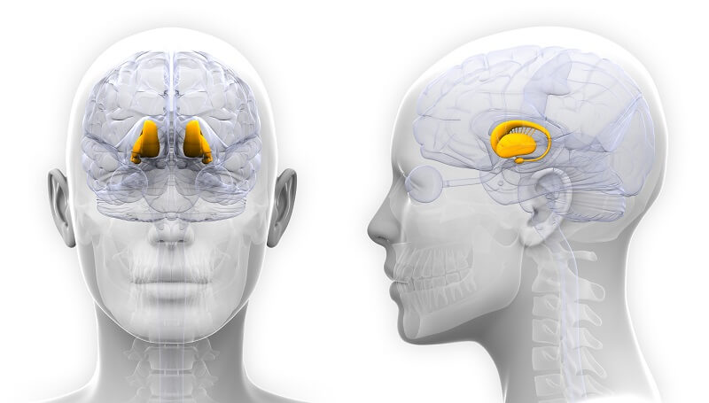
Diencephalon: The Thalamus
Relay center for sensory information to the cortex
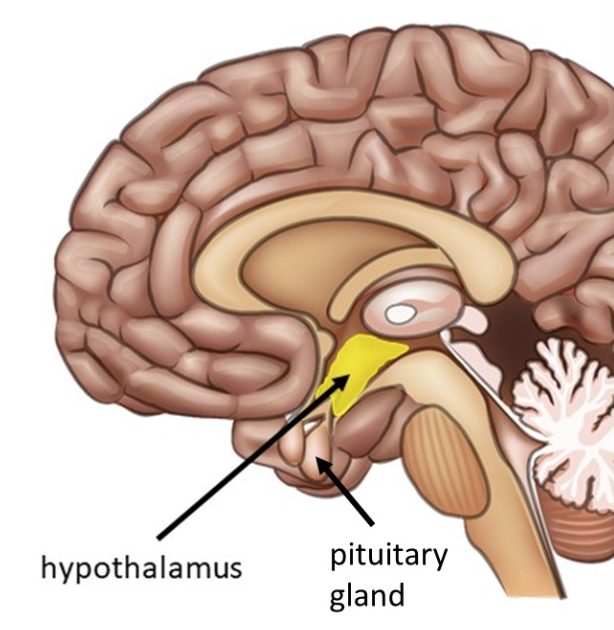
Diencephalon: The Hypothalamus
Controls:
Autonomic process (temperature, blood pressure, metabolism)
Controls hormones released by the pituitary gland
Part of the limbic system, regulates emotions and feelings of pleasure (hunger, thirst, sex, pain)
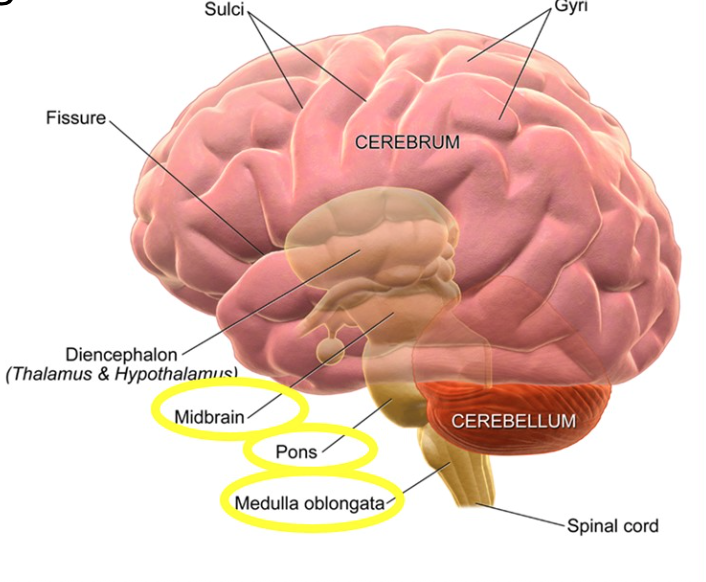
Brain Stem Structure
Located between the cerebrum and the spinal cord
It is broken into three regions: the midbrain, pons, and medulla oblongata
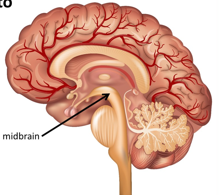
Brain Stem: The Midbrain
Also known as the mesencephalon
Relays information to cerebrum
Controls body movements and posture
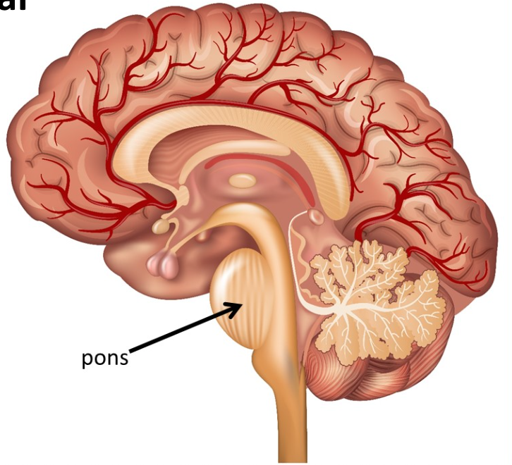
Brain Stem: The Pons
Almost completely made of white matter that links the cerebral cortex and the cerebellum
Carries information from one side of the brain to the other
Central control of breathing
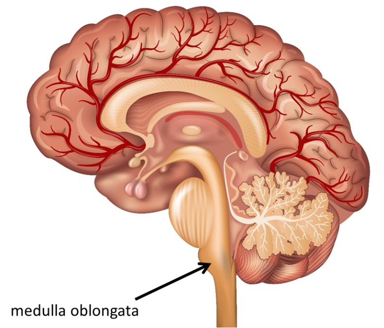
Brain Stem: The Medulla
The medulla oblongata is located just above the spinal cord.
Transmits impulses between the spinal cord and the brain.
Controls blood pressure, heart rate, swallowing, and coughing
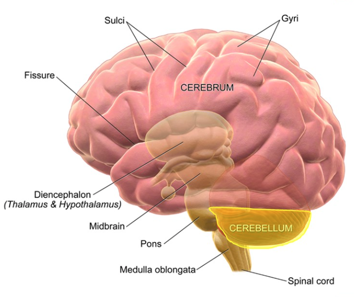
Function of the Cerebellum
Found near the back of the skull; its the second largest part of the brain
Coordinates voluntary movement for smooth, efficient motion.
Involved in motor learning and muscle memory.
Ensures movements are precise and balanced.
Structure of the Spinal Cord
Extends from the medulla oblongata to the lumbar vertebrae
Protected by meninges and the vertebral column
Contains 31 pairs of spinal nerves
Gray matter forms a “butterfly” shape, surrounded by white matter
Function of the Spinal Cord
The dorsal horns receive impulses from afferent nerves, then pass the impulses through the ventral horns to the efferent nerves
Dorsal and ventral roots merge to form spinal nerves
How does the Reflex Arc travel?
Reflex is a rapid, involuntary response to a stimulus
Pathway (reflex arc):
1. Receptor
2. Sensory neuron
3. Integration Center (CNS)
4. Motor neuron
5. Effector
Types of Reflex Arcs
Somatic reflexes: Involve skeletal muscles.
Autonomic reflexes: Involve smooth or cardiac muscle and glands