MRI Brain Pathology
 Call Kai
 Learn
Learn Practice Test
 Spaced Repetition
 Match
Match Flashcards
 Knowt Play
1/112

Earn XP
Description and Tags
From Brain Metastases to Pituitary Iron Overload
Name | Mastery | Learn | Test | Matching | Spaced | Call with Kai | Chat |
|---|
No analytics yet
Send a link to your students to track their progress
113 Terms
Brain Metastases
The spreading of a primary tumor to the brain.
Symptoms of metastases
vision, hearing, coordination, confusion, speech, nausea, vomiting, numbness, personality changes, weakness, seizures, fever
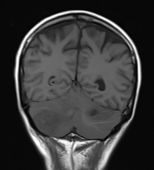
T1-weighted images (T1WI) of Brain meastases
appears hypointense, signal intensity is generally lower than that of surrounding normal tissues. Hypo intensity caused by increased cellularity and compactness of cancerous tissue in comparison to the brain parenchyma (functional substance)
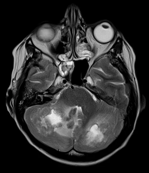
T2-weighted images (T2WI) of Brain metastases
may vary, often appearing hyperintense due to their relatively high water content. Hyperintensity is caused by the increased extracellular space and edema associated with the tumor.
Fluid-Attenuated Inversion Recovery (FLAIR) of Brain metastases
typically appear hyperintense due to increased water content surrounding the tumor. Effectively suppresses the cerebrospinal fluid signal, enhancing peritumoral edema.
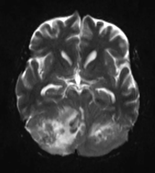
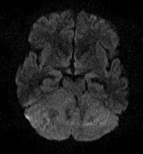
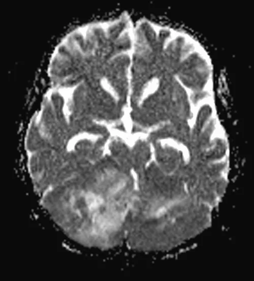
Diffusion-Weighted Imaging (DWI) of Brain Metastases
Lesions can show variable signal intensity on DWI depending on cellularity and restricted diffusion. May appear hyperintense due to reduced diffusion caused by increased cellularity and cellular membrane structures.
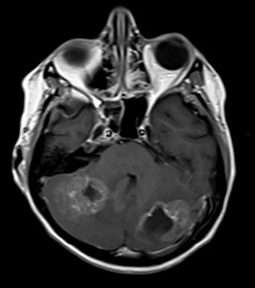
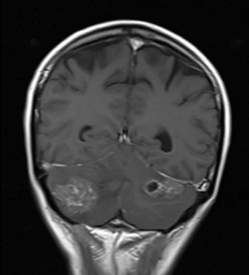
Post-Contrast T1-weighted images of Brain Metastases
After contrast administration (gadolinium-based) metastatic lesions typically enhance. Enhancements can vary due to permeability of blood vessels within the tumor.
T2 of Brain metastasis axial
T1 post contrast of Brain Metastasis axial
T1 of Brain Metastasis coronal
DWI b0 of Brain metastasis axial
DWI b1000 of Brain Metasiasis axial
ADC of Brain Metastasis axial
T1 post contrast of Brain Metastasis coronal
Pituitary Adenomas
non-cancerous tumors that develop in the pituitary gland
Pituitary adenoma classification
Functioning adenoma - produce excessive amounts of hormones, prolactinomas, somatotropinomas (growth hormones), corticotropinomas (ACTH leading to Cushing’s disease), and throtropinomas (thyroid)
Non-Functioning adenoma - Don’t produce significant amounts of hormones but can cause symptoms due to size.
as well as micro and macro adenomas
T1-weighted images (T1WI) of Pituitary adenomas
typically appear as iso- (same as) or hypointense compared to gray matter due to relatively higher fat content and variable protein content.
T2- weighted image (T2WI) of Pituitary adenomas
usually appear hyperintense due to high water content. Signal may vary based on tumors consistency and cystic components.
Post-Contrast T1-weighted image of Pituitary adenomas
After contrast administration (gadolinium-based) adenomas can enhance. enhancement can vary from rim to heterogenous (entire)
T1 of Pituitary Adenoma sagittal
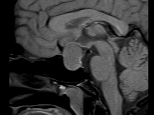
T1 of Pituitary Adenoma coronal
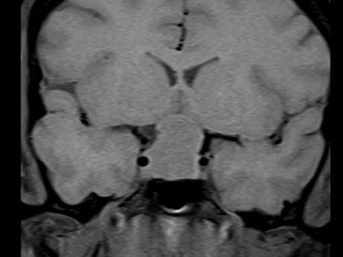
T2 of Pituitary Adenoma coronal
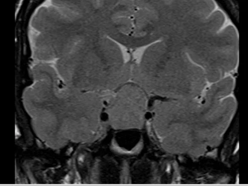
T1 post contrast of Pituitary Adenoma sagittal
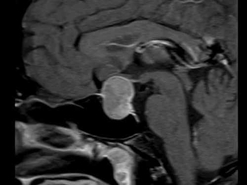
T1 post contrast of Pituitary Adenoma coronal
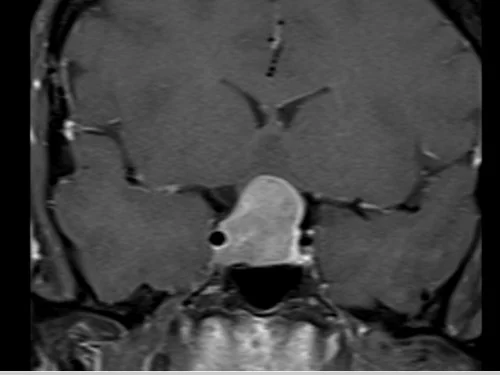
Subdural Hemorrhage
subdural hematoma, bleeding between the brain and the dura mater.
Subdural Hemorrhage symptoms
persistent headaches that don’t go away with medication, changes in behavior, difficulty speaking or moving, and even seizures
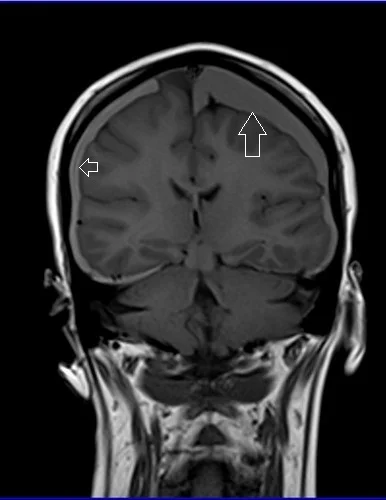
T1-weighted images (T1WI) of Subdural hemorrhage
may vary in levels of brightness, ranging from hyperintense to isointense or even hypointense, depending on the stage of the hemorrhage (acute, subacute, or chronic)
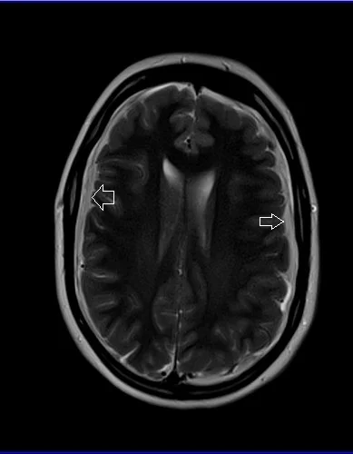
T2-weighted images (T2WI) of subdural hemorrhage
differing degrees of brightness appearing hyperintense or hypointense based on the stage
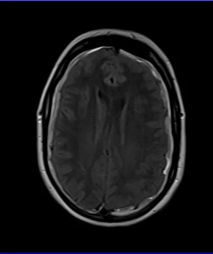
Fluid-Attenuated Inversion Recovery (FLAIR) of Subdural hemorrhage
to suppress the signal from the CSF flow, which enhances visibility of the lesions. showing up either hyer or hypointense, with each stage.
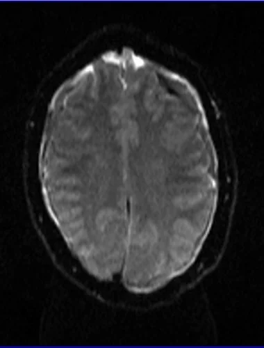
Diffusion-Weighted Image (DWI) of Subdural hemorrhage
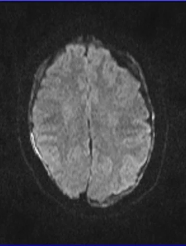
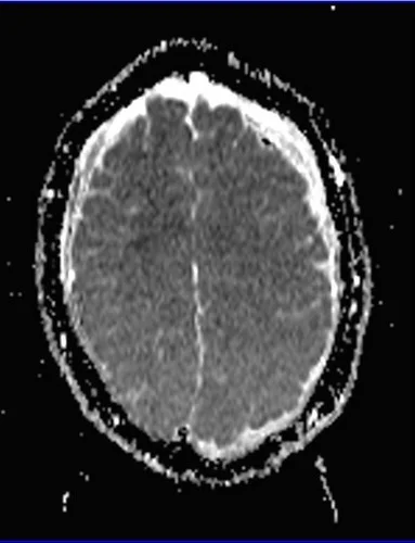
presents as hypointense, blood products hinder the movement of water molecules, leading to reduced signal intensity
Susceptibility-Weighted image (SWI) of Subdural hemorrhage
attuned to magnetic properties of blood products, especially those containing iron. Hypointense due to presence of deoxyhemoglobin and breakdown products of hemosiderin (insoluble protein containing iron)
T2 of Subdural hemorrhage axial
FLAIR of Subdural hemorrhage axial
T1 of Subdural hemorrhage coronal
DWI b0 of Subdural hemorrhage axial
DWI b1000 of Subdural hemorrhage axial
DWI ADC of Subdural hemorrhage axial
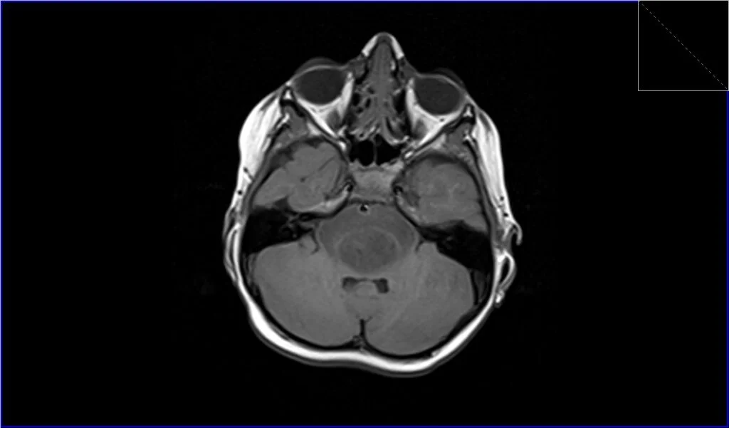
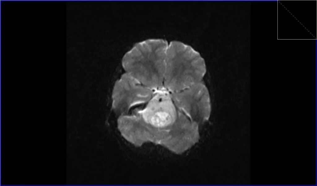
Pontine Glioma
AKA (DIPG) is a type of brain tumors that occurs in the pons. Primarily affects children, between 5-10. Aggressive nature and poor prognosis.
DIPG symptoms
Problems with eye movement and facial expressions, weakness in the arms and legs, difficulty in walking and coordination, changes in speech, headaches and nausea.
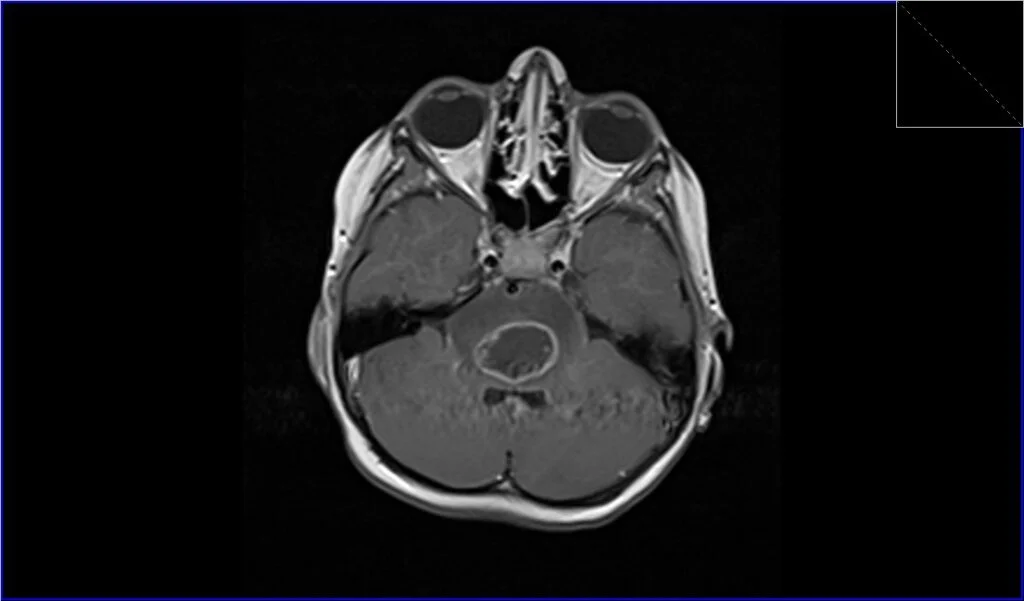
T1-weighted image (T1WI) of Pontine glioma
appear hypointense or iso intense.
T2-weighted image (T2WI) of Pontine glioma
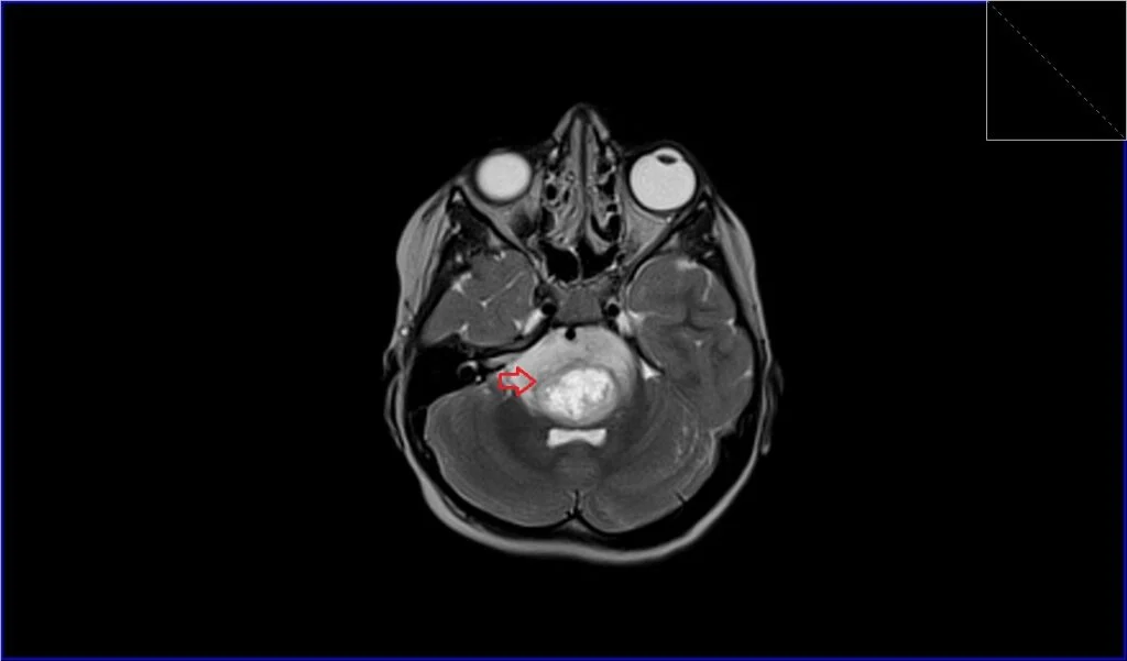
appear hyperintense due to higher water content
Fluid-Attenuated Inversion Recovery (FLAIR) of Pontine glioma
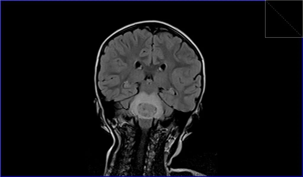
appear hyperintense, suppresses the signal from CSF allowing better visualization.
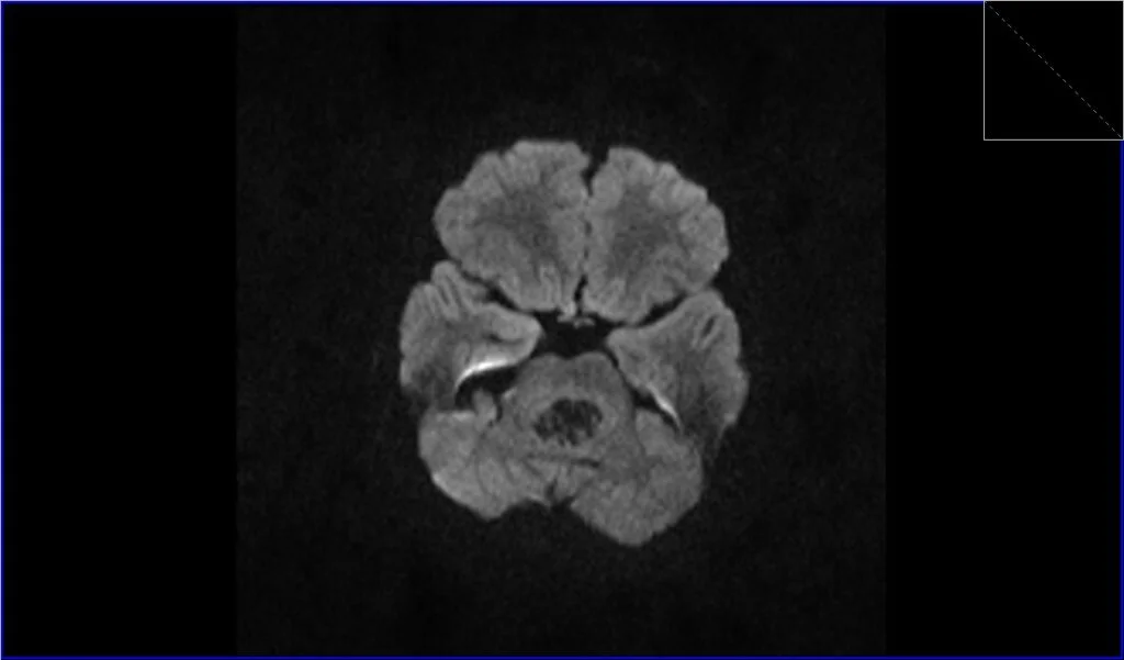
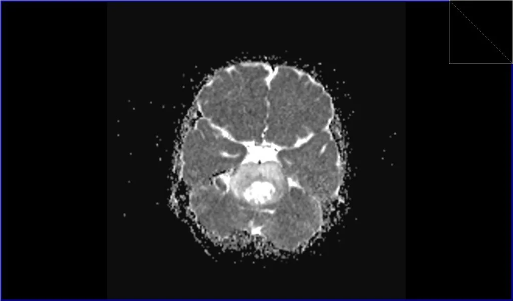
Diffusion Weighted Imaging (DWI) of Pontine glioma
may vary
Post-Contrast T1-weighted of Pontine glioma
generally does not enhance uniformly, heterogeneous (some does some don’t) nature
T2 of Pontine Glioma axial
FLAIR of Pontine Glioma
T1 of Pontine Glioma axial
DWI b0 of Pontine Glioma axial
DWI b1000 of Pontine Glioma axial
DWI ADC of Pontine Glioma axial
T1 post contrast of Pontine Glioma (shows medulloblastoma) axial
Herpes simplex encephalitis
serious and potentially life-threatening condition characterized by inflammation of the brain caused by HSV. relatively rare.
HSE symptoms
Fever, headache, neurological issues (confusion, seizures, personality changes, or hallucinations), sensitivity to light, stiff neck and back, and altered consciousness
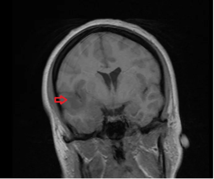
T1WI of HSE
slight hypointensity or normal signal in affected area for early stages. Later stages show pronounced hypointensity due to destruction and necrosis.
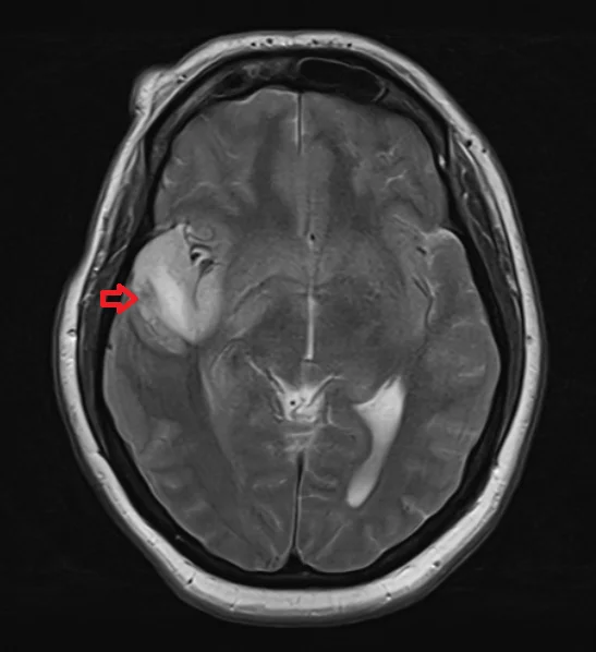
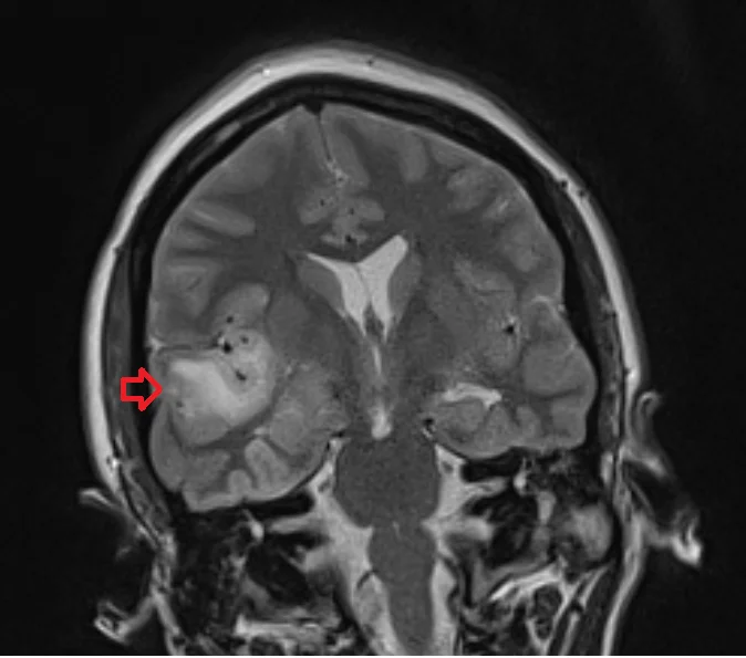
T2WI of HSE
show hyperintense signals in areas of inflammation and edema
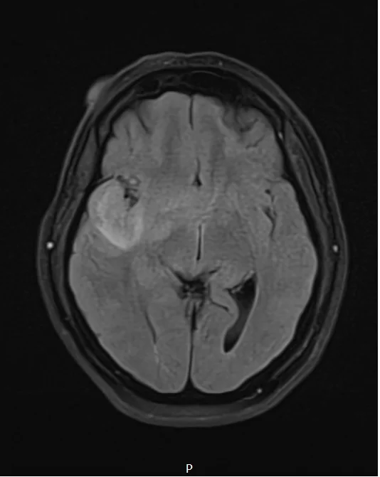
FLAIR of HSE
show hyperintense signals of inflammation and edema especially in white matter and cortical regions.
DWI of HSE
(b0, b1000) hyperintense signals in affected areas, indicating restricted diffusion due to cytotoxic edema
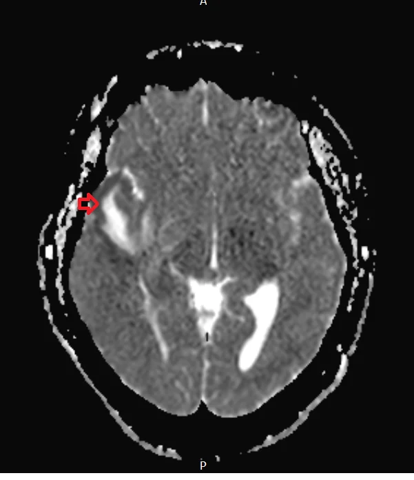
ADC of HSE
appear the opposite of DWI reading, hypointense confirming restricted diffusion.
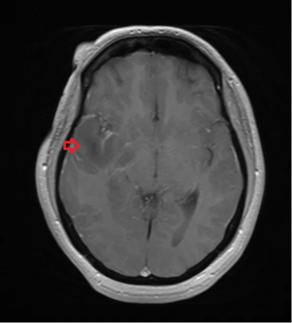
Post contrast T1 of HSE
meningeal (layer of dura mater) enhancement due to inflammation
T2 of HSE axial
FLAIR of HSE axial
T1 of HSE coronal
T2 of HSE sagittal
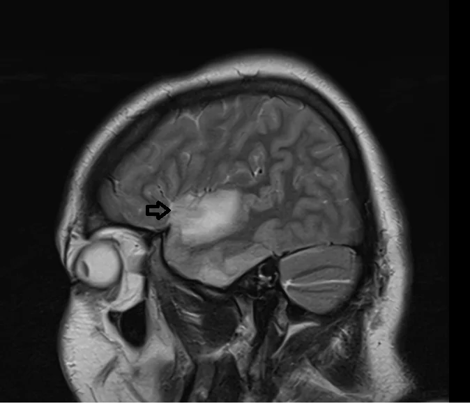
T2 of HSE coronal
DWI b0

DWI b1000 of HSE

DWI ADC of HSE
T1 post contrast of HSE axial
T1 post contrast of HSE coronal
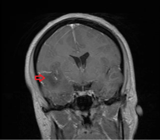
Dysplastic cerebellar gangliocytoma (Lhermitte-Duclos disease) LDD
rare, slow-growing, benign brain tumor that predominantly affects the cerebellum.
LDD symptoms
Headaches, nausea and vomiting, visual disturbances, ataxia (difficulty with coordination and balance), dizziness, tinnitus or hearing loss, cognitive impairments.
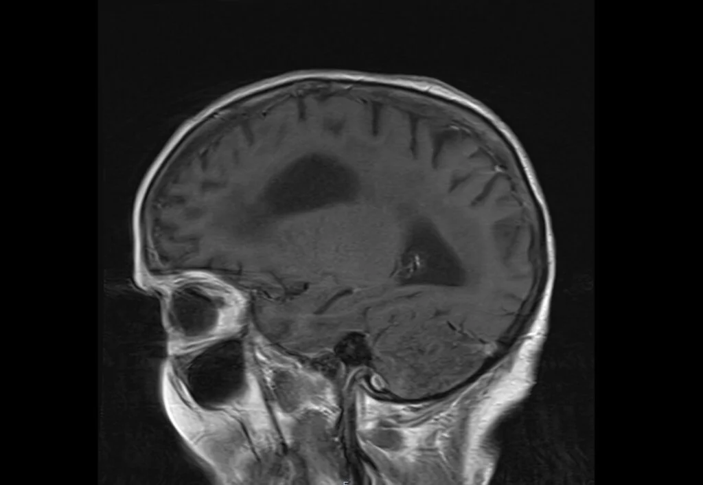
T1WI of LDD
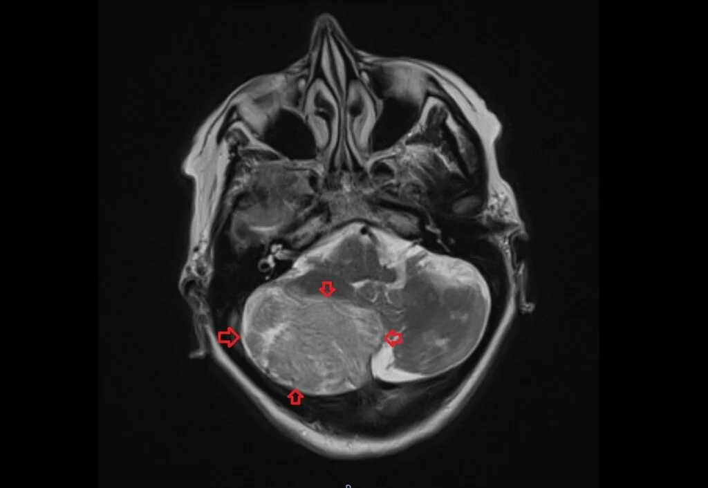
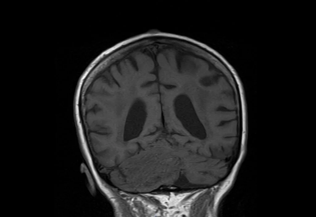
hypointense or isointense. reflects the dense fibrous tissue and abnormally large ganglion cells replacing the normal cerebellar architecture.
T1WI of LDD
striated appearance, alternating bands of high and low signal intensity. high signal correspond to myxoid changes and abnormal ganglion cells, whereas the low signal areas reflect fibrous bands.
FLAIR of LDD
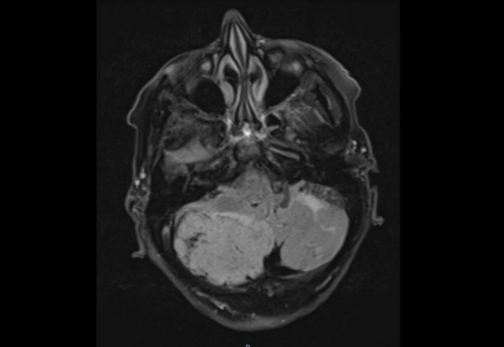
hyperintense with same striped pattern on T2WI. FLAIR suppresses CSF.
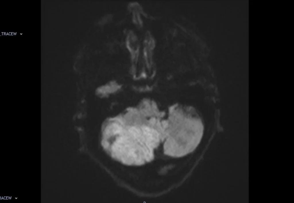
DWI of LDD
no restricted diffusion, lack of acute cell death or high cellularity often seen in more aggressive tumors.
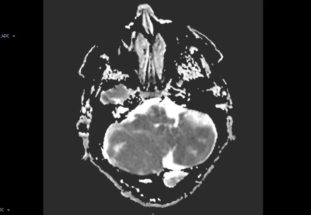
ADC of LDD
lesion usually exhibits increased diffusion, reflecting the non-restrictive, expanded extracellular space characteristic of the tumor.
Post-Contrast T1 of LDD
show no significant enhancement, helping differentiate from the neoplastic lesions that may enhance.
T2 image of LDD axial
FLAIR of LDD axial
T1 of LDD coronal
T2 of LDD sagittal
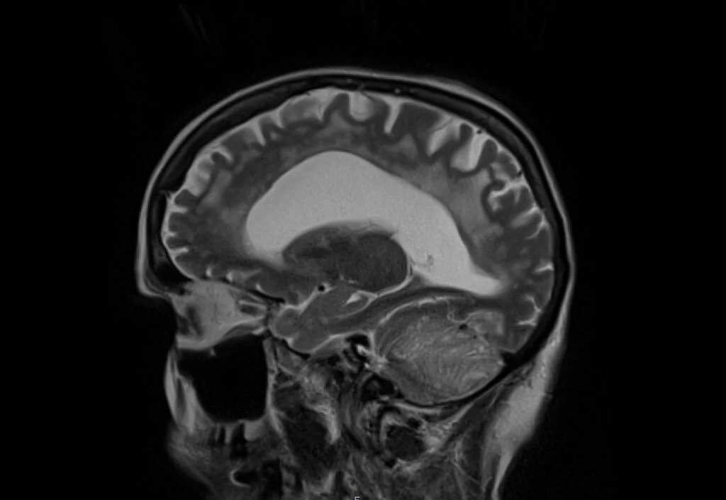
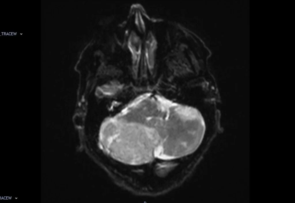
DWI b0 of LDD axial
DWI b1000 of LDD axial
DWI ADC of LDD axial
T1 post contrast of LDD axial
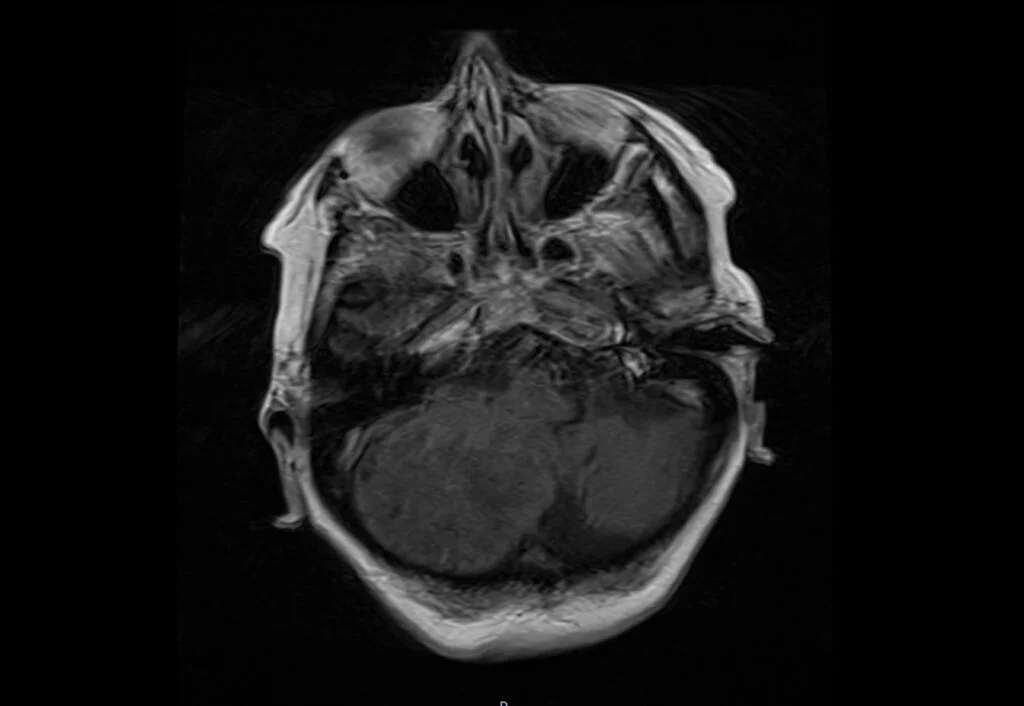
T1 post contrast of LDD sagittal
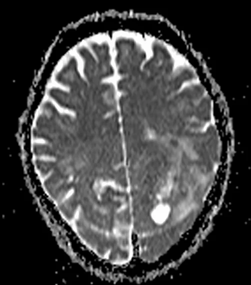
Glioblastomas
aggressive and highly invasive primary brain tumors. originate from glial cells, particularly astrocytes. Most common malignant brain tumors in adults.
Glioblastomas symptoms
nonspecific symptoms, headaches, seizures, cognitive decline, and focal neurological deficits.
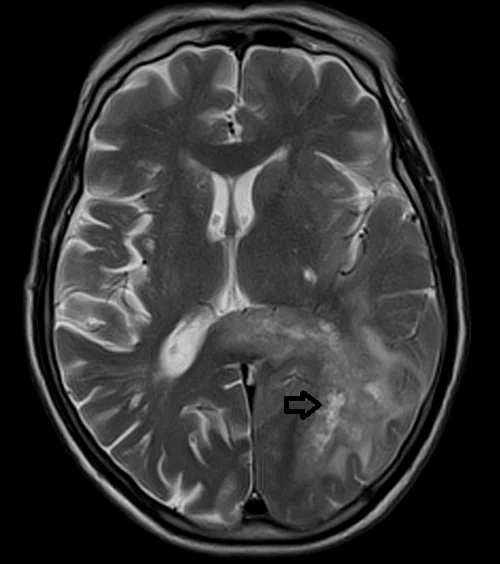
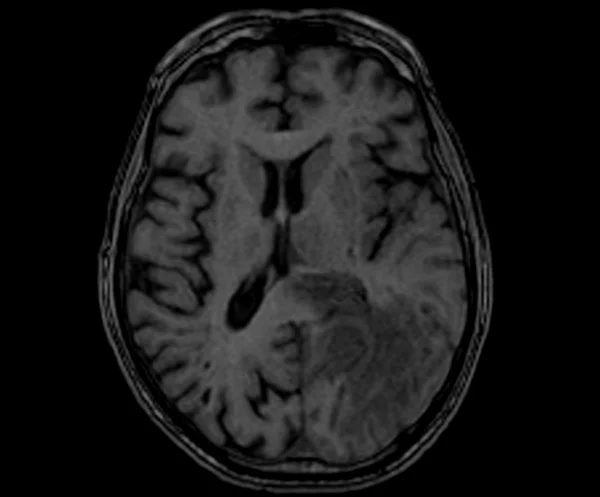
T2WI of Glioblastomas
hypointense due to high cellularity and presence of necrotic regions.
T2WI of Glioblastoma
hyperintense due to high water content, presence of necrosis, and edema.
FLAIR of Glioblastomas
appear hyperintense due to high water content, presence of necrosis, and edema. indicates the presence of extensive peritumoral edema.
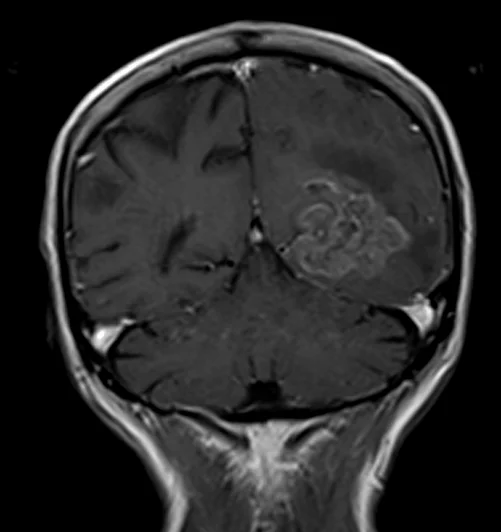
T1 post contrast of Glioblastomas
avid and diverse enhancement following gadolinium based contrast. reflects disruptions in the blood-brain barrier and increased vascular proliferation within the tumor.
T2 of Glioblastoma axial
T1 of Glioblastoma axial
DWI b0 of Glioblastoma
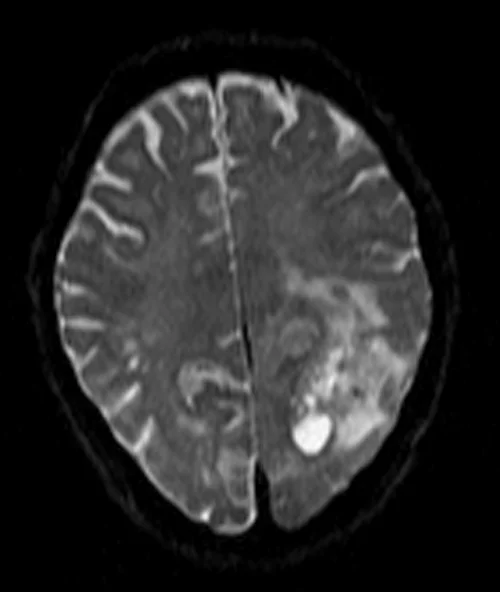
DWI b1000 of Glioblastoma
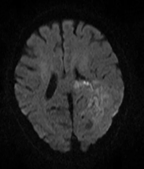
DW ADC of Glioblastoma
T1 post contrast of Glioblastoma coronal
T1 post contrast of Glioblastoma axial
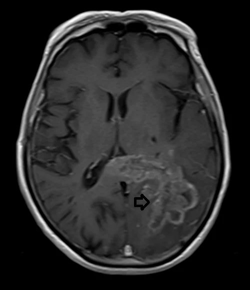
Acute MCA infarction (Stroke)
sudden neurological deficit that results from a cerebrovascular event. Middle cerebral artery is the largest cerebral artery and is most commonly affected by a cerebrovascular accident.