GABAergic Transmission
 Call Kai
 Learn
Learn Practice Test
 Spaced Repetition
 Match
Match Flashcards
 Knowt Play
1/37

There's no tags or description
Looks like no tags are added yet.
Name | Mastery | Learn | Test | Matching | Spaced | Call with Kai | Chat |
|---|
No analytics yet
Send a link to your students to track their progress
38 Terms
how diazapine works
- benzodiazepines enhance IPSPs, thereby making it even more difficult for the postsynaptic neuron to fire an action potential—this is clearly important for limiting the neuronal activity responsible for seizures.
amplifies inhibition

balacing excitation and inhibition
seizures are a good example of what happens when this balance is tipped in favor of excitation.
sedation and comas are examples of what happens when this balance is tipped in favor of inhibition
Gaba in the synaptic cleft
can go through reuptake, ionotropic and metabatropic receptors,

Gaba synthesis
γ-amino butyric acid (GABA) is the main inhibitory neurotransmitter in the brain.
GABA is actually a derivative of glutamate, and is synthesized from the decarboxylation of glutamate by glutamic acid decarboxylase (GAD).

GAD isoforms
- there are two isoforms of GAD—GAD65 and GAD67, which are derived from separate genes.
Glutamic acid decarboxylase (GAD)
the enzymes for synthesis are made in soma and transferred to to presynaptic terminal through axonal transport
localalized synthesis of GABA but not GAD

GAD67 v GAD65
- GAD67 is responsible for ~ 90% of the GABA production in the presynaptic terminal, whereas GAD65 is responsible for boosting GABA production when there is a high demand.
- palmitoylation of GAD65 targets it to synaptic vesicles, where it synthesizes GABA next to the transporter that loads the vesicle—GAD67 is thought to associate with synaptic vesicles through a different mechanism.
GAD staining
- because the GADs are only expressed in GABAegic synapses, they are a good marker of GABAergic (or inhibitory) neurons in the brain.
vesicular GABA transporter
- this is accomplished by the vesicular GABA transporter (vGAT), which uses the energy of the vesicle’s proton gradient to drive GABA into the vesicle against its concentration gradient.
- chloride ions are thought to be exchanged in order to preserve the charge balance, although the mechanism is unclear.

GABA recycling and degradation
- aside from binding to GABA receptors, GABA in the synaptic cleft is either transported back into the presynaptic terminal or neighboring astrocytes via GABA transporters (GAT).
- GABA can be converted into glutamate by GABA transaminase (GABA-T) and returned to the presynaptic neuron as glutamine

inhibitory post synaptic current
the reversal potential is below threshold
what ionic gradients have the potential to be inhibitory
Potassium and Chloride
how can we determine which ion mediates this IPSC?
- note that as there is no change in PSP when the membrane potential is held at the ECl (with a DC current injection)—this is consistent with an IPSC mediated by a chloride conductance.
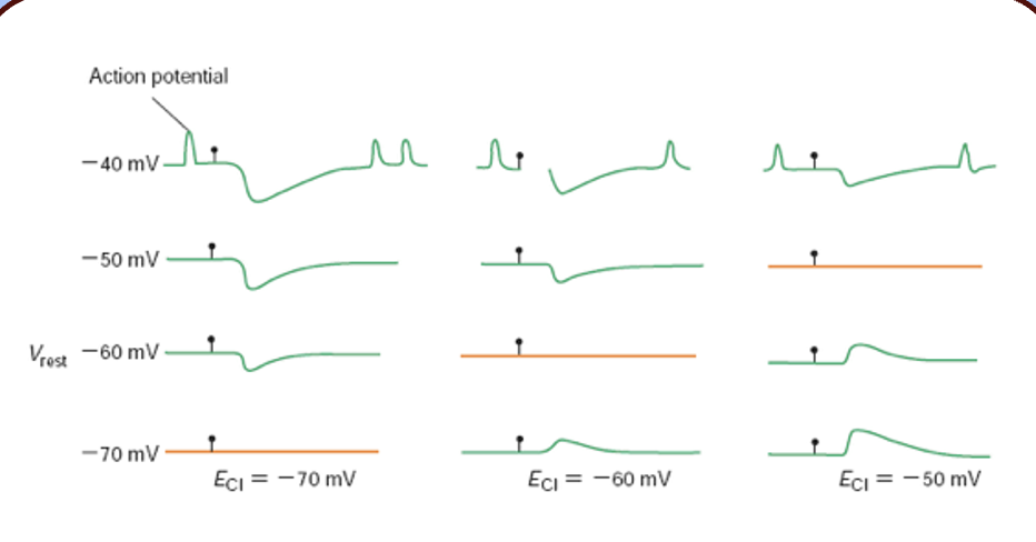
chloride-based IPSC is mediated by what
ionotropic GABA receptor called the GABAA receptor.
GABAa receptor
- the GABAAR is a pentameric channel, related to the nAChR, composed of a ligand binding domain and a transmembrane channel domain.

GABAaR subunit topology
- GABAA receptor subunits are four-pass transmembrane proteins (M1–4) with extracellular N- and C-termini.
- the N-terminus, which contains a cys-loop, forms the GABA binding site.
- the intracellular loop in between M3 and M4 is the site of intracellular regulation, mostly through phosphorylation.

GABAAR subunits
- although α, β, γ, δ, ρ, ε, θ, and π subunits have been described, most GABAARs are composed of two α subunits, two β subunits, and one of the other “modulatory” subunits (γ, δ, ε, and θ).
- ρ subunits tend to form homopentamers, and are considered a separate class of GABA receptor—the GABAC receptor.

most common species of GABAaR
is the 2α1,2β2,γ2 isoform.
alpha subunits of GABAaR
tend to form homopentamers, and are considered a separate class of GABA receptor—the GABAC receptor.
formation of the GABA receptor
- the GABAAR is formed when five subunits coalesce around a central axis to form a pore—these subunits are arranged so that their M2 segments line the channel’s pore.

binding GABA
- like its cousin the nAChR, the GABAA receptor binds its ligand (GABA) at the interfaces between the N-terminal ligand binding domains of its subunits.
- specifically, GABA binds to residues located in the α and β subunits—this is why there are always at least two α and β subunits in each receptor.
- this binding event is translated into the opening of the channel’s pore.

the GABAA receptor pore
- the M2 segment of the GABAA receptor lines the channel’s pore and contains residues responsible for chloride selectivity.
- the M2 segments of anion channels (the GABAAR and GlyR) are similar to their cation-passing cousins (the nAChR and 5-HT3α1) except that they have neutral residues at the -1 position—this region of the channel is thought to form a constriction that acts as a selectivity filter.

unitary GABAAR-mediated currents
in symmetrical chloride solutions, GABAAR-mediated currents reverse at 0 mV—the slope of the I–V plot reveals a single channel conductance of 30 pS.
- as the chloride concentration is changed to alter ECl, the reversal potential changes accordingly—this indicates that the GABAAR mediates a “pure” chloride conductance.

building the IPSC
the macroscopic IPSC is simple the summation of many unitary GABAAR-mediated currents.
here, we can see several spontaneous IPSCs, with one blown up on a much shorter time scale—note the discrete current steps.

the benzodiazepine binding site
however, benzodiazepines bind at the interface between the α and γ subunits.
furthermore, not all α subunits interact with benzodiazepines equally —only receptors containing α1,α2,α3, and α5 can be potentiated by benzodiazepines.
the effect of benzodiazepines
although they interact directly with GABAARs, benzodiazepines do not increase the single channel conductance or the dwell time in the open state.
instead, benzodiazepines increase the frequency that the channel opens in the presence of GABA—note that benzodiazepines don’t act as agonists, but instead potentiate the receptor’s response to GABA.

interneurons of the cortical microcircuit
inhibitory inputs, pyramidal cell, excitatory inputs

defining the interneurons
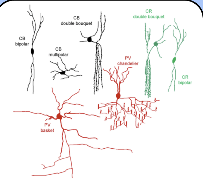
interneurons are commonly defined by the calcium-binding proteins that they express—calbindin (CB), calretinin (CR) and parvalbumin (PV).
interneurons are also categorized by their shape and the region of the postsynaptic cell that they innervate
variety of GABAergic interneurons of the neocortex
there is a tremendous variety of neurons in the brain—even a specific region such as the neocortex has many types of GABAergic interneurons.
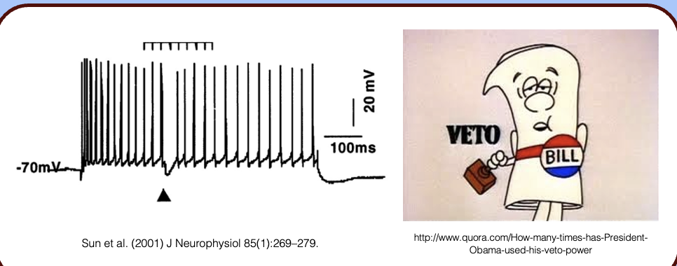
inhibitory veto power
- inhibitory synapses are often positioned so that they can override excitatory inputs—remember the synapses on the soma and AIS?
- in the above recording, an inhibitory neuron was stimulated (black triangle) while the neuron fired a train of action potentials—note the interruption in the train associated with stimulating the interneuron.
the symmetrical synapse
Gray’s type II, they lack the large postsynaptic density seen in glutamatergic synapses/excitatory synapses
also have pleomorphic or ‘flat’ synaptic vesicles
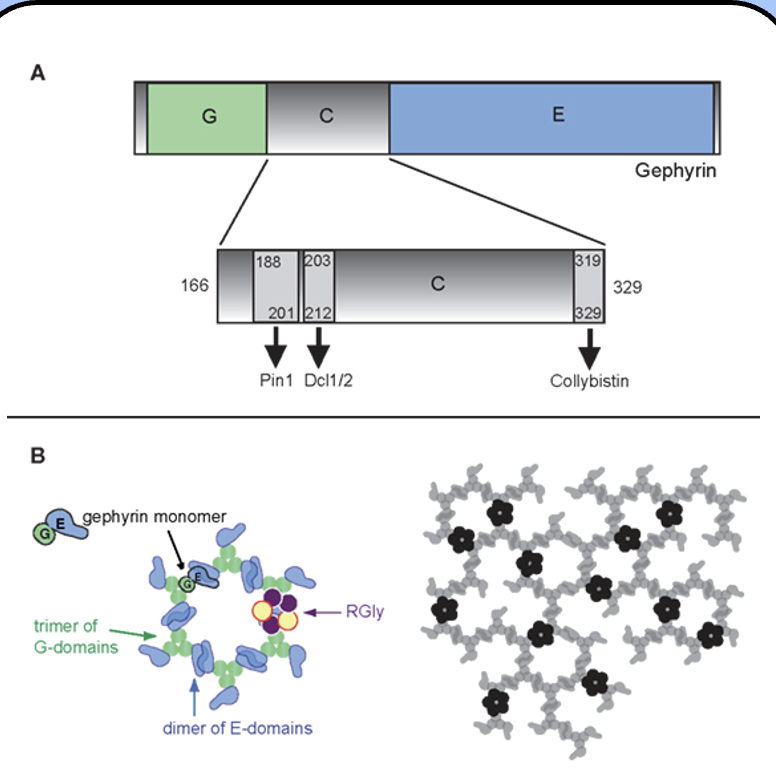
molecular organization of the inhibitory synapse
Gephyrin is the primary scaffolding protein

gephyrin
- the central scaffold of the GABAergic synapse (and glycinergic synapses)
- gephyrin forms a hexagonal, “honeycomb” lattice beneath the membrane that holds GABAA receptors in place, along with many other proteins.
types of inhibition
phasic and tonic inhibition
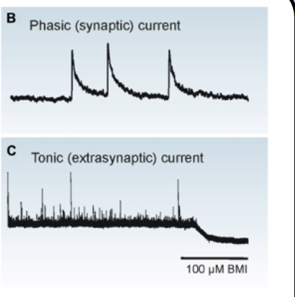
phasic inhibition
- the GABAA receptors bound to gephyrin across from the presynaptic terminal react to changes in the [GABA] due to synaptic release—this is called phasic inhibition.
tonic inhibition
there is a non-zero concnetration of ambient GABA present
- however, a separate, extrasynaptic population of GABAA receptors provides a steady inhibitory current due to ambient GABA in the extracellular space or spillover from the synapse—this is called tonic inhibition.
GAMMA subunit of GABA subunit
5th subunit (modulatory) of GABA subunit that allows benzodiazapine to bind
delta subunit of GAMMA receptor
associated with the uptake of ambient Gabba of tonic inhibition- ‘extra-synaptic GABA receptors’
