Male Reproductive System Grossing
 Call Kai
 Learn
Learn Practice Test
 Spaced Repetition
 Match
Match Flashcards
 Knowt Play
1/131

There's no tags or description
Looks like no tags are added yet.
Name | Mastery | Learn | Test | Matching | Spaced | Call with Kai | Chat |
|---|
No analytics yet
Send a link to your students to track their progress
132 Terms
What are the three segments of the male urethra?
Prostatic
Membranous (bulbomembranous)
Penile (spongy)

How does the epithelium of the urethra change along its length?
Proximal urethra — Urothelium
Distal urethra — Stratified squamous epithelium (SSE)
What are the two segments of the female urethra?
Anterior (distal 1/3)
Posterior (proximal 2/3)
What are the periurethral glands in males and females?
Male — Littré glands
Female — Skene glands
Both can be sites of periurethral gland adenocarcinoma
What is the most common overall urethral carcinoma?
Urothelial carcinoma
Commonly in the proximal urethra
Particularly in the:
Prostatic urethra
Bladder neck
What carcinoma is most common in the distal/penile urethra and female urethra?
Squamous cell carcinoma (SCC)
Where does urethral adenocarcinoma arise?
From the periurethral glands
Littré glands in males
Skene glands in females
What are other variants of tumor types of the male reproductive system?
Müllerian carcinoma
Neuroendocrine carcinoma
Sarcomatoid carcinoma
What types of urethral specimens may be received?
Biopsy/transurethral resection
Partial or total urethrectomy
Urethrectomy with radical cystectomy
Urethrectomy with radical cystoprostatectomy
Urethrectomy with penectomy
Anterior pelvic exenteration
What is the difference between partial and total penectomy?
Partial — Distal portion removed; shaft remains
Total— Entire penis removed
What should be documented when receiving a urethral specimen?
Nature of specimen received
Fresh vs. Fixative; What type?
Specimen orientation
Surgeon vs. anatomic landmarks vs. not orientated
Specimen integrity
Intact vs. disrupted
Ink key
Anatomic site of each ink color
NOTE: Also document uninvolved tissue/structures
What should be measured in a urethrectomy specimen?
Overall dimensions
3D, or length x diameter for a tubular segment
Measure every anatomic component present
Bladder wall / bladder neck
Prostate (lateral-lateral x apex-base x anterior-posterior), seminal vesicles, vasa deferentia
Corpus spongiosum, corpus cavernosum, periurethral muscle
Anterior vagina, rectum (female specimens)
What tumor characteristics should always be documented grossly?
Tumor site
Focality
Number of tumors
Size
Color/appearance
Configuration
Extent of invasion
What are possible gross appearances of urethral tumors?
Papillary
Polypoid
Verrucous
Ulcerated
Sessile
Pigmented
Solid/nodular
Flat
Infiltrative
What is important about taking gross photographs in urethral tumors?
They are:
Quick
Valuable
Assist in gross descriptions
What is the general progression of male urethral tumor invasion?
Subepithelial connective tissue → prostatic stroma → periprostatic fat → corpus spongiosum/periurethral muscle → corpus cavernosum → adjacent structures such as bladder wall or rectum
What makes up a urethrectomy with radical cystectomy?
Bladder with ureters
What makes up a urethrectomy with radical cystoprostatectomy?
Bladder with ureters + prostate with vas deferens and seminal vesicles
What are the two options of a urethrectomy with penectomy specimen?
Partial penectomy (distal portion removed, shaft intact)
Total penectomy (entire penis removed)
What makes up a anterior pelvic exenteration?
Bladder
Urethra (+ rectum)
In women:
Cervix
Vagina
Ovaries
Regional LNs
What structures should be evaluated for invasion in female urethral tumors?
Subepithelial connective tissue → Periurethral muscle (fibromuscular/adipose tissue) → Interior anterior vagina → Adjacent structures
(Document extent for every structure present in the specimen, even if UNINVOLVED)
What urethral margins are assessed?
Proximal margin
Distal margin
Soft tissue margins
(all three inked separately)
What should be reported regarding tumor and margins?
The distance from tumor to the closest margin
Each margin distinctly inked before sectioning
What is important to remember about lymph nodes in urethral specimens?
“No lymph nodes are identified” should still be documented
What should be recorded for identified lymph nodes?
Number examined
Number suspicious for tumor
Size
Site if known
Largest metastatic deposit
Macroscopic extranodal extension
What must be included in urethrectomy blocks/cassettes?
Proximal/distal margins
Tumor, especially deepest invasion
All uninvolved anatomic components
All identified lymph nodes
What are the major layers of the external penis from superficial to deep?
Skin → dartos fascia → Buck's fascia
What are the erectile bodies of the penis?
Two corpora cavernosa (dorsal)
Surrounded by the tunica albuginea
One corpus spongiosum (ventral)
Surrounds the urethra
What forms the glans?
The distal expansion of the corpus spongiosum
Cone-shaped
Where is Buck's fascia located?
Subjacent to the skin and dartos fascia
Superficial to the tunica albuginea
What is the most common non-neoplastic penile specimen?
Circumcision—removal of the foreskin
What is the most common malignancy of the penis?
Squamous cell carcinoma
What is the difference between partial and total penectomy?
Partial — Distal penis removed; shaft remains
Total — Entire penis removed, generally for proximal/extensive disease
What are some penile speciemens?
Incisional bx
Excisional bx
Circumcision
Partial penectomy
Total penectomy
How is foreskin length classified during circumcision grossing?
Short — Orifice between corona and coronal sulcus
Medium — Orifice between meatus and glans corona
Long — Orifice beyond the glans meatus, completely covers the glans
What measurements are taken on a circumcision specimen?
Length × width × thickness, OR
Length × diameter if received as a cylindrical cuff
What surfaces should be identified on a circumcision specimen?
Mucosal/inner surface
Cutaneous/outer skin surface
How should circumcision margins be inked?
Ink the:
Mucosal margin
Cutaneous margins
in different colors
What structures should be identified in a penectomy?
Shaft
Glans
Two corpora cavernosa
Corpus spongiosum
How should sections be taken for circumcisions?
Serially section perpendicular to the margins
Submit representative sections and all lesions
How should a penectomy specimen be measured?
Total specimen: length x diameter
Foreskin, if present: length x width x thickness
How should the foreskin be processed on a penectomy specimen?
Remove along the corona (if present)
Ink its resection margins on both sides
Mucosal side
Cutaneous side
What is the sectioning protocol for a penectomy?
Take a complete shave section of the proximal (amputation) margin
Bread-loaf the shaft with transverse sections
Proximal to distal
Open the urethra with scissors on the ventral surface
Use the opened urethra as a guide to bisect the glans longitudinally
Section the glans additionally, parallel to the midline, as needed
Document tumor size and depth of invasion
Why should friable penile tumors be handled carefully?
To avoid tissue loss and seeding of the grossing field
What is the progression of tumor invasion through the glans?
Lamina propria → corpus spongiosum → tunica albuginea → corpus cavernosum
What is the progression of tumor invasion through the foreskin?
Lamina propria → dartos → dermis → epidermis
What is the progression of tumor invasion through the penile shaft/body?
Dermis → dartos → Buck's fascia → tunica albuginea → corpus cavernosum
What structures can penile cancer invade beyond the penis?
Regional skin such as the pubis/inguinal region
Adjacent structures including:
Scrotum
Prostate
Pubic bone
What are the important penectomy margins?
Urethral margin
Periurethral tissues (lamina propria, corpus spongiosum, Buck’s fascia) margin
Corpus cavernosum margin
Skin margin
What are the important circumcision margins?
Coronal sulcus mucosal margin
Cutaneous margin
What should be reported for each penile margin?
Whether involved/uninvolved
Distance to the closest margin
What are the major lymph node groups relevant to penile cancer?
Sentinel nodes
Inguinal nodes
Pelvic/regional nodes
What is the general progression of penile lymphatic drainage?
Superficial inguinal → deep inguinal → pelvic nodes
What is this?

Penile SCC
What is the tunica albuginea?
A thick white fibrous capsule surrounding the testis
What is the tunica vaginalis?
A mesothelial-lined sac with visceral and parietal layers surrounding the tunica albuginea
Where is the epididymis located?
Posteriorly
Continuity with the spermatic cord
What is the rete testis?
A network at the testicular hilum connecting seminiferous tubules to the epididymis
A common route of extratesticular spread
What are the two major prognostic groups of testicular germ cell tumors?
Seminoma
Non-seminomatous germ cell tumor (NSGCT)
What is the classic presentation of a testicular germ cell tumor?
Painless enlargement of the testis
What is the characteristic lymphatic route of testicular cancer spread?
Retroperitoneal lymph nodes
Para-aortic lymph nodes (characteristic)
What are common pattern of spread in testicular cancers?
Lymphatics
Hematogenous
What is the primary hematogenous metastatic site for testicular germ cell tumors
Lungs
Additional sites including:
Liver
Brain
Bone
Which tumor marker is associated with choriocarcinoma?
β-hCG
Can also be elevated in some seminomas because of syncytiotrophoblasts
Which tumor marker is associated with yolk sac tumor?
AFP (alpha-fetoprotein)
What does LDH indicate in testicular germ cell tumors?
It is less specific than AFP or hCG
Correlates with:
Tumor burden
Stage
Treatment response
When should testicular tumor markers be obtained/trended?
Before and after orchiectomy to assess treatment response
What is the primary operation for a suspected testicular mass?
Radical orchiectomy
Through an inguinal approach
Is biopsy performed for a suspected testicular tumor?
No — Biopsy is performed only for infertility evaluation and never for a suspected tumor
When are lymphadenectomy specimens received?
Received for staging/post-chemotherapy resection
Retroperitoneal LN dissections
What should always be documented for an orchiectomy?
Laterality
Right, left, or not specified
When should the testis be weighed?
Before fixation
What dimensions are taken in an orchiectomy?
Testis: 3 dimensions
Epididymis: 3 dimensions
Spermatic cord: length × diameter
Why should the surgeon not bisect the testis before it reaches pathology?
It can obscure tunical invasion
It can compromise tissue needed for flow cytometry/karyotyping
If possible, when should testis specimens be grossed?
Fresh!
Prevents poor fixation
If delayed, fix in 10% buffered formalin
What is the overview for measurement and triage of testis specimens?
Weigh (prior to fixation)
Record dimensions
Asses integrity
Gross fresh whenever possible
How should the testis be sectioned after inking?
Bisect sagittally from the anterior aspect
What is the critical first step involving the spermatic cord?
Shave the spermatic cord margin before anything else
Then continue transverse sections along the cord first
What structure must be confirmed in the spermatic cord margin?
The vas deferens
A thin, firm, white tube and may retract
How should testis specimens be inked?
Ink per institutional protocol
Could be:
Tunica AND spermatic cord, OR
Tunica only where adhesions are found
What structures should be evaluated for testicular tumor extension?
Testis
Rete testis
Hilar soft tissue
Epididymis
Tunica albuginea/vaginalis
Spermatic cord
Scrotum
(Note any fluid within the TA sac)
What is the primary margin of interest in an orchiectomy?
The spermatic cord margin
What additional testicular margins may be evaluated?
Parietal tunica vaginalis margin
Scrotal skin margin
How should uninvolved structures be stated for testicular specimens?
Testis
Unremarkable, presence/absence of fine tubules, soft, fibrotic, hemorrhagic, infarcted, ischemic, ruptured
Epididymis
Unremarkable or describe abnormality
Tunica albuginea
Unremarkable or describe abnormality
Where are the major retroperitoneal lymph nodes relevant to testicular cancer?
Interaortocaval
Para-aortic
Paracaval
Preaortic
Precaval
Retroaortic
Retrocaval
What can post-chemotherapy retroperitoneal lymph nodes look like grossly?
They may be hemorrhagic, cystic, or necrotic
What is this?

Testis — Sebaceous cyst
What is this?
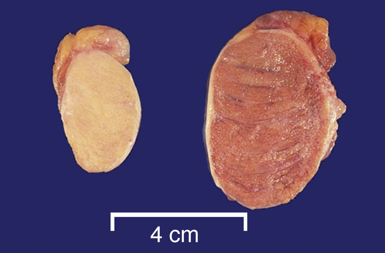
Testis — Atrophic

What is this?
Testis — Torsion
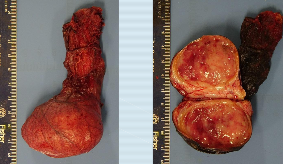
What is this?

Testis — Seminoma
What is this?

Testis — Embryonal carcinoma
What is this?
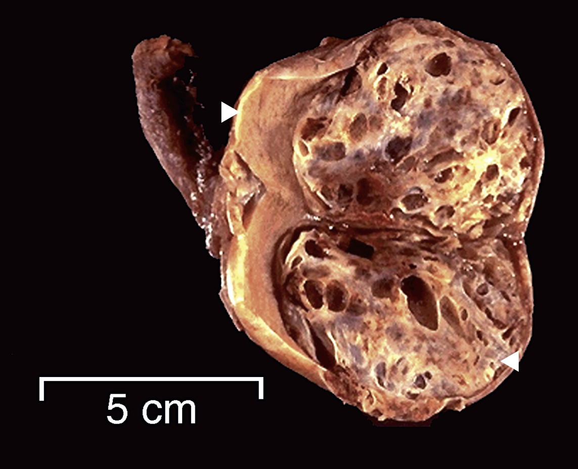
Testis — Embryonal carcinoma w/ teratoma
What is this?

Testis — Teratoma
What are the indications for prostate needle core biopsy?
Elevated PSA or a palpable/imaging-identified nodule
What is the purpose of a TURP?
To relieve obstruction from benign prostatic hyperplasia (BPH)
Carcinoma can be an incidental finding
What is the purpose of radical prostatectomy?
Definitive treatment for clinically localized adenocarcinoma
What is the purpose of a suprapubic prostatectomy/enucleation (simple or subtotal)?
To remove a large gland causing obstructive symptoms; benign disease
What measurements should be taken on a radical prostatectomy?
Overall prostate dimensions and weight
Right/left prostate gland — medial-lateral (R-L) x anterior-posterior x superior-inferior
Right/left vas deferens length × diameter
Right/left seminal vesicles in 3D
Extraprostatic soft tissue and its location
What landmarks must be identified before inking the prostate?
Apex
Base
Anterior
Posterior
Right
Left
How is each side (R/L) of the prostate divided in the 12-zone system?
Each side has six zones:
Anterior base
Anterior mid
Anterior apex
Posterior base
Posterior mid
Posterior apex