cell biology unit 2 ; microscopy, bacterial division, cell division and stem cells
1/27
There's no tags or description
Looks like no tags are added yet.
Name | Mastery | Learn | Test | Matching | Spaced | Call with Kai |
|---|
No analytics yet
Send a link to your students to track their progress
28 Terms
microscope
an instrument used to see things too small to be seen with the naked eye by magnification
magnification
resolution
how many times bigger the image is compared to the size of the real object
the ability to distinguish between two points ( higher resolution = sharper and detailed img)
how microscopy techniques have developed over time
techniques have developed over time as technology and knowledge improved - over the years light microscopes have been used a lot to study cells, allowed us to make important discoveries about the structures inside cells
Throughout their development, the magnification of light microscopes has increased, but very high magnifications are not possible. The maximum magnification with a light microscope is around ×1500.
this lead to the development of the electron microscope which uses a beam of electrons instead of light rays
limits of the light microscope
light microscopes have a limited magnification e.g. structures inside the nucleus wouldnt be easily observable with a light m. scope
limited resolution even if the image was magnified the image would still be blurred and fine detail wouldnt be able to be seen
so if scientists wanted to look at structures inside cells in detail then a light microscope was not that useful - so they invented the electron microscope
explain how electron microscopy has increased understanding of
sub-cellular structures.
An electron microscope has much higher magnification and resolving power than a light microscope. This means that it can be used to study cells in much finer detail. This has enabled biologists to see and understand many more sub-cellular structures.
adv and disadv. of (1.2.) light microscopes and (3.4.) electron microscopes
1. inexpensive, easy to use doesnt require training, can observe both living and dead specimens, can observe in colour
2. limited magnification and resolution
3. much greater magnification and resolution
4. large so less portable, only observe dead specimens, black and white, requires training to use
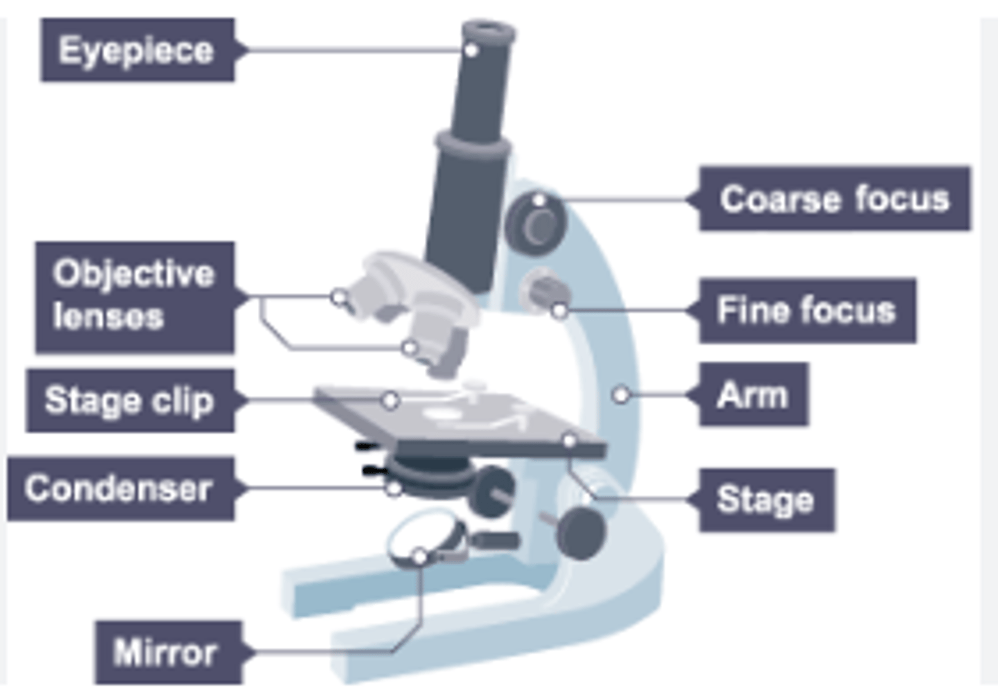
labelled light microscope
the centre of the microscope has a stage where we place the m. scope slide - stage has clips to hold the slide in place
- below the stage there is a lamp - light from the lamp passes up through the microscope slide
- the lenses above the stage are objective lenses (usually have a magnification of 4x 10x or 40x) -
top of the microscope is the eyepiece where we look through - eyepiece contains the eyepiece lens which has a magnification of 10x -
then coarse focusing dial and fine focusing dial
how to use an optical m. scope to view a prepared slide (steps 1 and 2)
1.) place the slide onto the stage and use the clips the hold the slide in place
2.) select the lowest power objective lens slowly turning the coarse focusing dial to position the objective lens so it almost touches the microscope slide - don't look from the eyepiece while you are doing this as if we look from it lens then there is a risk of damaging the slide
how to use an optical m. scope to view a prepared slide (steps 3,4,5)
3.) look down the eyepiece and , turn the coarse focusing dial to slowly move the stage away from the objective lens until the cells are roughly into focus
4.) then turn the fine focusing dial to bring the cells into a clear focus
5.) if a higher power magnification is needed, make sure there is enough distance and select higher powered objective lens and refocus
total magnification and magnification formulae
eyepiece lens mag. X objective lens mag. = (total magnification) have to be same unit
magnification = img size/ real size
processes needed for preparing a slide
thin layer of tissue to help see individual cells and to allow light to penetrate
iodine to stain to see parts of the cell more clearly and to highlight objects in a cell by adding cololur
cover slip is lowered at a tilted angle to prevent and reduce the number of air bubbles forming
drawing observations of. microscopy
no shading or colouring, structures should be drawn in proportion to other structures , include magnification and title , should take at least half of the space available include title no arrows and in pencil
mag scale - place clear plastic ruler over the stage and measure the diameter of the FOV in mm and draw a scale bar.
bacterial division and growth and why uncontaminated cultures of microorganisms are required
Bacteria multiply by simple cell division (binary fission) as often as once every 20 minutes if they have enough nutrients (and water) and suitable temperature.
Bacteria can be grown in a nutrient broth solution or as colonies on an agar gel plate.
Uncontaminated cultures of microorganisms are required for
investigating the action of disinfectants and antibiotics.
calculate population of bacteria after a given time
2^n (n = no of divisions) divisions if a bacteria can multiply once every 20 min - undergo simple cell division - binary fission
aseptic techniques for preparing an uncontaminated culture
Petri dishes and culture media must be sterilised before use
• inoculating loops used to transfer microorganisms to the media must be sterilised by passing them through a flame
• the lid of the Petri dish should be secured with adhesive tape and stored upside down
• in school laboratories, cultures should generally be incubated at 25°C.
effects of aseptic techniques
sterilisation - kills any unwanted microorganisms and prevents contamination
tape - stops the lid from falling off and unwanted microorganisms from entering
storing upside down - stops moisture from dripping down onto the bacteria and disrupting the colonies and stops MGs in the air from entering
incubation @ 25°C - reduces the risk that harmful bacteria will grow
required practical - effect of antibiotics on the growth of bacteria (steps 1,2,3)
1.Clean the bench with disinfectant solution.
This kills microorganisms that could contaminate our culture.
2. Sterilise an inoculating loop by passing it through a Bunsen burner flame.
3. Open a sterile agar gel plate near a Bunsen burner flame. The flame kills bacteria in the air.
required practical - effect of antibiotics on the growth of bacteria (steps 4,5,6)
4. Now use the loop to spread the chosen bacteria evenly over the plate.
5. Place sterile filter paper discs containing antibiotic onto the plate.
6. Incubate the plate at 25°C.
inhibition zone
Around the antibiotic discs, we have a region where the bacteria have not grown. This is called the zone of inhibition.
bigger inhib zone = better effectiveness of the antibiotic
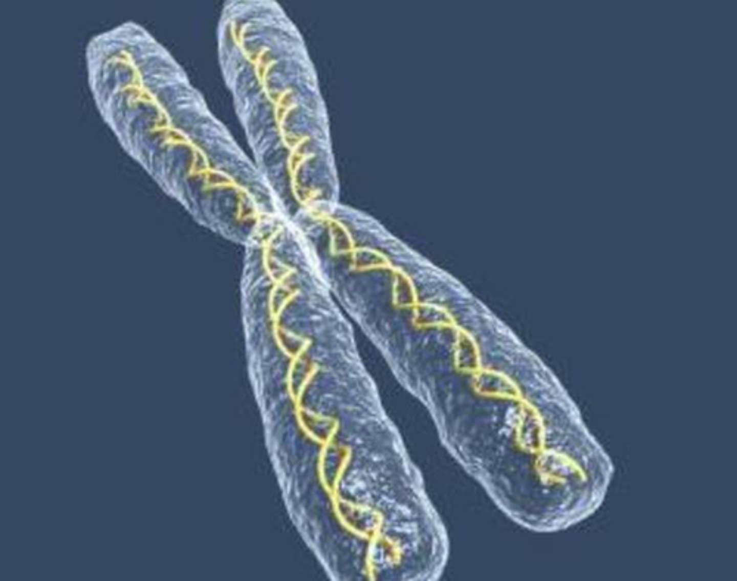
chromosomes
in the nucleus of cells there are structures called chromosomes which are made up of DNA molecules, chromosomes carries a large number of genes - short sections of DNA that determine many of our features. In body cells, there are two of each chromosome (23 pairs) (just not in gametes where there are only 23 single chromosomes) - most human cells contain 100's of chromosomes
Cells divide in a series of stages called the cell cycle.
the cell cycle - consists of growth and DNA replication and then mitosis
During the cell cycle the genetic material is doubled and then divided
into two identical cells.
Before a cell can divide it needs to grow and increase the number of sub-cellular structures such as ribosomes and mitochondria. The DNA replicates to form two copies of each chromosome - growth and DNA replication
In mitosis one set of chromosomes is pulled to each end of the cell and the nucleus divides. Finally the cytoplasm and cell membranes divide to form two identical cells.
functions and importance of mitosis
1. Mitosis is essential for growth and development of multicellular organisms (eg plants and animals).
2. Mitosis takes place when an organism repairs itself (eg when a broken bone heals).
3. Mitosis happens during asexual reproduction.
stem cell
A stem cell is an undifferentiated cell of an organism which is capable of giving rise to many more cells of the same type, and from which certain other cells can arise from differentiation.
human embryonic stem cells
humans start when a sperm cell joins with an ovum (fertilisation). The fertilised ovum now undergoes mitosis and forms a ball of cells called an embryo. overtime these cells continue to undergo mitosis and also differentiate into specialised cells
Cells in the early stage embryo have not differentiated. Any cell is capable of differentiating into any type of body cell - why they are stem cells
stem cells in adult organisms bone marrow and bone marrow transplant
contains stem cells that can only differentiate to blood cells (RBC, WBC, Platelets) not other cell types
Leukaemia is a cancer of the bone marrow.
To treat this, first the patient's existing bone marrow is destroyed using radiation.
The patient then recieves a transplant of bone marrow from a donor.
The stem cells in the bone marrow now divide and form new bone marrow. They also differentiate and form blood cells.
problems with bone marrow transplant
The donor has to be compatible with the patient. Otherwise the white blood cells produced by the donated bone marrow could attack the patient's body.
There is a risk that viruses can be passed from the donor to the patient.
Therapeutic cloning
In therapeutic cloning, an embryo is produced with the same genes as the patient.
Stem cells from the embryo can be transplanted into the patient without being rejected by the patient's immune system.
Once inside the patient, the stem cells can then differentiate to replace cells which have stopped working correctly.
This technique could be useful for a range of medical conditions such as diabetes or paralysis.
however there are ethical or religious objections to this procedure
plant stem cells
Roots and buds contain meristem tissue. These stem cells can differentiate into any type of plant tissue, at any point in the life of the plant.
Stem cells from meristems in plants can be used to produce clones of plants quickly and economically.
• Rare species can be cloned to protect from extinction.
• Crop plants with special features such as disease resistance can be cloned to produce large numbers of identical plants for farmers.